. 2019 Jul 23;116(30):15042-15051.
doi: 10.1073/pnas.1901318116. Epub 2019 Jul 9.

Structure of KAP1 tripartite motif identifies molecular interfaces required for retroelement silencing

Affiliations

Structure of KAP1 tripartite motif identifies molecular interfaces required for retroelement silencing

Guido A Stoll et al. Proc Natl Acad Sci U S A. .

Abstract

Transcription of transposable elements is tightly regulated to prevent genome damage. KRAB domain-containing zinc finger proteins (KRAB-ZFPs) and KRAB-associated protein 1 (KAP1/TRIM28) play a key role in regulating retrotransposons. KRAB-ZFPs recognize specific retrotransposon sequences and recruit KAP1, inducing the assembly of an epigenetic silencing complex, with chromatin remodeling activities that repress transcription of the targeted retrotransposon and adjacent genes. Our biophysical and structural data show that the tripartite motif (TRIM) of KAP1 forms antiparallel dimers, which further assemble into tetramers and higher-order oligomers in a concentration-dependent manner. Structure-based mutations in the B-box 1 domain prevent higher-order oligomerization without significant loss of retrotransposon silencing activity, indicating that, in contrast to other TRIM-family proteins, self-assembly is not essential for KAP1 function. The crystal structure of the KAP1 TRIM dimer identifies the KRAB domain binding site in the coiled-coil domain near the dyad. Mutations at this site abolished KRAB binding and transcriptional silencing activity of KAP1. This work identifies the interaction interfaces in the KAP1 TRIM responsible for self-association and KRAB binding and establishes their role in retrotransposon silencing.

Keywords: endogenous retrovirus; epigenetic silencing; transcriptional repressor; transposable element; ubiquitin E3 ligase.


Conflict of interest statement

The authors declare no conflict of interest.

Figures

Fig. 1.
Fig. 1.
Self-assembly in solution of KAP1 RBCC and its subcomponent domains. (A) Domain organization of KAP1. B1, B-box 1; B2, B-box 2. (BD) SEC-MALS data for the RING (B), RING-B-box 1 (C), and RBCC (D). (E) SE-AUC analysis of RBCC molecular weight as a function of protein concentration. The average molecular weight isotherm from individual fits at different concentrations was fitted to an isodesmic self-association model (black line) yielding dissociation constants of Kd2,4 = 9 µM [with a 1-σ (68.3%) CI of 7–11 µM] and Kdiso = 19 µM [1-σ CI, 16–23 µM] for the dimer–tetramer and isodesmic equilibria, respectively. An alternative fit to a dimer–tetramer–octamer model (gray line) yielded dissociation constants of Kd2,4 = 12.6 µM [1-σ CI, 7.4–23.5 µM] and Kd4,8 = 6 µM [1-σ CI, 3–11 µM] for the dimer–tetramer and tetramer–octamer equilibria, respectively. (F) SEC-MALS of full-length KAP1.
Fig. 2.
Fig. 2.
Crystal structure of KAP1 RBCC. (A) Domain organization of the crystallized construct. B1, B-box 1; B2, B-box 2; CC, coiled-coil; T4L, T4 lysozyme. (B) Overall structure of the RBCC homodimer. Three views along or perpendicular to the dyad are shown. The components are colored as in A. Zn atoms are shown as magenta spheres. (C) View of the RBCC dimer perpendicular to the dyad, with 1 subunit shown as a cartoon and the other as a surface.
Fig. 3.
Fig. 3.
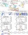
Self-assembly properties and KRAB binding activity of KAP1 RBCC mutants. (A) Positions of the mutations in the RING and B-box 2 domains. A reference RBCC dimer is colored as in Fig. 2. Adjacent RBCC dimers forming crystal packing contacts are shown in gray with their residue numbers followed by an asterisk. (B) Model of a KAP1/TRIM28 B-box 1 dimer based on the TRIM19 B-box 1 dimer structure (29) with selected residues forming dimer contacts shown. An alignment of B-box 1 sequences (Right), the TRIM28 B-box 1 model, and the TRIM19 B-box 1 structure were used to identify mutations in KAP1 B-box 1 likely to disrupt dimer contacts. Residues known or predicted to participate in dimer contacts are shown in bold typeface in the sequence alignment. (CF) SEC-MALS data for (C) RBCC with RING domain mutations, (D) RBCC with the B-box 2 mutations, (E) RING-B-box 1 with B-box 1 mutations, (F) and RBCC (black curve, WT; red curve, B-box 1 mutant A160D/T163A/E175R). (G) Model for oligomerization of KAP1 via B-box 1.
Fig. 4.
Fig. 4.
Transcriptional silencing assays with KAP1 mutants. Data are presented as fold repression of reporter luciferase luminescence in KAP1 KO HEK293T cells transfected with a KAP1 variant. (A) SVA reporter repression with the oligomerization-deficient B-box 1 mutant (B1mut), the KRAB binding-deficient coiled-coil mutant (CCmut), an HP1-box mutant (HP1mut), and a PHD mutant (PHDmut). (B) LINE-1 reporter repression with the same set of mutants as in A. Data were normalized to KAP1 KO cells transfected with an empty vector (EV). Error bars represent SEM between measurements (n = 3). Statistical significance was assigned as follows: not significant (n.s.), P > 0.05; *, 0.05 > P > 0.01. (Lower) Western blots of cell lysates from WT or KAP1 KO HEK293T cells transfected with each of the variants (WT, CC, B1, HP1, PHD) or empty vector (−).
Fig. 5.
Fig. 5.
Formation of a 2:1 KAP1:KRAB complex and identification of KRAB binding residues in the KAP1 coiled-coil domain required for silencing. (A) SEC-MALS of full-length KAP1 bound to ZNF93 MBP-KRAB. The expected molecular weights of a KAP1 dimer and for 2:1 and 2:2 KAP1:KRAB complexes are indicated with dashed lines. The total protein concentration of each analyte was 1.2 g L−1. (B) Close-up of the cluster of solvent-exposed hydrophobic residues near the dyad. The variant V293S/K296A/M297A/L300S (CC mutant) was generated to test for KRAB binding. (C) Pulldown KAP1-KRAB binding assay. KAP1 RBCC was incubated with Twin-StrepII-MBP-ZNF93 KRAB, and the mixture was loaded on Strep-Tactin Sepharose. Bound proteins were detected by SDS/PAGE/Coomassie. (D) SPR KAP1-KRAB binding assay. MBP-KRAB was immobilized on the chip. WT or CC mutant KAP1 RBCC were flowed over the chip. Binding kinetics of WT RBCC: kon = 3.6 ± 0.96 × 10⁴ M−1 s−1; koff = 2.7 ± 0.1 × 10⁻⁴ s−1. (E) Model for binding of KAP1 dimers to KRAB-ZFPs.

References

    1. Friedli M., Trono D., The developmental control of transposable elements and the evolution of higher species. Annu. Rev. Cell Dev. Biol. 31, 429–451 (2015). - PubMed
    1. Goodier J. L., Restricting retrotransposons: A review. Mob. DNA 7, 16 (2016). - PMC - PubMed
    1. Chuong E. B., Elde N. C., Feschotte C., Regulatory evolution of innate immunity through co-option of endogenous retroviruses. Science 351, 1083–1087 (2016). - PMC - PubMed
    1. Zhou L., et al. , Transposition of hAT elements links transposable elements and V(D)J recombination. Nature 432, 995–1001 (2004). - PubMed
    1. Dupressoir A., Lavialle C., Heidmann T., From ancestral infectious retroviruses to bona fide cellular genes: Role of the captured syncytins in placentation. Placenta 33, 663–671 (2012). - PubMed

Publication types

MeSH terms

Associated data