

Multifocal electroretinogram findings in sickle cell maculopathy
- PMID: 31289356
- PMCID: PMC7002728
- DOI: 10.1038/s41433-019-0499-7
Multifocal electroretinogram findings in sickle cell maculopathy
Abstract
Background: The aim of the present work was to describe and compare multifocal electroretinogram findings (mfERG) between patients with sickle cell disease (SCD) without clinical sign of maculopathy and controls (HbAA).
Methods: Both HbSS (homozygous SCD) and HbSC (compound heterozygous SCD) patients, the two most frequent SCD genotypes, were included. All individuals underwent a full ophthalmologic examination (with a fundoscopy), a spectral domain ocular coherence tomography (SD-OCT) and a mfERG.
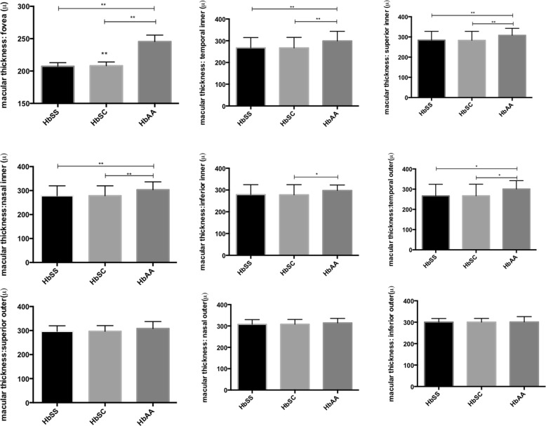
Results: A total of 86 subjects were included: 54 SCD patients (107 eyes) with 32 HbSS (63 eyes) and 22 HbSC (44 eyes) and 32 controls (64 eyes). None of the eyes showed retinal clinical abnormalities. SD-OCT analysis showed that macular thickness was statistically lower in SCD eyes than in controls. mfERG analysis demonstrated a significant reduction of N1 (initial-negative deflection), and P1 (positive peak) response amplitude densities of HbSS eyes compared to HbAA eyes from the centre (<2°) and to the periphery (>15°). Implicit time response was also reduced in the centre (<2°). N1 and P1 response amplitude densities of HbSC eyes were significantly lower than those of HbAA eyes from the centre (<2°) to the periphery (>15°). N1 implicit time was statistically reduced in HbSS compared to HbSC eyes.
Conclusion: Our study is the first one to describe macular electrophysiological dysfunction in SCD patients. Moreover, we confirm that SCD maculopathy is equally frequent in HbSS and HbSC.
Conflict of interest statement
The authors declare that they have no conflict of interest.
Figures
References
-
- Connes P, Renoux C, Romana M, Abkarian M, Joly P, Martin C, et al. Blood rheological abnormalities in sickle cell anemia. Connes P, editor. Clin Hemorheol Microcirc. 2018;68:165–72. - PubMed
Publication types
MeSH terms
LinkOut - more resources
Full Text Sources
Medical
