Review
. 2019 Sep;20(9):514-532.
doi: 10.1038/s41583-019-0197-2.

Optical control of neuronal ion channels and receptors

Affiliations
Review

Optical control of neuronal ion channels and receptors

Pierre Paoletti et al. Nat Rev Neurosci. 2019 Sep.

Abstract

Light-controllable tools provide powerful means to manipulate and interrogate brain function with relatively low invasiveness and high spatiotemporal precision. Although optogenetic approaches permit neuronal excitation or inhibition at the network level, other technologies, such as optopharmacology (also known as photopharmacology) have emerged that provide molecular-level control by endowing light sensitivity to endogenous biomolecules. In this Review, we discuss the challenges and opportunities of photocontrolling native neuronal signalling pathways, focusing on ion channels and neurotransmitter receptors. We describe existing strategies for rendering receptors and channels light sensitive and provide an overview of the neuroscientific insights gained from such approaches. At the crossroads of chemistry, protein engineering and neuroscience, optopharmacology offers great potential for understanding the molecular basis of brain function and behaviour, with promises for future therapeutics.


Figures

Fig. 1 |:
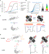
Fig. 1 |:. Main strategies to endow light-sensitivity to neuronal receptors and ion channels.
a | Chemical approaches are based exclusively on modified ligands that are rendered responsive to light (hν). No modification of the protein target is required. With caged compounds, light triggers the release of biologically-active ligands. Photoswitchable ligands reversibly alternate between active and inactive forms using two different wavelengths of light. Photolabels bind a target receptor and upon light irradiation from a covalent link with the protein binding site. Contrasting with photoswitchable ligands, caged compounds and photolabels act irreversibly. b |Genetic approaches are based on the insertion of genetically encoded light-sensitive protein modules in the target protein of interest. Color coding: light and dark grey represent two different states (e.g. inactive and active) of the protein. Light-sensitivity is endowed by photosensitive co-factors (such as retinal or flavin) that are endogenously present in mammalian cells. Opsin chimeras consist in the fusion between a light-sensitive opsin (grey) and a light-insensitive G-protein coupled neurotransmitter receptor (black). Illumination triggers the conversion from 11-cis to 11-trans retinal, which causes conformational change in the fusion protein and its activation. In LOV-domain chimeras, light triggers unfolding of the LOV domain, which directly or indirectly modulates protein function. In CALI, light activates the flavin chromophore and generates reactive oxygen species, which results in irreversible inactivation of nearby proteins (shown in white with dashed borders). c | Hybrid or chemogenetic approaches are based on a photosensitive synthetic chemical and its genetic attachment to or incorporation within the target protein. Different types of light-sensitive unnatural amino-acids (UAAs) can be incorporated into proteins: caged, photocrosslinking (i.e. photolabels), or photoswitchable (i.e. alternating between two configurations upon illumination with different wavelenghts) as depicted. Photoswitchable tethered ligands can be covalently attached to receptors in two ways: either to a cysteine-substituted site usually through maleimide-sulfhydryl chemistry (as depicted), or to a self-labeling protein tag (not depicted), resulting in both cases in reversible control of protein activity. In nanotweezers, a bis-maleimide photoswitch bridges two cysteine mutants. Light-induced conformational changes of the photosensitive moiety exert mechanical forces on the protein, potentially triggering its activation in the absence of ligand. Note that caged compounds, photolabels and CALI are unidirectional while photoswitches (both synthetic and natural) allow for bidirectionality.
Fig. 2 |
Fig. 2 |. Photocontrol of ion channel and receptor biophysics and pharmacology.
a | With appropriate optopharmacological tools, light can be used to directly activate receptors or ion channels (agonist), to inhibit them (antagonist or negative allosteric modulator (NAM)), or to positively modulate their function (positive allosteric modulator (PAM)). b | Activity of the target receptor or ion channel can be adjusted in a graded manner using different light intensity and/or wavelength. The graph illustrates various levels of photo-antagonism associated with changes in light intensity or wavelength. c | Photomodulation of NMDA receptors (NMDARs) using photoswitchable amino acids (PSAAs). Left: Chemical structures of trans and cis PSAA in the context of a protein. The azobenzene moiety is highlighted in shaded grey. Right, upper part: Schematic representation of the mechanism of NMDAR photomodulation. The PSAA is incorporated in the GluN1 subunit (dark grey), close to the GluN2 subunit (light grey). GluN1 binds glycine (orange), whereas GluN2 binds glutamate (red). Right, lower part: Illumination with 365 nm light isomerizes PSAA to cis, a conformational change sufficient to destabilize glycine binding (that is, leading to a decrease in glycine affinity). This change results in a reduction of current amplitude during agonist applications (Glu, glutamate; Gly, glycine) (left trace) and in an acceleration in deactivation kinetics upon glycine washout (right trace; current normalized). I, current amplitude; Inorm., normalized current amplitude; t, time. d | Photocontrol of kainate receptors using photoswitchable tethered ligands (PTLs). Left: Chemical structures of the PTL maleimide azobenzene glutamate (MAG) in the trans and cis configurations. The azobenzene moiety is highlighted in shaded grey. Right, upper part: Schematic representation of heteromeric GluK2/K5 kainate receptors. MAG is covalently attached to an engineered cysteine residue on the GluK2 subunit (light grey). Under 380 nm light, MAG adopts its cis configuration allowing the glutamate moiety to dock in the agonist binding pocket. Right, lower part: Heteromeric kainate receptors conjugated with two PTLs can be directly activated with 380 nm light which isomerizes the photoswitch to cis, and deactivated with 500 nm light which reverts the photoswitch to trans (left trace). In such conditions, no or little desensitization is observed. In contrast, when two agonists (pale red) are pre-bound selectively to GluK5 subunits, photoswitching leads to full receptor occupancy and almost complete receptor desensitization (right trace). Part c is adapted from ref.. Part d is adapted from ref..
Figure 3:
Figure 3:. Optopharmacology for subcellular neuronal studies.
a. Functional receptor mapping. Left: Chemical structure of MNI glutamate and photorelease of glutamate following either one-photon (1P) or two-photon (2P) illumination. Right: Two-photon uncaging of glutamate at different spots along the dendrite for functional glutamate receptor mapping. Receptor activity is revealed using whole-cell patch-clamp recordings in voltage-clamp mode. I, current amplitude; t, time. b. Compartment-specific blockade of HCN channels using the photoswitch AAQ. Left: Chemical structures of trans and cis AAQ. Middle: Schematic representation of photoreversible HCN blockade using AAQ. Right: Probability of spiking is increased when HCN channels are blocked in the axon initial segment (AIS), but probability of spiking is decreased when these channels are blocked in the soma. Neuronal excitability is measured using whole-cell patch-clamp recordings in current-clamp mode. Vm, membrane potential; t, time. c. Single-spine structural LTP induced using two-photon (2P) uncaging of glutamate or one-photon (1P) activation of light-controllable NMDA receptors (LiGluN) expressed in transfected neurons (green). The left and middle cartoons show how illumination leads to LiGluN activation, while the right cartoon depicts the resulting long-lasting changes in dendritic spine morphology. The graph on the right illustrates the evolution of the spine volume as a function of time before and after LTP induction (red arrow). Part a is adapted from ref.. Part b is adapted from ref.. Part c is adapted from ref. and ref..
Figure 4:
Figure 4:. Optopharmacology for cell- and receptor-specific interrogation of synaptic physiology.
In all panels, the cell selectively expressing the photosensitized receptors is shown in green. a | Optical control of neurotransmitter release using light-controllable metabotropic glutamate receptors (LimGluRs) expressed in axon terminals. Photo-antagonizing LimGluR with 380 nm light decreases neurotransmitter release and post-synaptic currents (bottom traces). I, current amplitude; t, time. b | Optical control of post- and extra-synaptic currents. Phasic (synaptic) and tonic (extra-synaptic) GABAA receptor-mediated inhibitory currents (bottom traces) can be photo-antagonized under 380 nm light using α1- and α5-containing light-inhibitable GABAA receptors (LiGABARs), respectively. I, current amplitude; t, time. c | Optical control of gliotransmission using light-activatable glutamate receptors (LiGluRs) expressed in astrocytes. When LiGluRs are activated with pulses of 380 nm light, intracellular calcium concentration increases (bottom trace) triggering non-vesicular release of glutamate. ΔF/F, changes in calcium-dependent fluorescence; t, time. Part a is adapted from ref.. Part b is adapted from ref.. Part c is adapted from ref.
Fig. 5 |
Fig. 5 |. Optopharmacology for behavioral studies
a | In vivo optical manipulation of GPCR signaling with opto-XRs. A guide cannula for concomitant photocontrol and electrical recording is depicted. Inset: Light-stimulation of opto-β2AR activates downstream Gαs pathway in transduced neurons, leading to an increase in intracellular cAMP and Ca2+ concentrations, to the phosphorylation of ERK, and eventually to an increase in cellular excitability. b | In vivo optical control of neurotransmitter receptors and associated behaviors with PTLs. The cannula guide allows for local delivery of the photoswitch and light as well as for electrical recordings. Inset: Schematic showing conditional expression of LinAChRs in dopamine (DA) neurons of the ventral tegmental area (VTA) using a Cre-dependent expression system (AM, unpublished data). In this scenario, LinAChRs are absent in other neurons of the VTA or in cholinergic afferents from extra VTA regions, allowing acute disruption of nicotinic transmission at the post-synaptic level. The cell selectively expressing LinAChRs is shown in green. In the original study, LinAChRs were non-selectively expressed in both DA and non-DA cells of the VTA. Top right: Spontaneous activity of VTA DA neurons is reduced under 380 nm light, when LinAChRs are photo-antagonized. Bottom right: Behavioral experiment using the nicotine-induced conditional place preference test. Preference to nicotine is reversibly disrupted under 380 nm light. Part a is adapted from ref.. Part b is adapted from ref..

References

    1. Hille B Ionic Channels of Excitable Membranes. Third Edition Sinauer Associates, Inc. (2001).
    1. Lemoine D et al. Ligand-gated ion channels: new insights into neurological disorders and ligand recognition. Chem Rev 112, 6285–318 (2012). - PubMed
    1. Smart TG & Paoletti P Synaptic neurotransmitter-gated receptors. Cold Spring Harb Perspect Biol 4(2012). - PMC - PubMed
    1. Foster DJ & Conn PJ Allosteric Modulation of GPCRs: New Insights and Potential Utility for Treatment of Schizophrenia and Other CNS Disorders. Neuron 94, 431–446 (2017). - PMC - PubMed
    1. Rask-Andersen M, Almen MS & Schioth HB Trends in the exploitation of novel drug targets. Nat Rev Drug Discov 10, 579–90 (2011). - PubMed

Publication types

MeSH terms

Substances