

Palmitate up-regulates laminin expression via ROS/integrin αvβ3 pathway in HLSECs
- PMID: 31289608
- PMCID: PMC6609245
- DOI: 10.18632/oncotarget.26937
Palmitate up-regulates laminin expression via ROS/integrin αvβ3 pathway in HLSECs
Abstract
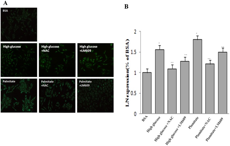
Aims/introduction: To investigate the roles of reactive oxygen species (ROS) and integrin αvβ3 in palmitate-induced laminin expression of human liver sinusoidal endothelial cells (HLSECs).
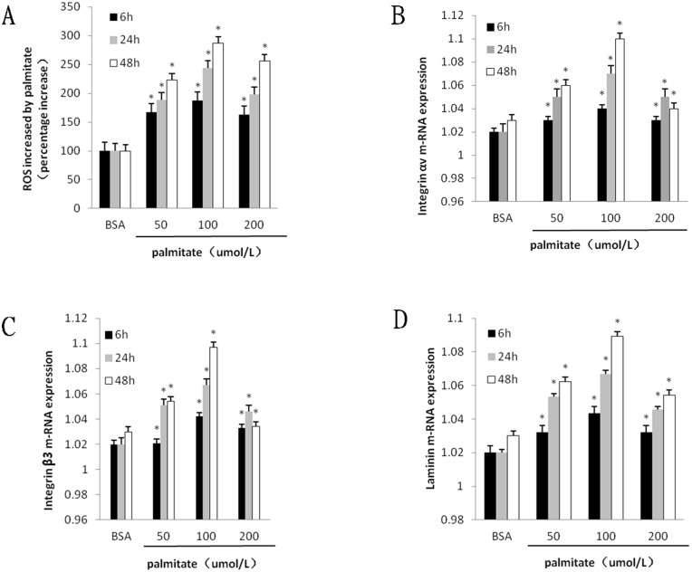
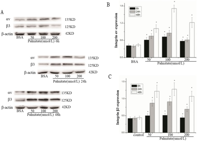
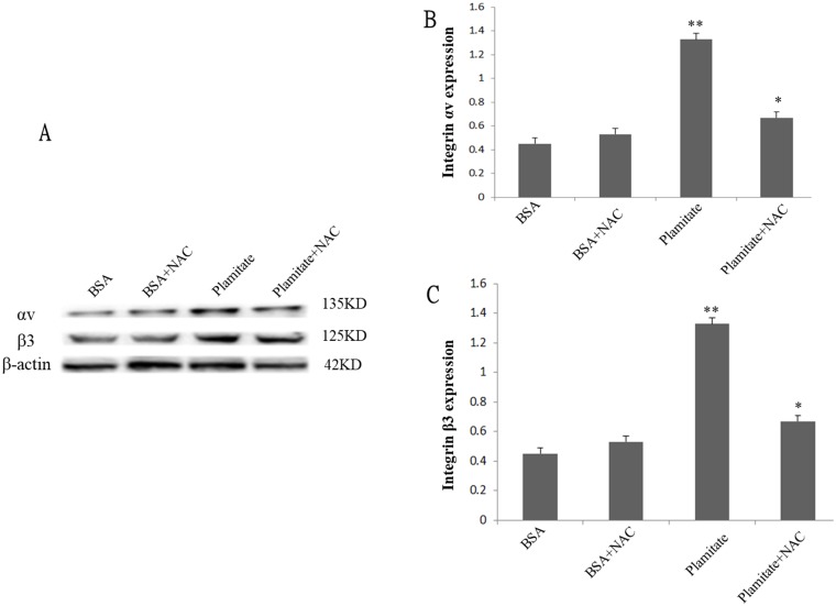
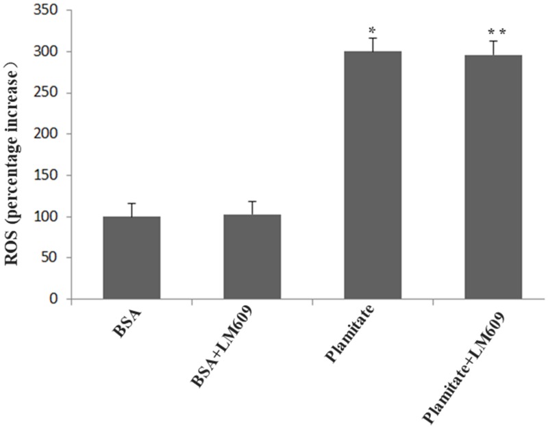
Results: The protein expression of integrin αv, integrin β3 and laminin are increased by palmitate in HLSECs in a time- and dose-dependent manner. NAC, the ROS inhibitor, significantly inhibited the up-regulation of protein expression of integrin αv, integrin β3 and laminin by palmitate (P < 0.05). Palmitate markedly enhanced ROS formation (P < 0.05), which was not inhibited by LM609, the antibody of integrin αvβ3. Palmitate significantly increased laminin synthesis (P < 0.05), which was attenuated by LM609 and NAC (P < 0.05).
Materials and methods: HLSECs were treated with palmitate in the presence or absence of LM609 (10 μg/ml) or N-acetylcysteine (NAC) (2 mM). Expression of integrin αv, integrin β3 and laminin were measured by RT-PCR and Western blot. Immunocytochemistry were used for examining laminin expression. The generation of ROS was measured using the fluorescent signal 2',7' dichloro-fluorescein diacetate (DCFH-DA).
Conclusions: The results suggested that palmitate increases laminin expression through ROS/integrin αv/β3 pathway.
Keywords: human liver sinusoidal endothelial cells; integrin αvβ3; laminin.
Conflict of interest statement
CONFLICTS OF INTEREST The authors declare no conflicts of interest.
Figures

Similar articles
-
High glucose regulates LN expression in human liver sinusoidal endothelial cells through ROS/integrin αvβ3 pathway.Environ Toxicol Pharmacol. 2016 Mar;42:231-6. doi: 10.1016/j.etap.2016.01.021. Epub 2016 Jan 29. Environ Toxicol Pharmacol. 2016. PMID: 26896612
-
Kaempferol inhibits the production of ROS to modulate OPN-αvβ3 integrin pathway in HUVECs.J Physiol Biochem. 2016 Jun;72(2):303-13. doi: 10.1007/s13105-016-0479-3. Epub 2016 Mar 21. J Physiol Biochem. 2016. PMID: 27000882
-
Hepatic Sinusoid Capillarizate via IGTAV/FAK Pathway Under High Glucose.Appl Biochem Biotechnol. 2024 Mar;196(3):1241-1254. doi: 10.1007/s12010-023-04605-8. Epub 2023 Jun 29. Appl Biochem Biotechnol. 2024. PMID: 37382792
-
Antisense integrin alphaV and beta3 gene therapy suppresses subcutaneously implanted hepatocellular carcinomas.Dig Liver Dis. 2007 Jun;39(6):557-65. doi: 10.1016/j.dld.2007.01.025. Epub 2007 Mar 19. Dig Liver Dis. 2007. PMID: 17374519
-
The Therapeutic Antibody LM609 Selectively Inhibits Ligand Binding to Human αVβ3 Integrin via Steric Hindrance.Structure. 2017 Nov 7;25(11):1732-1739.e5. doi: 10.1016/j.str.2017.09.007. Epub 2017 Oct 12. Structure. 2017. PMID: 29033288 Free PMC article.
Cited by
-
Role of liver sinusoidal endothelial cells in liver diseases.Nat Rev Gastroenterol Hepatol. 2021 Jun;18(6):411-431. doi: 10.1038/s41575-020-00411-3. Epub 2021 Feb 15. Nat Rev Gastroenterol Hepatol. 2021. PMID: 33589830 Review.
References
-
- Rösen P, Nawroth PP, King G, Möller W, Tritschler HJ, Packer L. The role of oxidative stress in the onset and progression of diabetes and its complications: asummary of a Congress Series sponsored byUNESCO-MCBN, the American Diabetes Association and the German Diabetes Society. Diabetes Metab Res Rev. 2001; 17:189–212. 10.1002/dmrr.196. - DOI - PubMed
-
- Castro Cabezas M, Erkelens DW, Van Dijk H. [Free fatty acids: mediators of insulin resistance and atherosclerosis]. [Article in Dutch]. Ned Tijdschr Geneeskd. 2002; 146:103–109. - PubMed
LinkOut - more resources
Full Text Sources
