. 2019 Jun 25:9:51.
doi: 10.1186/s13578-019-0316-9. eCollection 2019.

Regulation of platelet-activating factor-mediated interleukin-6 promoter activation by the 48 kDa but not the 45 kDa isoform of protein tyrosine phosphatase non-receptor type 2

Affiliations

Regulation of platelet-activating factor-mediated interleukin-6 promoter activation by the 48 kDa but not the 45 kDa isoform of protein tyrosine phosphatase non-receptor type 2

Geneviève Hamel-Côté et al. Cell Biosci. .

Abstract

Background: An underlying state of inflammation is thought to be an important cause of cardiovascular disease. Among cells involved in the early steps of atherosclerosis, monocyte-derived dendritic cells (Mo-DCs) respond to inflammatory stimuli, including platelet-activating factor (PAF), by the induction of various cytokines, such as interleukin 6 (IL-6). PAF is a potent phospholipid mediator involved in both the onset and progression of atherosclerosis. It mediates its effects by binding to its cognate G-protein coupled receptor, PAFR. Activation of PAFR-induced signaling pathways is tightly coordinated to ensure specific cell responses.

Results: Here, we report that PAF stimulated the phosphatase activity of both the 45 and 48 kDa isoforms of the protein tyrosine phosphatase non-receptor type 2 (PTPN2). However, we found that only the 48 kDa PTPN2 isoform has a role in PAFR-induced signal transduction, leading to activation of the IL-6 promoter. In luciferase reporter assays, expression of the 48 kDa, but not the 45 kDa, PTPN2 isoform increased human IL-6 (hIL-6) promoter activity by 40% after PAF stimulation of HEK-293 cells, stably transfected with PAFR (HEK-PAFR). Our results suggest that the differential localization of the PTPN2 isoforms and the differences in PAF-induced phosphatase activation may contribute to the divergent modulation of PAF-induced IL-6 promoter activation. The involvement of PTPN2 in PAF-induced IL-6 expression was confirmed in immature Mo-DCs (iMo-DCs), using siRNAs targeting the two isoforms of PTPN2, where siRNAs against the 48 kDa PTPN2 significantly inhibited PAF-stimulated IL-6 mRNA expression. Pharmacological inhibition of several signaling pathways suggested a role for PTPN2 in early signaling events. Results obtained by Western blot confirmed that PTPN2 increased the activation of the PI3K/Akt pathway via the modulation of protein kinase D (PKD) activity. WT PKD expression counteracted the effect of PTPN2 on PAF-induced IL-6 promoter transactivation and phosphorylation of Akt. Using siRNAs targeting the individual isoforms of PTPN2, we confirmed that these pathways were also active in iMo-DCs.

Conclusion: Taken together, our data suggest that PTPN2, in an isoform-specific manner, could be involved in the positive regulation of PI3K/Akt activation, via the modulation of PKD activity, allowing for the maximal induction of PAF-stimulated IL-6 mRNA expression.

Keywords: GPCR; IL-6; PTPN2; Platelet-activating factor; Protein tyrosine phosphatase; TC-PTP.


Conflict of interest statement

Competing interestsThe authors declare that they have no competing interests.

Figures

Fig. 1
Fig. 1
Differential effects of PTPN2 isoforms on PAF-induced IL-6 expression. a HEK-PAFR were transiently co-transfected with the hIL-6-luc, control vector (pcDNA3) or phosphatase constructs a WT and b D182A mutants. Cells were incubated overnight in DMEM-0.2% BSA and stimulated with a, b PAF (100 nM), vehicle or a TNF-α (20 ng/ml) for 6 h and luciferase activity was measured. The data presented are a mean ± SEM of 6–16 independent experiments performed in triplicate. Significance was established with two-way ANOVA with Sidak post-test. *p < 0.05. c iMo-DCs were either not transfected (NT) or transfected with siRNAs: either control (siCTRL) or against isoforms of PTPN2 (siPTPN2 48 kDa or 45 kDa) on day 4 and 5 before being collected on day 7 for experiments. iMo-DCs were starved for 3 h in RPMI 0.2% BSA and stimulated with PAF (1 nM) for 5 h. Cells were lysed in Trizol and RNA was extracted and converted to cDNA. IL-6 mRNA was quantified by real-time PCR. Data are presented as mean ± SEM of mRNA expression calculated by the delta–delta (ΔΔ)Ct method on their respective unstimulated control for 4 independent experiments, using RPL13A and GAPDH as housekeeping genes. Significance was established with two-way ANOVA with Sidak post-test, *p < 0.05
Fig. 2
Fig. 2
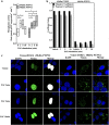
PAF modulates 45 kDa and 48 kDa PTPN2 activity but not the 45 kDa PTPN2 localization. a Graph represents the phosphatase activity determined by the hydrolysis rate of pNPP by Flag-tagged PTPN2, immunoprecipitated from HEK-PAFR which had been starved overnight in DMEM 0.2% BSA before being stimulated with 100 nM PAF, for indicated times. After PTP experiments, proteins were separated by SDS-PAGE, transferred to nitrocellulose membranes and blotted overnight with anti-PTPN2 antibodies for normalization of pNPP rates. Data are presented as low–high graphs (with lines at mean) of normalized pNPP hydrolysis rates for 3 independent experiments. Significance was established with paired two-way ANOVA with Sidak post-test. *p < 0.05. b, c HEK-PAFR, grown on poly-l-lysine coated coverslips, were transfected with Venus-tagged D182A-PTPN2 then stimulated, 30 h later, with vehicle or 100 nM PAF for 2, 5 or 10 min before permeabilization with 0.5% Triton X-100. Nuclei were stained with DAPI and cells were analyzed by confocal microscopy. Results are representative of 4 independent experiments, with at least 28 pictures analyzed from different fields per condition. Scale bar = 5 µm. b Compilation of Pearsons’ and Manders’ Coefficients for PTPN2. Data presented are mean ± SEM of 4 independent experiments and c representative pictures of Venus-tagged D182A 45 kDa or 48 kDa-D182A PTPN2 isoforms
Fig. 3
Fig. 3
Modulation of PAF-induced IL-6 promoter activity by 48 kDa PTPN2 depends on multiple signaling pathways. HEK-PAFR were transiently co-transfected with the hIL-6-luc, control vector (pcDNA3), or the 48 kDa or 45 kDa PTPN2 constructs. Cells were incubated overnight in DMEM-0.2% BSA and ae pre-treated for 30 min in DMEM-0.2%BSA with DMSO as vehicle control or a MEK 1/2 inhibitor U-0126, 5 µM, b JNK inhibitor SP600125, 1 µM, c pan PKC inhibitor GF109203x 2 µM or d Src kinase family inhibitor PP2, 10 nM and e Jak2 inhibitor AG490 5 µM. Stimulation with PAF (100 nM) or vehicle was for 6 h and luciferase activity was then measured. The data presented are a mean ± SEM of at least 4 independent experiments performed in triplicate. Significance was established with two-way ANOVA with Sidak post-test. *p < 0.05, **p < 0.01
Fig. 4
Fig. 4
48 kDa PTPN2 modulation of PAF-induced hIL-6 promoter activity does not depend on G-protein activation. HEK-293 were transiently co-transfected with the hIL-6-luc, wild type or mutant PAFRs and control vector (pcDNA3) or the 48 kDa PTPN2 constructs. Cells were incubated overnight in DMEM-0.2% BSA, stimulated with PAF (100 nM) or vehicle for 6 h and luciferase activity was measured. The data presented are mean ± SEM of 4–5 independent experiments performed in triplicate. Significance was established with paired Student’s t test. **p < 0.01
Fig. 5
Fig. 5
48 kDa PTPN2 modulates PAF-induced Akt activation in iMoDCs. Immature monocyte-derived dendritic cells (iMo-DCs) were transfected with control siRNAs (siCTRL) or against isoforms of PTPN2 (siPTPN2 48 kDa or 45 kDa) on day 4 and 5 before being collected on day 7 for experiments. iMo-DCs were starved for 5 h in RPM1 + 0.2% BSA and stimulated with PAF (1 nM) for indicated times. Whole cell lysates were separated by SDS-PAGE, transferred to nitrocellulose membranes and blotted overnight with anti-pSer473Akt, anti-Akt and anti-actin antibodies. a Representative blots and b compilations of at least 4 independent experiments are shown. The data presented are a mean ± SEM. Significance was established with two-way ANOVA with Sidak post-test. *p < 0.05, **p < 0.01
Fig. 6
Fig. 6
48 kDa PTPN2 modulates PAF-induced PKD activation in HEK-PAFR. a HEK-PAFR were transiently transfected with 48 kDa or 45 kDa PTPN2 cDNAs or control vector. Cells were incubated overnight in DMEM-0.2% BSA, then stimulated with PAF (100 nM) or vehicle for indicated times. Whole cell lysates were separated by SDS-PAGE, transferred to nitrocellulose membranes and blotted overnight with anti-pSer910 PKD then anti-PKD antibodies, after stripping. a Representative blots and b compilations of 3 independent experiments are shown. The data presented are a mean ± SEM. Significance was established with two-way ANOVA with Sidak post-test. *p < 0.05
Fig. 7
Fig. 7
48 kDa PTPN2 modulates PAF-induced PKD activation in iMoDCs. Immature monocyte-derived dendritic cells (iMo-DCs) were transfected with control siRNAs (siCTRL) or siRNAs against isoforms of PTPN2 (siPTPN2 48 kDa or 45 kDa) on day 4 and 5 before being collected on day 7 for experiments; or not transfected (NT). iMo-DCs were starved for 5 h in RPM1 + 0.2% BSA and stimulated with PAF (1 nM) for indicated times. Whole cell lysates were separated by SDS-PAGE, transferred to nitrocellulose membranes and blotted overnight with anti-pSer910 PKD, anti-PKD and anti-actin antibodies. NT samples have been loaded on the same blots as PTPN2 samples, to serve as controls. a Representative blots and b compilations of at least 3 independent experiments are shown. Significance was established with two-way ANOVA with Sidak post-test. *p < 0.05, **p < 0.01
Fig. 8
Fig. 8
48 kDa PTPN2 forms a signaling complex with PKD and Akt. HEK-PAFR were transiently transfected with D182A-48 kDa or − 45 kDa PTPN2 constructs tagged with Flag or control vector (Ctrl). Cells were incubated overnight in DMEM-0.2% BSA, then stimulated with PAF (100 nM) or vehicle for indicated times. Stimulation was stopped on ice, cells were scraped and lysed. Whole cell lysates were submitted to immunoprecipitation using anti-FLAG antibodies coupled to agarose beads. a Precipitates were separated by SDS-PAGE, transferred to nitrocellulose membranes and blotted overnight with anti-Akt, anti-PKD or anti-Flag antibodies. Lysate samples have been loaded onto the same blots as immunoprecipitated PTPN2 samples and serve as loading controls. a Representative blots and b compilations of at least 3 independent experiments are shown. The data presented are mean ± SEM. Significance was established with two-way ANOVA with Sidak post-test. *p < 0.05
Fig. 9
Fig. 9
48 kDa PTPN2 modulates Akt activation via a PKD-dependent pathway. a, b HEK-PAFR were transiently transfected with 48 kDa or 45 kDa PTPN2 constructs tagged with FLAG, PKD tagged with GST or control vectors (pcDNA3 or pcDNA3-GST). Cells were incubated overnight in DMEM-0.2% BSA, then stimulated with PAF (100 nM) or vehicle for indicated times. Stimulation was stopped on ice, cells were scraped and lysed. Whole cell lysates were separated by SDS-PAGE, transferred to nitrocellulose membranes and blotted overnight with anti-pSer473Akt, anti-GST, anti-actin, anti-FLAG and anti-Akt antibodies. Results shown are a compilation of 3 independent experiments, expressed as variations of Akt phosphorylation due to PTPN2 overexpression where values of pAkt levels of control cells (without PTPN2) are set to 1 (Dotted line) for each indicated stimulation time. c HEK-PAFR were transiently co-transfected with the hIL-6-luc, control vector pcDNA3, 48 kDa PTPN2 or PKD constructs. Cells were incubated overnight in DMEM-0.2% BSA and stimulated with PAF (100 nM) or vehicle for 6 h and luciferase activity was measured. The data presented are mean ± SEM of at least 3 experiments performed in triplicate. ac The data presented are mean ± SEM. Significance was established with two-way ANOVA with Sidak post-test. *p < 0.05
Fig. 10
Fig. 10
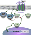
Schematic representation of pathways involved in PAF-induced PTPN2 modulation of IL-6 expression

Similar articles

Cited by

References

    1. Naugler WE, Karin M. The wolf in sheep’s clothing: the role of interleukin-6 in immunity, inflammation and cancer. Trends Mol Med. 2008;14(3):109–119. doi: 10.1016/j.molmed.2007.12.007. - DOI - PubMed
    1. Galkina E, Ley K. Immune and inflammatory mechanisms of atherosclerosis. Annu Rev Immunol. 2009;27:165–197. doi: 10.1146/annurev.immunol.021908.132620. - DOI - PMC - PubMed
    1. Demopoulos CA, Karantonis HC, Antonopoulou S. Platelet activating factor—a molecular link between atherosclerosis theories. Eur J Lipid Sci Technol. 2003;105(11):705–716. doi: 10.1002/ejlt.200300845. - DOI
    1. Montrucchio G, Alloatti G, Camussi G. Role of platelet-activating factor in cardiovascular pathophysiology. Physiol Rev. 2000;80(4):1669–1699. doi: 10.1152/physrev.2000.80.4.1669. - DOI - PubMed
    1. Brown SL, Jala VR, Raghuwanshi SK, Nasser MW, Haribabu B, Richardson RM. Activation and regulation of platelet-activating factor receptor: role of G(i) and G(q) in receptor-mediated chemotactic, cytotoxic, and cross-regulatory signals. J Immunol Baltim Md 1950. 2006;177(5):3242–3249. - PubMed