. 2020 Jul 7;41(26):2487-2497.
doi: 10.1093/eurheartj/ehz459.

Single systemic transfer of a human gene associated with exceptional longevity halts the progression of atherosclerosis and inflammation in ApoE knockout mice through a CXCR4-mediated mechanism

Affiliations

Single systemic transfer of a human gene associated with exceptional longevity halts the progression of atherosclerosis and inflammation in ApoE knockout mice through a CXCR4-mediated mechanism

Annibale Alessandro Puca et al. Eur Heart J. .

Abstract

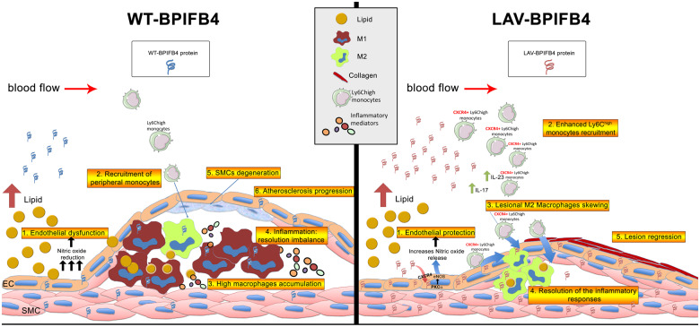
Aims: Here, we aimed to determine the therapeutic effect of longevity-associated variant (LAV)-BPIFB4 gene therapy on atherosclerosis.

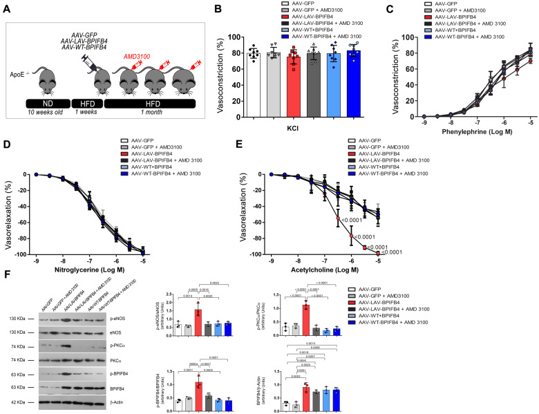
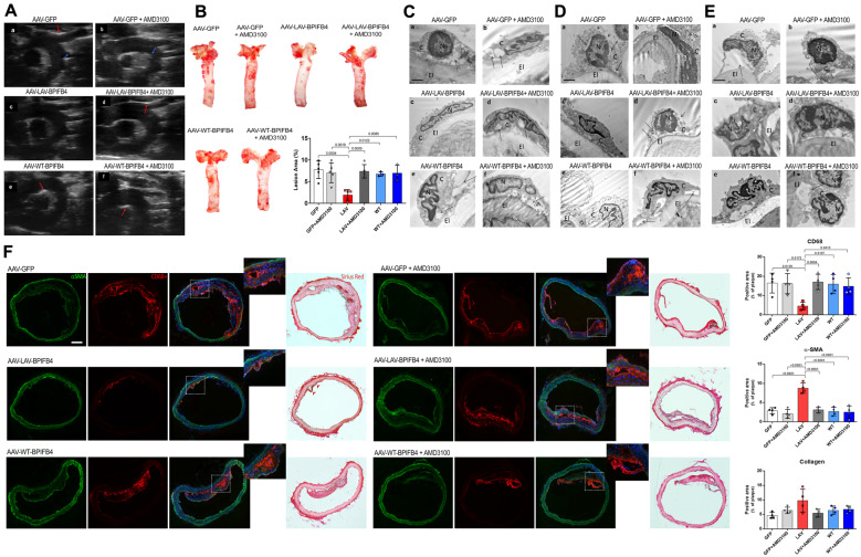
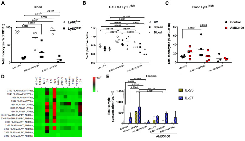
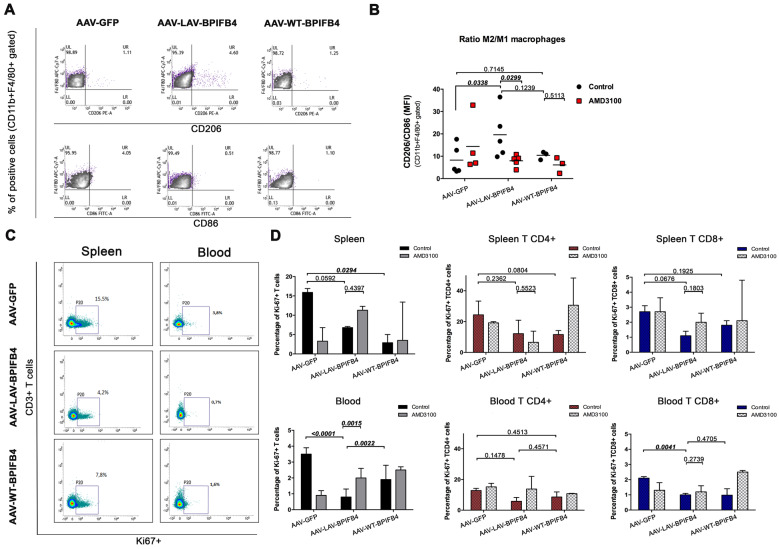
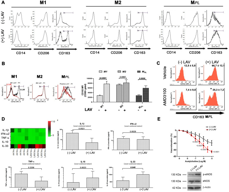
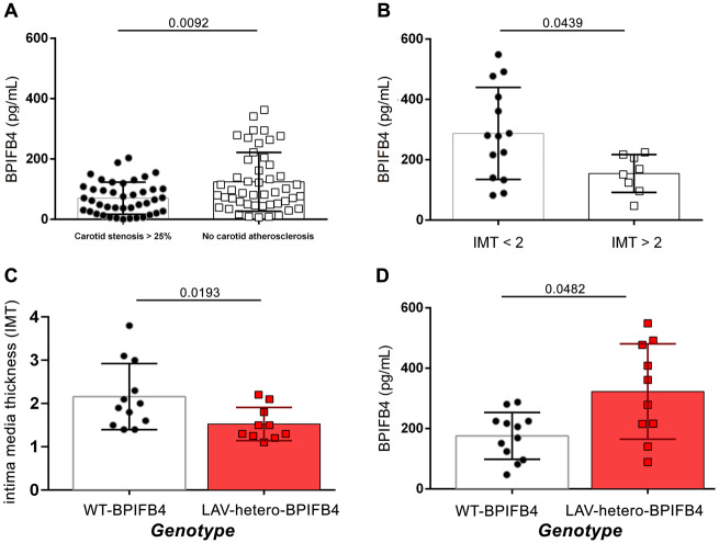
Methods and results: ApoE knockout mice (ApoE-/-) fed a high-fat diet were randomly allocated to receive LAV-BPIFB4, wild-type (WT)-BPIFB4, or empty vector via adeno-associated viral vector injection. The primary endpoints of the study were to assess (i) vascular reactivity and (ii) atherosclerotic disease severity, by Echo-Doppler imaging, histology and ultrastructural analysis. Moreover, we assessed the capacity of the LAV-BPIFB4 protein to shift monocyte-derived macrophages of atherosclerotic mice and patients towards an anti-inflammatory phenotype. LAV-BPIFB4 gene therapy rescued endothelial function of mesenteric and femoral arteries from ApoE-/- mice; this effect was blunted by AMD3100, a CXC chemokine receptor type 4 (CXCR4) inhibitor. LAV-BPIFB4-treated mice showed a CXCR4-mediated shift in the balance between Ly6Chigh/Ly6Clow monocytes and M2/M1 macrophages, along with decreased T cell proliferation and elevated circulating levels of interleukins IL-23 and IL-27. In vitro conditioning with LAV-BPIFB4 protein of macrophages from atherosclerotic patients resulted in a CXCR4-dependent M2 polarization phenotype. Furthermore, LAV-BPIFB4 treatment of arteries explanted from atherosclerotic patients increased the release of atheroprotective IL-33, while inhibiting the release of pro-inflammatory IL-1β, inducing endothelial nitric oxide synthase phosphorylation and restoring endothelial function. Finally, significantly lower plasma BPIFB4 was detected in patients with pathological carotid stenosis (>25%) and intima media thickness >2 mm.

Conclusion: Transfer of the LAV of BPIFB4 reduces the atherogenic process and skews macrophages towards an M2-resolving phenotype through modulation of CXCR4, thus opening up novel therapeutic possibilities in cardiovascular disease.

Keywords: Atherosclerosis; Immune system; Low-density lipoprotein; Vascular function.


Figures

Figure 1
Figure 1
Overexpression of LAV-BPIFB4 improves the vascular reactivity of ApoE null mice fed, a high-fat diet, and a CXCR4 inhibitor abolishes this protective effect. (A) Experimental protocol; (B) vascular response of ex vivo mesenteric arteries from ApoE knockout mice to potassium (80 mmol/L KCl) and (C) the dose–responses to phenylephrine, (D) acetylcholine, and (E) nitroglycerine after 1 month of AAV-LAV-BPIFB4 treatment. AAV-GFP was used as a control. Values are mean ± standard deviation of eight independent experiments. B, C, D, E two-way ANOVA followed Tukey’s multiple comparisons test. Numbers next to the curve show adjusted P-values. (F) Representative western blot (left) and densitometric analysis (right) conducted on mesenteric artery lysates. Values are mean ± standard deviation (N = 3). One-way ANOVA followed Tukey’s multiple comparisons test. Numbers above square brackets show adjusted P-values.
Figure 2
Figure 2
LAV-BPIFB4 gene therapy hinders plaque formation, preserves vascular endothelium integrity, and reduces monocyte/macrophages infiltration. (A) Ultrasound scanning of aortic arch with epi-aortic vessels in AAV-treated ApoE−/− mice fed a high-fat diet. LAV-BPIFB4-treated mice did not have any plaques, whereas all those on the other treatments had calcified plaques (red arrows) and lipidic plaques (blue arrows). (B) Oil Red O staining and quantitative analysis of atherosclerotic lesion size in the aorta. Oil Red O staining was quantified using ImageJ software. One-way ANOVA followed by Tukey’s multiple comparisons test. Numbers above square brackets show adjusted P-values. (CE) Representative micrographs of endothelial cells from aorta (C), mesenteric (D), and femoral (E) arteries: (a) Vascular endothelium of the AAV-GFP group showed cytosolic derangement (arrows) and broken plasma membranes (arrowheads); (b) these alterations were also evident in the group treated with the CXCR4 inhibitor. Diluted cytoplasm (arrow) and markedly condensed chromatin in the nucleus (N) was observed. (c) The endothelium was preserved by gene therapy with LAV-BPIFB4. (d) AMD3100 contrasted the benefit of LAV-BPIFB4, with the endothelial cells showing a diluted cytoplasm (arrows) and disruption of the plasma membrane (arrowheads); (e) The AAV-WT-BPIFB4-treated group presented with severely altered ultrastructure of the endothelium; (f) AMD3100 administration conferred additional ultrastructural damage, with endothelial cell detachment from the elastic membrane lamina (arrows). (F) Immunofluorescence staining of aortic arches from treated ApoE knockout mice, using the monocyte/macrophage marker CD68+, the smooth muscle cell marker αSMA, and Sirius Red staining to evaluate collagen (N = 4 per group). One-way ANOVA followed by Tukey’s multiple comparisons test. Numbers above square brackets show adjusted P-values.
Figure 3
Figure 3
LAV-BPIFB4 modulates monocyte dynamics in ApoE knockout mice. (A) Relative abundance of Ly6Chigh and Ly6Clow subsets in the peripheral blood of treated mice (AAV-GFP, N = 5; AAV-LAV-BPIFB4, N = 5; AAV-WT-BPIFB4, N = 3). (B) Percentage of CD11b+Ly6G-Ly6C+ monocytes expressing CXCR4 in bone marrow, spleen, and blood (AAV-GFP, N = 5; AAV-LAV-BPIFB4, N = 5; AAV-WT-BPIFB4, N = 3). (C) Effect of CXCR4 inhibitor on the frequency of peripheral blood Ly6Chigh monocytes (AAV-GFP, N = 5; AAV-LAV-BPIFB4, N = 5; AAV-WT-BPIFB4, N = 3; GFP-AMD3100, N = 5; LAV-BPIFB4-AMD3100, N = 5; WT-BPIFB4-AMD3100, N = 3). (D and E) Plasma collected from all the mice groups indicated above was assessed for circulating cytokine levels with bead-based multiplex ELISA. The relative concentrations of analytes are presented as a heat map (D) or expressed as the mean ± standard deviation of each sample determination conducted in duplicate (E). Statistical analysis by two-way ANOVA with post hoc Fisher’s Least Significant Difference (LSD) test was conducted. Numbers above square brackets show unadjusted LSD P-values.
Figure 4
Figure 4
Enhanced enrichment of M2 macrophages in the spleen of AAV-LAV-BPIFB4 ApoE knockout mice fed a high-fat diet. (A and B) CD45hi CD11b+ F4/80 + splenic macrophages were additionally stained with flow cytometric markers CD206+ or CD86+ to discern the CD11b+ F4/80 +CD206 M2 type from CD11b+ F4/80 +CD86 M1 type of splenic macrophages. A representative dot plot panel (left) is presented. The graph on the right reports the mean ± standard deviation of ratios of M2 vs. M1 splenic macrophages (CD206/CD86 ratio) from all recipient mice (N = 3–5 per group). MFI stands for mean fluorescence intensity of selected markers. (C) Analysis of the proliferation of splenic and blood CD3+ T cells in ApoE−/− mice infected with AAV-LAV-BPIFB4, AAV-WT-BPIFB4 or AAV-empty vector, treated or not with AMD3100. Representative dot plot showing changes of percentage Ki-67 expression in both spleen and blood TCD3+ cells. (D) Bar graph reporting the percentage ± standard deviation of Ki67+, CD3+ gated T cells. Percentage of Ki-67+ expression in CD4+ and CD8+ T cell subsets from the spleen and the blood of recipient mice were determined. The bar graph reports the percentage ± standard deviation. Statistical analysis by two-way ANOVA with post hoc Fisher’s Least Significant Difference (LSD) test was conducted. Numbers above square brackets show unadjusted LSD P-values.
Figure 5
Figure 5
In vitro conditioning with recombinant LAV-BPIFB4 protein leads to polarization of patient macrophages towards M2 phenotypes and reduces the inflammatory milieu in the vessel wall. (A) Macrophages generated from peripheral blood monocytes from atherosclerotic patients (N = 5) were cultured with or without recombinant LAV-BPIFB4 (18 ng/mL) for the last 72 h. Representative cytofluorimetric histogram profiles of CD14, CD206, and CD163 protein levels at the cell surface of macrophages (viable gated CD68+ cells) polarized towards M1, M2, or MPL (autologous plasma) phenotypes. (B) (left panel) Histogram overlay for CD163 M2 markers in M1, M2, or MPL macrophages from control, untreated- (red curve), and LAV-BPIFB4-treated cells (black curve) from a representative atherosclerotic patient. (right panel) The bar graph reports the mean fluorescence intensity (MFI) values ± standard deviation of CD163 on viable CD68+ gated cells. Two-way ANOVA followed by Tukey's multiple comparisons test. Numbers above square brackets show adjusted P-values. (C) Representative histogram profiles of CD163 protein level on MPL cell surface in controls and in cells treated with LAV-BPIFB4 (18 ng/mL) in the presence or absence of AMD3100 (20 μM) for 72 h. Percentages of positive cells are indicated in the upper right corner and are representative of four independent experiments. (D) Vessels from atherosclerotic patients (N = 5) were incubated for 24 h at 37°C in the presence or absence of 18 ng/mL LAV-BPIFB4. Conditioned medium was then assessed for cytokine secretion with bead-based multiplex ELISA. The relative concentrations of analytes detected is presented as a heat map. Statistical comparisons were performed with two-tailed Student’s t-test for simple comparisons. Numbers above square brackets show unadjusted P-values. (E) Vascular response of ex vivo human atherosclerotic femoral arteries (upper) to increasing doses of acetylcholine, before and after treatment for 1 h with LAV-BPIFB4 recombinant protein (18 ng/mL) (N = 5); two-way ANOVA followed Tukey's multiple comparisons test. Numbers reported on the curves show adjusted P-values. (Bottom) representative western blots conducted on the studied human vessels.
Figure 6
Figure 6
High plasma BPIFB4 detected in LAV-carrier patients is associated with reduced carotid stenosis and IMT < 2mm. Graphs showing correlation between: (A) BPIFB4 plasma level in patients with carotid stenosis >25% (N = 48) and in patients with no carotid atherosclerosis (N = 42). (B) BPIFB4 level in patients stratified for IMT <2 mm (N = 14) vs. IMT >2 mm (N = 8). (C and D) Correlation between BPIFB4 protein levels with intima media thickness (C) and with plasma concentration (D) after genotype stratification of WT- or LAV-carrier patients. Results are presented as mean ± standard deviation and were analysed by two-tailed non-parametric Mann–Whitney test. Numbers above lines show unadjusted P-values.
Take home figure
Take home figure
LAV-BPIFB4 exerts protective effects against the onset and progression of the atherogenic process. LAV-BPIFB4 modulates both endothelial function and the immune compartment through a CXCR4-dependent mechanism. In endothelial cells, LAV-BPIFB4 promotes nitric oxide production, ensuring endothelial protection. At the same time, it is able to enhance Ly6Chigh monocyte recruitment, directing them towards the pro-resolving M2 macrophage phenotype, driving atherosclerotic regression at the early stage of the disease and favouring resolution of the inflammatory responses.
None

Comment in

References

    1. Ridker PM, Everett BM, Thuren T, MacFadyen JG, Chang WH, Ballantyne C, Fonseca F, Nicolau J, Koenig W, Anker SD, Kastelein JJP, Cornel JH, Pais P, Pella D, Genest J, Cifkova R, Lorenzatti A, Forster T, Kobalava Z, Vida-Simiti L, Flather M, Shimokawa H, Ogawa H, Dellborg M, Rossi PRF, Troquay RPT, Libby P, Glynn RJ, Group CT.. Antiinflammatory therapy with canakinumab for atherosclerotic disease. N Engl J Med 2017;377:1119–1131. - PubMed
    1. Jin DK, Shido K, Kopp HG, Petit I, Shmelkov SV, Young LM, Hooper AT, Amano H, Avecilla ST, Heissig B, Hattori K, Zhang F, Hicklin DJ, Wu Y, Zhu Z, Dunn A, Salari H, Werb Z, Hackett NR, Crystal RG, Lyden D, Rafii S.. Cytokine-mediated deployment of SDF-1 induces revascularization through recruitment of CXCR4+ hemangiocytes. Nat Med 2006;12:557–567. - PMC - PubMed
    1. Shantsila E, Tapp LD, Wrigley BJ, Montoro-García S, Lip GYH.. CXCR4 positive and angiogenic monocytes in myocardial infarction. Thromb Haemost 2013;109:255–262. - PubMed
    1. Chong SZ, Evrard M, Devi S, Chen J, Lim JY, See P, Zhang Y, Adrover JM, Lee B, Tan L, Li JL, Liong KH, Phua C, Balachander A, Boey A, Liebl D, Tan SM, Chan JK, Balabanian K, Harris JE, Bianchini M, Weber C, Duchene J, Lum J, Poidinger M, Chen Q, Renia L, Wang CI, Larbi A, Randolph GJ, Weninger W, Looney MR, Krummel MF, Biswas SK, Ginhoux F, Hidalgo A, Bachelerie F, Ng LG.. CXCR4 identifies transitional bone marrow premonocytes that replenish the mature monocyte pool for peripheral responses. J Exp Med 2016;213:2293–2314. - PMC - PubMed
    1. Rahman K, Vengrenyuk Y, Ramsey SA, Vila NR, Girgis NM, Liu J, Gusarova V, Gromada J, Weinstock A, Moore KJ, Loke P, Fisher EA.. Inflammatory Ly6Chi monocytes and their conversion to M2 macrophages drive atherosclerosis regression. J Clin Invest 2017;127:2904–2915. - PMC - PubMed

Publication types