. 2019 Jul 9;36(9):133.
doi: 10.1007/s11095-019-2665-9.

Targeting KRAS Mutant Lung Cancer Cells with siRNA-Loaded Bovine Serum Albumin Nanoparticles

Affiliations

Targeting KRAS Mutant Lung Cancer Cells with siRNA-Loaded Bovine Serum Albumin Nanoparticles

Aditi Mehta et al. Pharm Res. .

Abstract

Purpose: KRAS is the most frequently mutated gene in human cancers. Despite its direct involvement in malignancy and intensive effort, direct inhibition of KRAS via pharmacological inhibitors has been challenging. RNAi induced knockdown using siRNAs against mutant KRAS alleles offers a promising tool for selective therapeutic silencing in KRAS-mutant lung cancers. However, the major bottleneck for clinical translation is the lack of efficient biocompatible siRNA carrier systems.

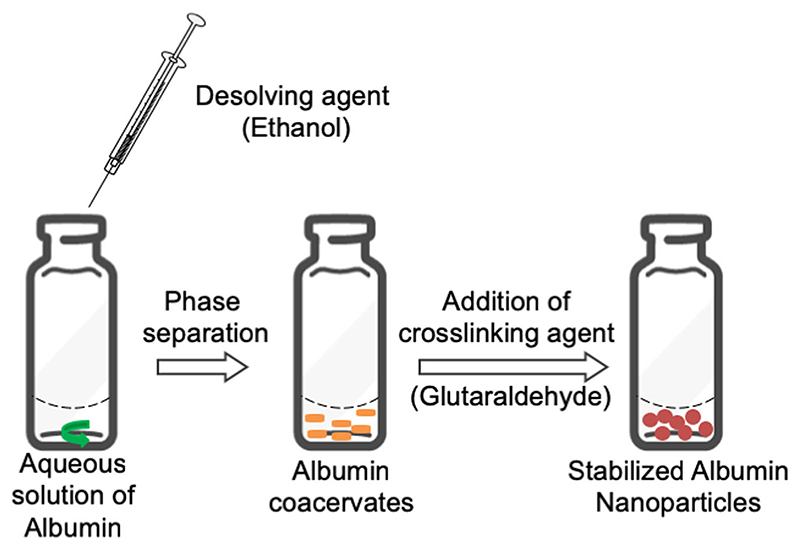
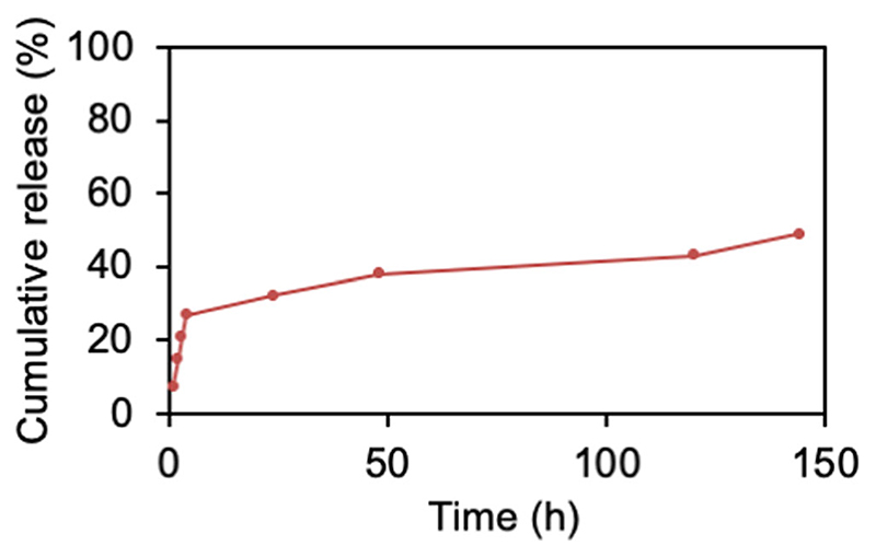
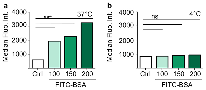
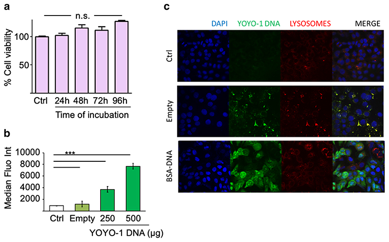
Methods: Bovine serum albumin (BSA) nanoparticles were prepared by desolvation method to deliver siRNA targeting the KRAS G12S mutation. The BSA nanoparticles were characterized with respect to their size, zeta potential, encapsulation efficiency and nucleic acid release. Nanoparticle uptake, cellular distribution of nucleic acids, cytotoxicity and gene knock down to interfere with cancer hallmarks, uncontrolled proliferation and migration, were evaluated in KRAS G12S mutant A459 cells, a lung adenocarcinoma cell line.

Results: BSA nanoparticles loaded with siRNA resulted in nanoparticles smaller than 200 nm in diameter and negative zeta potentials, displaying optimal characteristics for in vivo application. Encapsulating and protecting the siRNA payload well, the nanoparticles enabled transport to A549 cells in vitro, could evade endosomal entrapment and mediated significant sequence-specific KRAS knockdown, resulting in reduced cell growth of siRNA transfected lung cancer cells.

Conclusions: BSA nanoparticles loaded with mutant specific siRNA are a promising therapeutic approach for KRAS-mutant cancers.

Keywords: BSA; KRAS; lung cancer; siRNA delivery.


Figures

Fig. 1
Fig. 1. Schematic representation of the desolvation method used to prepare BSA nanoparticles.
Fig. 2
Fig. 2. DNA release profile of bulk DNA loaded nanoparticles prepared following Method 3.
Released DNA (%, Y axis) was measured by SYBR Gold assay and plotted against time (hours, X-axis). Data points indicate mean ±SD, n = 3.
Fig. 3
Fig. 3. Cellular uptake of FITC-BSA nanoparticles in A549 cells at (a) 37°C and (b) 4°C measured by flow cytometry represented as median fluorescence intensity (MFI, Y axis) Data points indicate mean ± SD, n = 3; One-way ANOVA, ***, p < 0.001, n.s., not significant).
Fig. 4
Fig. 4. BSA nanoparticles can safely and efficiently deliver DNA to A549 cells.
(a) Cell viability (% Y-axis) as measured by MTS assay in A549 cells treated with 200 μg of BSA nanoparticles for 24, 48, 72 and 96 h (Time, X-axis). (b) Transfection of A549 cells with BSA nanoparticles loaded with a YOYO-1 labelled DNA measured by flow cytometry as median fluorescence intensity (MFI, Y-axis). μg DNA is DNA amount used for transfection, taking into account encapsulation efficiency for each preparation. (c) Confocal images of A549 cells 24 h after transfecting with BSA nanoparticles loaded with YOYO-1 labelled DNA (green) and staining with DAPI (blue, depicting the cell nuclei) and LysoTracker Red DND-99(red, representing the lysosomes). Empty nanoparticles exhibit auto fluorescence on excitation at 488 nm. Ctrl, untreated cells. Data points indicate mean ± SD, n = 3; One-way ANOVA, ***, p < 0.001; ns, not significant.
Fig. 5
Fig. 5. Transfection of A549 cells with BSA nanoparticles loaded with a GFP expressing plasmid measured by flow cytometry as median fluorescence intensity (MFI) ofGFP (Y-axis).
Ctrl, untreated cells. μg pDNA is pDNAamount used for transfection, taking into account encapsulation efficiency for each preparation. Data points indicate mean ± SD, n = 3; One-way ANOVA, ***, p < 0.001; ns, not significant.
Fig. 6
Fig. 6. Characterization of siRNA loaded BSA nanoparticles.
(a) Hydrodynamic diameters (left y-axis, bars) and polydispersity indices (PDI, right y-axis, dots) (b) zeta potentials of the nanoparticles (c) Scanning electron microscope (SEM) micrographs of empty (left) and siRNA loaded (right) BSA nanoparticles. (d) Electrophoretic mobility of naked siRNA (siRNA), siRNA loaded BSA nanoparticles (–) following incubation with FBS (+ FBS) or RNase (+ RNase) or both (+ +). L, ultra-low range DNA ladder. (e) Cellular uptake of BSA nanoparticles loaded with Alexa 350 labelled siRNA in A549 cells measured by flow cytometry and represented as median fluorescence intensity (MFI, Y axis). Data points indicate mean ± SD, n = 3.
Fig. 7
Fig. 7. Mutation specific knock of KRAS after transfection with siRNA loaded BSA nanoparticles.
(a) Protein extracts from A549 cells transfected with BSA nanoparticles loaded with 30 nM of scrambled control siRNA (siCtrl, Lane I), siRNA against KRAS GI2S (siKRAS GI2S, Lane 2) or siKRAS against both WT and mutant allele (siKRAS Total, Lane 3) were analysed by western blot using the indicated antibodies. Histone H3 (H3) was used as loading control. (b) Quantification of the KRAS knockdown observed in A. (c) Cell viablilty (% Y-axis) as measured by MTS assay in A549 cells transfected with siCtrl, siKRAS GI2S and siKRAS total. (d) Dot plot showing the percentage of Annexin V- or propidium iodide positive A549 cells after transfection with siCtrl, siKRAS GI2S or siKRAS Total. x and y axes denote Propidium iodide and Annexin V signals respectively, (e) Quantitation of Annexin V-positive cells represented as percentage of apoptotic cells. (f) A549 were subjected to transwell migration assays 24 h aftertransfection as indicated. The crystal violet dye staining images of the membranes are shown. (g) Percentage of migrated cells was quantified by counting three fields (20x magnification) per chamber and compared with controls. Magnification, X20X. Data points indicate mean ± SD, n = 3. One-way ANOVA, **, p < 0.01, ***, p < 0.001.

References

    1. Siegel RL, Miller KD, Jemal A. Cancer statistics, 2018. CA Cancer J Clin. 2018;68(1):7–30. - PubMed
    1. Molina JR, Yang P, Cassivi SD, Schild SE, Adjei AA. Non-small cell lung cancer: epidemiology, risk factors, treatment, and survivorship. Mayo Clin Proc. 2008;83(5):584–94. - PMC - PubMed
    1. Herbst RS, Heymach JV, Lippman SM. Lung cancer. N Engl J Med. 2008;359(13):1367–80. - PMC - PubMed
    1. Dogan S, Shen R, Ang DC, Johnson ML, D’Angelo SP, Paik PK, et al. Molecular epidemiology of EGFR and KRAS mutations in 3, 026 lung adenocarcinomas: higher susceptibility of women to smoking-related KRAS-mutant cancers. Clin Cancer Res. 2012;18(22):6169–77. - PMC - PubMed
    1. ElOsta B, Behera M, Kim S, Berry LD, Sica G, Pillai RN, et al. Characteristics and outcomes of patients with metastatic KRAS-mutant lung adenocarcinomas: the lung Cancer mutation consortium experience. J Thorac Oncol. 2019 - PMC - PubMed