. 2019 Jul 9;14(1):228.
doi: 10.1186/s11671-019-3049-0.

A Dual Targeting Magnetic Nanoparticle for Human Cancer Detection

Affiliations

A Dual Targeting Magnetic Nanoparticle for Human Cancer Detection

Siwen Wu et al. Nanoscale Res Lett. .

Abstract

Malignant tumors are a major threat to human life and high lymphatic vessel density is often associated with metastatic tumors. With the discovery of molecules targeted at the lymphatic system such as lymphatic vessel endothelial hyaluronan receptor 1 (LYVE-1) and Podoplanin, many studies have been performed to determine the role of lymphatic endothelial cells (LECs) in tumor metastasis. However, disadvantages such as non-specificity and high cost limit their research and diagnostic applications. In this study, Fe3O4@KCTS, a core-shell type of magnetic nanoparticles, was prepared by activating Fe3O4 with carbodiimide and cross-linking it with α-ketoglutarate chitosan (KCTS). The LYVE-1 and Podoplanin antibodies were then incorporated onto the surface of these magnetic nanoparticles and as a result, dual-targeting magnetic nanoprobes were developed. The experimental tests of this study demonstrated that a dual-targeting magnetic nanoprobe with high-purity LECs from tumor tissues was successfully developed, providing a basis for clinical application of LECs in colorectal cancer treatment as well as in early clinical diagnosis using bimodal imaging.

Keywords: LYVE-1; Lymphatic endothelial cells; Magnetic nanoparticles; Podoplanin.


Conflict of interest statement

The authors declare that they have no competing interests.

Figures

Scheme 1
Scheme 1
Schematic illustration of Fe3O4@KCTS-LECs-double antibody magnetic nanoparticles
Fig. 1
Fig. 1
Characterization of magnetic nanoprobe. a Transmission electron micrograph image of Fe3O4 nanoprobe (scale bar, 100 nm). b Transmission electron micrograph image of Fe3O4@KCTS nanoprobe (scale bar, 100 nm). c Transmission electron micrograph image of Fe3O4@KCTS-LECs-double antibody magnetic nanoprobe (scale bar, 100 nm). d The particle size was detected by size analysis, and the average size of the probe was 91.28 nm. e Zeta potential was about − 32.8 mv. f The Fe3O4@KCTS-LECs-double antibody nanoprobe has emission peaks under excitation light of 488 and 545
Fig. 2
Fig. 2
Expression of lymphatic vessels in colorectal cancer tissues. a Dilated vessels in the tissue sections were positive for LYVE-1 on endothelial cell membrane by immunohistochemistry (scale bar, 100 μm). On the right is a magnified picture. b Dilated vessels in the tissue sections were positive for LYVE-1 and podoplanin on endothelial cell membrane as revealed by immunofluorescence (scale bar, 50 μm)
Fig. 3
Fig. 3
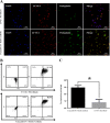
Analysis of the purity of lymphatic endothelial cells. a Fluorescence micrographs double-color imaging of LYVE-1 (red) and podoplanin (green)/nuclei [4,6-diamidino-2-phenylindole (DAPI, blue) in the isolated human colorectal cancer LECs sorted using LYVE-1 Microbeads and Fe3O4@KCTS-LECs-double antibody (scale bar, 100 μm). b The co-expression of LYVE-1 (APC) and podoplanin (FITC) as revealed by flow cytometric detection. c Results of flow cytometry, *P < 0.05
Fig. 4
Fig. 4
Comparison of biological functions of LECs. a Determination of in vitro LECs tube formation ability by Calcein AM (green) in vitro (scale bar, 50 μm). b Endothelial cell phagocytosis assay (DiI-labeled, red; DAPI, blue) of endothelial cell function using isolated LECs by LYVE-1 Microbeads and Fe3O4@KCTS-LECs-Double antibody (scale bar, 100 μm)
Fig. 5
Fig. 5
Fluorescence imaging and MRI of colorectal cancer subcutaneous xenografting tumor models of NOD/SCID mice. a NOD/SCID mice were anesthetized and imaged with fluorescence imaging system at preinjection and post-injection 0.5 h, 12 h, and 24 h. b NOD/SCID mice were anesthetized and imaged with 3.0T MRI scanner at pre injection and post injection 0.5 h, 12 h, and 24 h
Fig. 6
Fig. 6
Toxicity of Fe3O4@KCTS-LECs-double antibody magnetic nanoprobe. a LECs were incubated with various concentrations of magnetic nanoprobe, and cell viability was measured at 24 h and 48 h. b NOD/SCID mice were treated with PBS or magnetic nanoprobe, and sections from major organs were stained with hematoxylin-eosin and examined by light microscopy (scale bar, 100 μm)

References

    1. Siegel RL, Miller KD, Jemal A. Cancer statistics. CA Cancer J Clin. 2019;69:7–34. doi: 10.3322/caac.21551. - DOI - PubMed
    1. Onder L, Morbe U, Pikor N, Novkovic M, Cheng HW, Hehlgans T, Pfeffer K, Becher B, Waisman A, Rulicke T, gommerman J, Mueller CG, Sawa S, Scandella E, Ludewig B. Lymphatic endothelial cells control initiation of lymph node organogenesis. Immunity. 2017;47:80–92.e4. doi: 10.1016/j.immuni.2017.05.008. - DOI - PubMed
    1. Yang J, Zhang S, Zhang L, Xie X, Wang H, Jie Z, Gu M, Yang JY, Cheng X, Sun SC (2018) Lymphatic endothelial cells regulate B-cell homing to lymph nodes via a NIK-dependent mechanism. Cell Mol Immunol. 10.1038/cmi - PMC - PubMed
    1. Lee S, Choi I, Hong YK. Heterogeneity and plasticity of lymphatic endothelial cells. Semin Thromb Hemost. 2010;36:352–361. doi: 10.1055/s-0030-1253457. - DOI - PMC - PubMed
    1. Banerji S, Ni J, Wang SX, Clasper S, Su J, Tammi R, Jones M, Jackson DG. LYVE-1, a new homologue of the CD44glycoprotein, is a lymph-specific receptor for hyaluronan. J Cell Biol. 1999;144:789–801. doi: 10.1083/jcb.144.4.789. - DOI - PMC - PubMed

LinkOut - more resources