

Obtaining Informed Consent Using Patient Specific 3D Printing Cerebral Aneurysm Model
- PMID: 31290295
- PMCID: PMC6616983
- DOI: 10.3340/jkns.2019.0092
Obtaining Informed Consent Using Patient Specific 3D Printing Cerebral Aneurysm Model
Abstract
Objective: Recently, three-dimensional (3D) printed models of the intracranial vascular have served as useful tools in simulation and training for cerebral aneurysm clipping surgery. Precise and realistic 3D printed aneurysm models may improve patients' understanding of the 3D cerebral aneurysm structure. Therefore, we created patient-specific 3D printed aneurysm models as an educational and clinical tool for patients undergoing aneurysm clipping surgery. Herein, we describe how these 3D models can be created and the effects of applying them for patient education purpose.
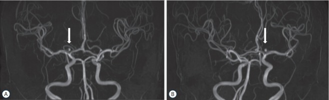
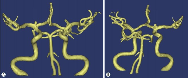
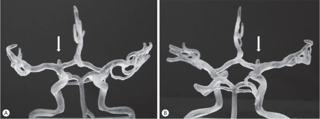
Methods: Twenty patients with unruptured intracranial aneurysm were randomly divided into two groups. We explained and received informed consent from patients in whom 3D printed models-(group I) or computed tomography angiography-(group II) was used to explain aneurysm clipping surgery. The 3D printed intracranial aneurysm models were created based on time-offlight magnetic resonance angiography using a 3D printer with acrylonitrile-butadiene-styrene resin as the model material. After describing the model to the patients, they completed a questionnaire about their understanding and satisfaction with aneurysm clipping surgery.
Results: The 3D printed models were successfully made, and they precisely replicated the actual intracranial aneurysm structure of the corresponding patients. The use of the 3D model was associated with a higher understanding and satisfaction of preoperative patient education and consultation. On a 5-point Likert scale, the average level of understanding was scored as 4.7 (range, 3.0-5.0) in group I. In group II, the average response was 2.5 (range, 2.0-3.0).
Conclusion: The 3D printed models were accurate and useful for understanding the intracranial aneurysm structure. In this study, 3D printed intracranial aneurysm models were proven to be helpful in preoperative patient consultation.
Keywords: Informed consent; Intracranial aneurysm; Printing, Three-dimensional.
Conflict of interest statement
No potential conflict of interest relevant to this article was reported.
Figures
Similar articles
-
3D-Printed Head Model in Patient's Education for Micro-Neurosurgical Aneurysm Clipping Procedures.World Neurosurg. 2023 Jul;175:e1069-e1074. doi: 10.1016/j.wneu.2023.04.070. Epub 2023 Apr 21. World Neurosurg. 2023. PMID: 37087042
-
Three-dimensional intracranial middle cerebral artery aneurysm models for aneurysm surgery and training.J Clin Neurosci. 2018 Apr;50:77-82. doi: 10.1016/j.jocn.2018.01.074. Epub 2018 Feb 10. J Clin Neurosci. 2018. PMID: 29439905
-
Comparison of Two Three-Dimensional Printed Models of Complex Intracranial Aneurysms for Surgical Simulation.World Neurosurg. 2017 Jul;103:671-679. doi: 10.1016/j.wneu.2017.04.098. Epub 2017 Apr 24. World Neurosurg. 2017. PMID: 28450234
-
Development of three-dimensional hollow elastic model for cerebral aneurysm clipping simulation enabling rapid and low cost prototyping.World Neurosurg. 2015 Mar;83(3):351-61. doi: 10.1016/j.wneu.2013.10.032. Epub 2013 Oct 16. World Neurosurg. 2015. PMID: 24141000 Review.
-
Intracranial vasculature 3D printing: review of techniques and manufacturing processes to inform clinical practice.3D Print Med. 2020 Aug 6;6(1):18. doi: 10.1186/s41205-020-00071-8. 3D Print Med. 2020. PMID: 32761490 Free PMC article. Review.
Cited by
-
Utility of 3-dimensionally printed models for parent education in pediatric plagiocephaly.PEC Innov. 2022 Aug 20;1:100077. doi: 10.1016/j.pecinn.2022.100077. eCollection 2022 Dec. PEC Innov. 2022. PMID: 37213734 Free PMC article.
-
Clinical situations for which 3D printing is considered an appropriate representation or extension of data contained in a medical imaging examination: neurosurgical and otolaryngologic conditions.3D Print Med. 2023 Nov 27;9(1):33. doi: 10.1186/s41205-023-00192-w. 3D Print Med. 2023. PMID: 38008795 Free PMC article. Review.
-
Shared Decision-Making in Surgery-The Role of 3D-Printed Models.JAMA Netw Open. 2025 Jun 2;8(6):e2513204. doi: 10.1001/jamanetworkopen.2025.13204. JAMA Netw Open. 2025. PMID: 40459897 Free PMC article. No abstract available.
-
Exploring the Influence of Concurrent Nutritional Therapy on Children with Spinal Muscular Atrophy Receiving Nusinersen Treatment.Children (Basel). 2024 Jul 23;11(8):886. doi: 10.3390/children11080886. Children (Basel). 2024. PMID: 39201821 Free PMC article.
-
Academics versus the Internet: Evaluating the readability of patient education materials for cerebrovascular conditions from major academic centers.Surg Neurol Int. 2022 Sep 2;13:401. doi: 10.25259/SNI_502_2022. eCollection 2022. Surg Neurol Int. 2022. PMID: 36128118 Free PMC article.
References
-
- Aoun RJ, Hamade YJ, Zammar SG, Patel NP, Bendok BR. Futuristic three-dimensional printing and personalized neurosurgery. World Neurosurg. 2015;84:870–871. - PubMed
-
- Kimura T, Morita A, Nishimura K, Aiyama H, Itoh H, Fukaya S, et al. Simulation of and training for cerebral aneurysm clipping with 3-dimensional models. Neurosurgery. 2009;65:719–725. - PubMed
-
- Knoedler M, Feibus AH, Lange A, Maddox MM, Ledet E, Thomas R, et al. Individualized physical 3-dimensional kidney tumor models constructed from 3-dimensional printers result in improved trainee anatomic understanding. Urology. 2015;85:1257–1261. - PubMed
-
- Mashiko T, Kaneko N, Konno T, Otani K, Nagayama R, Watanabe E. Training in cerebral aneurysm clipping using self-made 3-dimensional models. J Surg Educ. 2017;74:681–689. - PubMed
Grants and funding
LinkOut - more resources
Full Text Sources
