

doi: 10.4103/aja.aja_73_19.
Funneling of the bladder neck - radiological appearance after radical retropubic prostatectomy and clinical relevance
Affiliations
- PMID: 31290410
- PMCID: PMC7155796
- DOI: 10.4103/aja.aja_73_19
Item in Clipboard
Funneling of the bladder neck - radiological appearance after radical retropubic prostatectomy and clinical relevance
Asian J Androl.
2020 Mar-Apr.
No abstract available
Conflict of interest statement
None
Figures

(a) Preoperative MRI – flat and well-supported appearance of the bladder base. Well-developed levator sling (arrows). (b) Postprostatectomy MRI – typical tapered, funneled appearance of the bladder neck. (c) Typical fluoroscopic appearance of a funneled bladder neck after RP with the vesicourethral anastomosis just below it (arrow). (d) Descending urethrogram showing a funneled bladder neck (A) which is contracted and rigid down to the anastomosis (B) and across into the membranous urethra (C) and the proximal part of the urethral sphincter (D). (e) Funneling of the bladder neck not so prominent because the entire funnel is obliterated by the fibrosis giving rise to an extensive contracture (A). Close examination actually shows the proximal end of the funnel terminating abruptly at the proximal end of the contracture (B). RP: radical prostatectomy; MRI: magnetic resonance imaging.
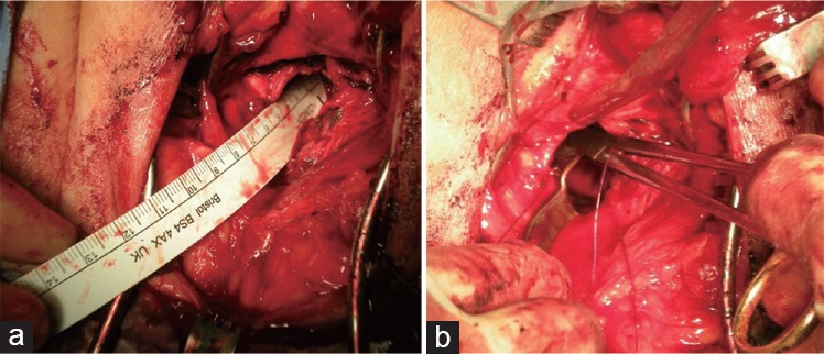
Redo vesicourethral anastomosis. (a) The neobladder neck lies deep in the perineum (12 cm from skin level in this case) after excising the extensive “funneled” bladder neck contracture. (b) Consequently fashioning the anastomosis at that depth is technically very difficult.
References
-
- Allen SD, Thompson A, Aslam Sohaib S. The normal post-surgical anatomy of the male pelvis following radical prostatectomy as assessed by magnetic resonance imaging. Eur Radiol. 2008;18:1281–91. - PubMed
-
- Summers RM, Korobkin M, Quint LE, Ellis JH, Grossman HB, et al. Pelvic CT findings after radical prostatectomy. J Comput Asist Tomogr. 1993;17:767–71. - PubMed
-
- Kordan Y, Alkibay T, Sozen S, Bozkurt Y, Acar C, et al. Is there an impact of postoperative urethral and periurethral anatomical features in post-radical retropubic prostatectomy incontinence? Urol Int. 2007;78:208–13. - PubMed
-
- Tuygun C, Imamoglu A, Keyik B, Alisir I, Yorubulut M. Significance of fibrosis around and/or at external urinary sphincter on pelvic magnetic resonance imaging in patients with postprostatectomy incontinence. Urology. 2006;68:1308–12. - PubMed
MeSH terms
LinkOut - more resources
Full Text Sources
