

doi: 10.1001/jamadermatol.2019.1668.
Distinct Histopathologic Patterns of Finger Eruptions in Dermatomyositis Based on Myositis-Specific Autoantibody Profiles
Affiliations
- PMID: 31290941
- PMCID: PMC6624797
- DOI: 10.1001/jamadermatol.2019.1668
Item in Clipboard
Distinct Histopathologic Patterns of Finger Eruptions in Dermatomyositis Based on Myositis-Specific Autoantibody Profiles
JAMA Dermatol.
.
Abstract
This histologic analysis of cutaneous manifestations of dermatomyositis examines whether skin eruptions can be histopathologically classified into myositis-specific autoantibodies-associated groups.
Conflict of interest statement
Figures
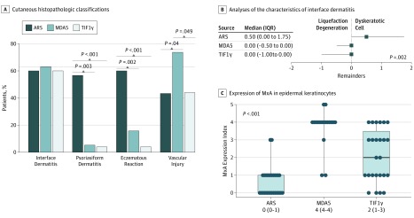
The percentages of patients grouped into anti–aminoacyl-transfer RNA synthetase (ARS, dark blue bars), antimelanoma differentiation-associated protein 5 (MDA5, blue bars), and antitranscriptional intermediary factor 1γ (TIF1γ, light blue bars) antibodies-associated dermatomyositis groups showing interface dermatitis, psoriasiform dermatitis, eczematous reaction, and vascular injury in the upper dermis, respectively. Interface dermatitis was determined using a total score of 2 or greater, which was evaluated with liquefaction degeneration of the basal layer and dyskeratotic cells (0, absent; 1, <5 cells per field [ × 100]; and 2, ≥5 cells per field, respectively). Psoriasiform dermatitis was determined by a total score of 2 or greater, which was evaluated with psoriasiform acanthosis (0, absent; 1, present) and parakeratosis (0, absent; 1, <50% of field; and 2, ≥50% of field). Eczematous reaction was determined by the presence or absence of spongiosis (absent, <50% of field; and present, ≥50% of field), and vascular injury was assessed by inflammatory cell infiltration into vessel walls and bleeding. B, The remainders (score for dyskeratotic cells minus the score for liquefaction degeneration of the basal layer) for each group were calculated. The squares and bars represent medians and interquartile ranges (IQRs), respectively. C, The MxA expression index in each group was evaluated by calculating the percentage of MxA-expressing epidermal areas (0, 0%; 1, 1%-24%; 2, 25%-49%; 3, 50%-74%; 4, 75%-99%; 5, 100%). The boxes containing lines and bars represent the IQRs with medians and full ranges, respectively. The dots indicate individual MxA values.
References
-
- Concha JSS, Merola JF, Fiorentino D, Werth VP. Re-examining mechanic’s hands as a characteristic skin finding in dermatomyositis. J Am Acad Dermatol. 2018;78(4):769-775.e2, e762. - PubMed
-
- Zhang SH, Zhao Y, Xie QB, Jiang Y, Wu YK, Yan B. Aberrant activation of type I interferon system may contribute to the pathogenesis of anti-MDA5 dermatomyositis. Br J Dermatol. 2018;180(5):1090-1098. - PubMed
