. 2019 Jul 10;14(7):e0215883.
doi: 10.1371/journal.pone.0215883. eCollection 2019.

Innate CD8αα+ cells promote ILC1-like intraepithelial lymphocyte homeostasis and intestinal inflammation

Affiliations

Innate CD8αα+ cells promote ILC1-like intraepithelial lymphocyte homeostasis and intestinal inflammation

Ali Nazmi et al. PLoS One. .

Abstract

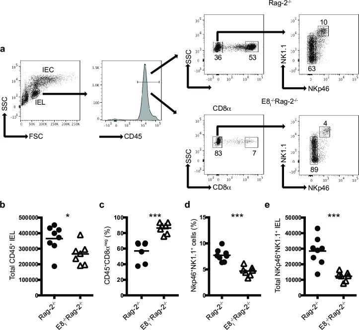
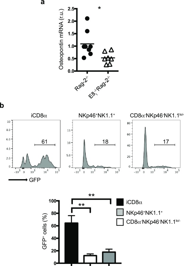
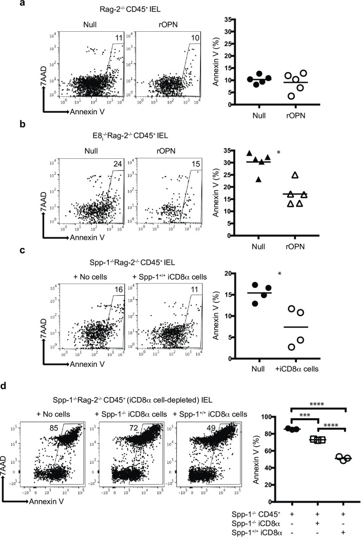
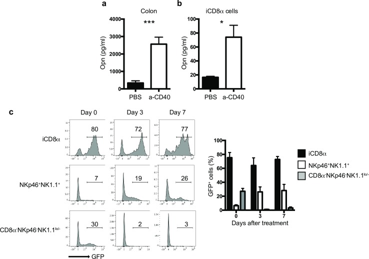
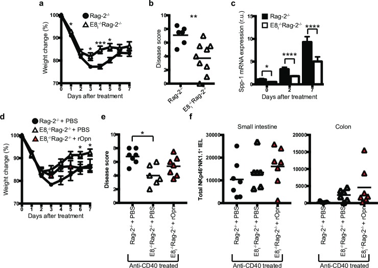
Innate CD8αα+ cells, also referred to as iCD8α cells, are TCR-negative intraepithelial lymphocytes (IEL) possessing cytokine and chemokine profiles and functions related to innate immune cells. iCD8α cells constitute an important source of osteopontin in the intestinal epithelium. Osteopontin is a pleiotropic cytokine with diverse roles in bone and tissue remodeling, but also has relevant functions in the homeostasis of immune cells. In this report, we present evidence for the role of iCD8α cells in the homeostasis of TCR-negative NKp46+NK1.1+ IEL (ILC1-like). We also show that the effect of iCD8α cells on ILC1-like IEL is enhanced in vitro by osteopontin. We show that in the absence of iCD8α cells, the number of NKp46+NK1.1+ IEL is significantly reduced. These ILC1-like cells are involved in intestinal pathogenesis in the anti-CD40 mouse model of intestinal inflammation. Reduced iCD8α cell numbers results in a milder form of intestinal inflammation in this disease model, whereas treatment with osteopontin increases disease severity. Collectively, our results suggest that iCD8α cells promote survival of NKp46+NK1.1+ IEL, which significantly impacts the development of intestinal inflammation.


Conflict of interest statement

The authors have declared that no competing interests exist.

Figures

Fig 1
Fig 1. iCD8α cell deficiency results in decreased NKp46+NK1.1+IEL numbers.
Total small intestine IEL from Rag-2-/- and E8I-/-Rag-2-/- mice were analyzed for the presence of CD45+ and CD45+CD8α-NKp46+NK1.1+ IEL. (a) Gating strategy for the analysis of the IEL compartment used throughout this report. Dead cells were excluded using a viability dye. (b) Total CD45+ IEL. (c) Frequencies of CD45+CD8αneg IEL. (d) frequencies and (e) total cell numbers of CD45+CD8αneg IEL derived from gating in the CD45+CD8αneg IEL population. Each symbol represents an individual mouse (n = 7 to 8). Data are representative of at least two independent experiments. *p<0.05, ***p<0.001 using unpaired two-tailed Student’s T test.
Fig 2
Fig 2. Osteopontin expression in the IEL compartment is primarily associated with iCD8α cells.
(a) Osteopontin mRNA expression in the intestine of the indicated mice. Expression levels in E8I-/-Rag-2-/- mice were compared to the average expression levels observed in Rag-2-/- mice. Each symbol represents an individual mouse (n = 7 to 8). Data are the combination of two independent experiments. (b) Osteopontin expression in naïve IEL from the small intestine (similar results were obtained with colon, not shown) of Rag-2-/-Spp-1-EGFP-KI mice. Cells were gated as in Fig 1. Histogram is a representative mouse (n = 4). Data are representative of at least two independent experiments. **p<0.01, using unpaired two-tailed Student’s T test for (a) and non-parametric one-way ANOVA for (b).
Fig 3
Fig 3. iCD8α cells and osteopontin promote survival of NKp46+NK1.1+ IEL.
Enriched CD45+ IEL from small intestine and colon of Rag-2-/- (a) or E8I-/-Rag-2-/- (b) mice were incubated in the presence or absence of recombinant osteopontin (2μg/ml final concentration). Cells were recovered 4 hours later, gated as described in the Materials and methods section, and analyzed for annexin V staining on gated NKp46+NK1.1+ IEL. Data are representative of at least two independent experiments (n = 4). (c) Enriched CD45+ cells from small intestine and colon of Spp-1-/-Rag-2-/- mice were incubated in the presence or absence of iCD8α cells derived from Rag-2-/- mice, and analyzed as described above. In order to obtain enough iCD8α cells, 2–3 mice were pooled and counted as one sample (n = 4). Data is representative of at least 2 experiments. (d) Enriched CD45+ cells depleted from iCD8α cells from small intestine and colon of Spp-1-/-Rag-2-/- mice were incubated in the presence or absence of iCD8α cells derived from small intestine and colon of Rag-2-/- or Spp-1-/-Rag-2-/- mice, and analyzed as described above. *p<0.05; ***p<0.001; ****p<0.0001 using unpaired two-tailed Student’s T test.
Fig 4
Fig 4. Osteopontin kinetics during intestinal inflammation.
Osteopontin protein concentration in the supernatants of (a) whole colon tissue or (b) enriched iCD8α cell cultures from naïve and anti-CD40-treated Rag-2-/- mice. Data is representative of at least 2 experiments (n = 5). In order to obtain enough iCD8α cells, 2–3 mice were pooled and counted as one sample (n = 3). (c) GFP expression in the small intestine (similar results were obtained with colon, not shown) of Rag-2-/-Spp-1-EGFP-KI mice treated with anti-CD40 and analyzed at the indicated time points. Cells were gated as indicated in Fig 1A. Histograms are from a representative sample (n = 3). Bar graph shows data summary. *p<0.05, ***p<0.001 using unpaired two-tailed Student’s T test.
Fig 5
Fig 5. Decreased intestinal inflammation in mice deficient in iCD8α cells.
Rag-2-/- and E8I-/-Rag-2-/- mice were treated with anti-CD40 and monitored for 7 days for weight change (a). (b) At the endpoint, colons were harvested for pathological analysis. (c) Osteopontin mRNA expression from the colons of anti-CD40 treated Rag-2-/- and E8I-/-Rag-2-/- mice at the indicated time points. Data is representative of at least two independent experiments (n = 6 to 8). (d) E8I-/-Rag-2-/- mice were treated with recombinant osteopontin or PBS at day -2, -1 and 1 before and after disease induction with 70μg of anti-CD40 antibodies, and their weights monitored for 7 days. (e) At the endpoint, colons were harvested for pathological analysis. Data is representative of at least 3 independent experiments (n = 6 to7). (f) Total NKp46+NK1.1+ IEL in the indicated mice after day 7 of anti-CD40 treatment. Data is representative of at least 3 independent experiments (n = 6 to7). Each symbol represents and individual mouse. *p<0.05, **p<0.01, ***p<0.01 using two-way ANOVA (a, d), unpaired two-tailed Student’s T test (b, c) or one-way ANOVA (e).

Similar articles

Cited by

References

    1. Van Kaer L, Olivares-Villagomez D. Development, Homeostasis, and Functions of Intestinal Intraepithelial Lymphocytes. J Immunol. 2018;200(7):2235–44. Epub 2018/03/21. 10.4049/jimmunol.1701704 - DOI - PMC - PubMed
    1. Cheroutre H. Starting at the beginning: new perspectives on the biology of mucosal T cells. Annu Rev Immunol. 2004;22:217–46. 10.1146/annurev.immunol.22.012703.104522 . - DOI - PubMed
    1. Olivares-Villagomez D, Van Kaer L. Intestinal Intraepithelial Lymphocytes: Sentinels of the Mucosal Barrier. Trends Immunol. 2017. Epub 2017/12/10. 10.1016/j.it.2017.11.003 . - DOI - PMC - PubMed
    1. Chen Y, Chou K, Fuchs E, Havran WL, Boismenu R. Protection of the intestinal mucosa by intraepithelial gamma delta T cells. Proc Natl Acad Sci U S A. 2002;99(22):14338–43. 10.1073/pnas.212290499 . - DOI - PMC - PubMed
    1. Das G, Augustine MM, Das J, Bottomly K, Ray P, Ray A. An important regulatory role for CD4+CD8 alpha alpha T cells in the intestinal epithelial layer in the prevention of inflammatory bowel disease. Proc Natl Acad Sci U S A. 2003;100(9):5324–9. PMC1535703. 10.1073/pnas.0831037100 . - DOI - PMC - PubMed

Publication types

MeSH terms