. 2019 Oct;33(10):11096-11108.
doi: 10.1096/fj.201802663R. Epub 2019 Jul 10.

Doxorubicin-induced cardiomyocyte death is mediated by unchecked mitochondrial fission and mitophagy

Affiliations

Doxorubicin-induced cardiomyocyte death is mediated by unchecked mitochondrial fission and mitophagy

Michael P Catanzaro et al. FASEB J. 2019 Oct.

Abstract

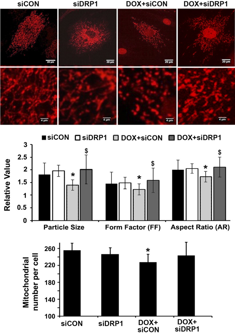
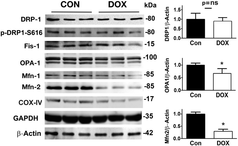
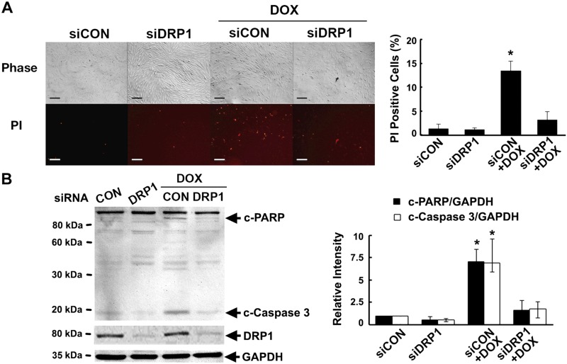
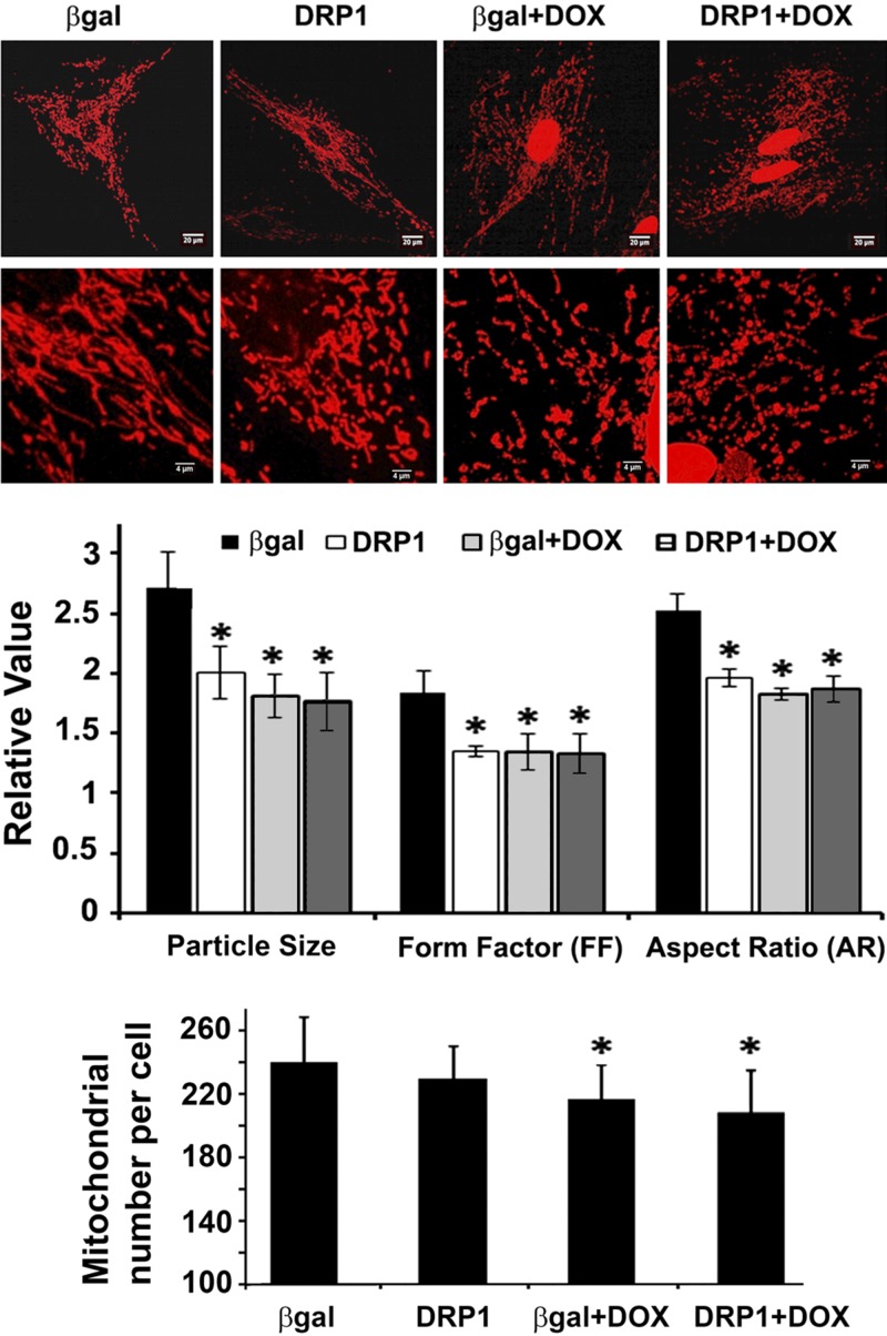
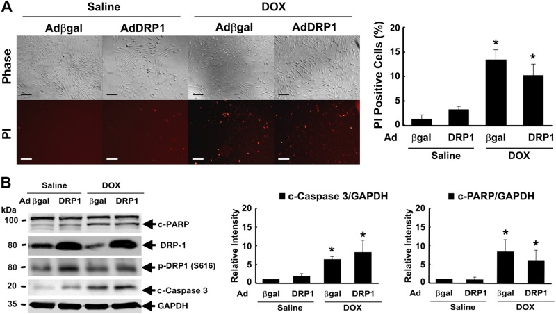
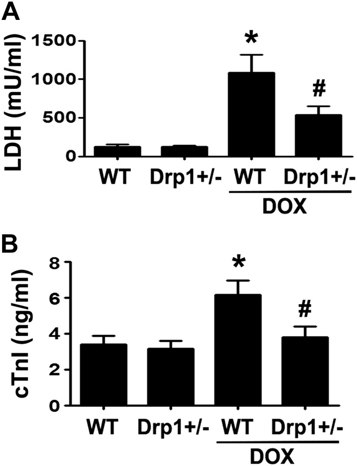
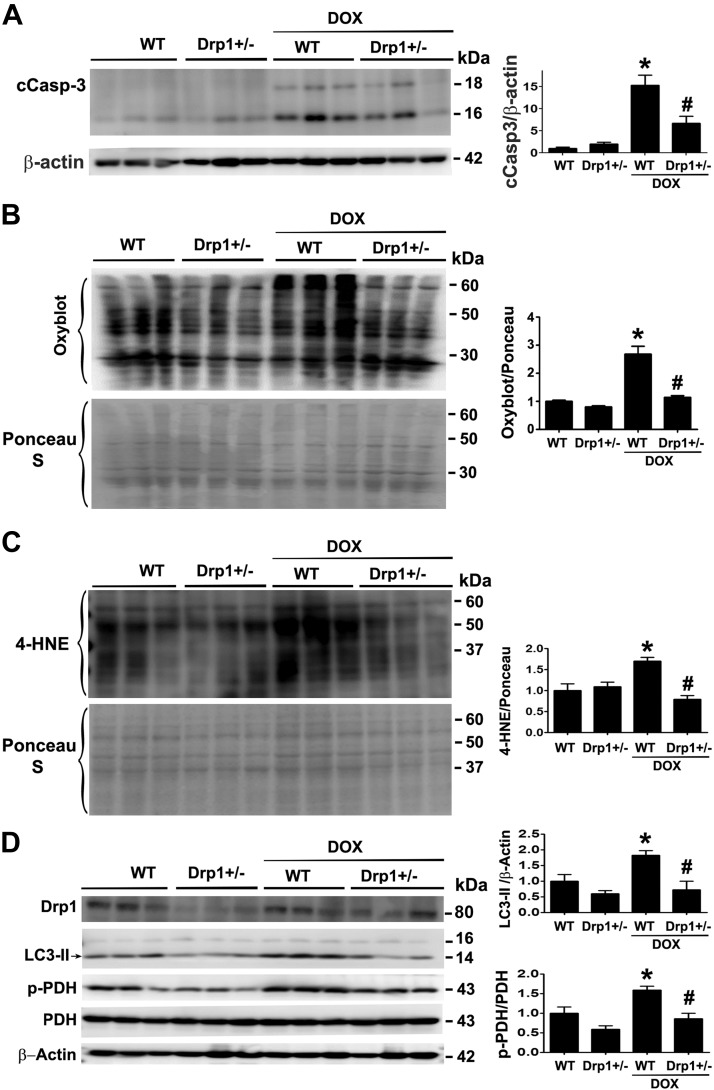
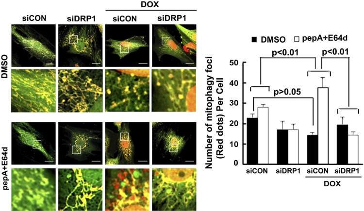
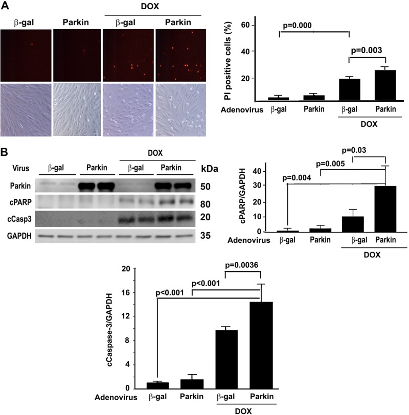
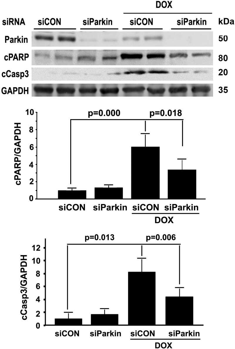
Doxorubicin (Dox) is a widely used antineoplastic agent that can cause heart failure. Dox cardiotoxicity is closely associated with mitochondrial damage. Mitochondrial fission and mitophagy are quality control mechanisms that normally help maintain a pool of healthy mitochondria. However, unchecked mitochondrial fission and mitophagy may compromise the viability of cardiomyocytes, predisposing them to cell death. Here, we tested this possibility by using Dox-treated H9c2 cardiac myoblast cells expressing either the mitochondria-targeted fluorescent protein MitoDsRed or the novel dual-fluorescent mitophagy reporter mt-Rosella. Dox induced mitochondrial fragmentation as shown by reduced form factor, aspect ratio, and mean mitochondrial size. This effect was abolished by short interference RNA-mediated knockdown of dynamin-related protein 1 (DRP1), a major regulator of fission. Importantly, DRP1 knockdown decreased cell death as indicated by the reduced number of propidium iodide-positive cells and the cleavage of caspase-3 and poly (ADP-ribose) polymerase. Moreover, DRP1-deficient mice were protected from Dox-induced cardiac damage, strongly supporting a role for DRP1-dependent mitochondrial fragmentation in Dox cardiotoxicity. In addition, Dox accelerated mitophagy flux, which was attenuated by DRP1 knockdown, as assessed by the mitophagy reporter mt-Rosella, suggesting the necessity of mitochondrial fragmentation in Dox-induced mitophagy. Knockdown of parkin, a positive regulator of mitophagy, dramatically diminished Dox-induced cell death, whereas overexpression of parkin had the opposite effect. Together, these results suggested that Dox cardiotoxicity was mediated, at least in part, by the increased mitochondrial fragmentation and accelerated mitochondrial degradation by the lysosome. Strategies that limit mitochondrial fission and mitophagy in the physiologic range may help reduce Dox cardiotoxicity.-Catanzaro, M. P., Weiner, A., Kaminaris, A., Li, C., Cai, F., Zhao, F., Kobayashi, S., Kobayashi, T., Huang, Y., Sesaki, H., Liang, Q. Doxorubicin-induced cardiomyocyte death is mediated by unchecked mitochondrial fission and mitophagy.

Keywords: anti-tumor agents; cardiotoxicity; heart failure; mitochondrial quality control.


Conflict of interest statement

Q.L. was supported by U.S. National Institutes of Health, National Heart, Lung, and Blood Institute Grant 1R15HL137130-01A1, and S.K. was supported by American Heart Association Grant 15SDG25080077. The authors declare no conflicts of interest.

Figures

Figure 1
Figure 1
Drp1 siRNA attenuated Dox-induced mitochondrial fragmentation. H9c2 cells were cultured, infected with AdDsRed, transfected with siDRP1 or control siRNA (siCon), and then exposed to either Dox (750 nM) or saline for 24 h. Mitochondrial morphology was analyzed using ImageJ and the following parameters were calculated: mitochondrial particle number and size, mean FF, and AR. At least 5 images (each containing 5–15 cells) were captured per treatment over 3 separate experiments. Data are expressed as means ± se and were analyzed by ANOVA. *P < 0.01 vs. control (n = 3–4).
Figure 2
Figure 2
Dox altered the protein expressions of mitochondrial fission and fusion factors. H9c2 cells were treated with either Dox (750 nM) or saline for 24 h. Mitochondrial fission and fusion factors were determined by Western blot analysis. GAPDH, glyceraldegyde 3-phosphate dehydrogenase. Data are expressed as means ± se and were analyzed by 1-way ANOVA. *P < 0.05 (n = 6 for Drp1 and n = 3 for Opa1 and Mfn2).
Figure 3
Figure 3
DRP1 knockdown protected against Dox-induced cardiomyocyte death. H9c2 cells were pretreated with siDRP1 or control siRNA (siCon) for 24 h and then exposed to either Dox (750 nM) or saline for an additional 24 h. Cell death was determined by PI staining (A) and cleavage of caspase-3 and PARP (B). Scale bars, 200 μm. Data are expressed as means ± se and were analyzed by 1-way ANOVA. *P < 0.05 vs. control (n = 3–4).
Figure 4
Figure 4
DRP1 overexpression did not aggravate Dox-induced mitochondrial fragmentation. H9c2 cells were infected with AdDsRed for 48 h, followed by the infection of AdDRP1 or adenovirus expressing β-galactosidase (Adβ-gal) for 48 h. The cells were then exposed to either Dox (750 nM) or saline for 24 h. Mitochondrial morphology was observed with confocal microscopy and analyzed using ImageJ. The following parameters were calculated: mitochondrial particle number and size, mean FF, and AR. At least 5 images (each containing 5–15 cells) were captured per treatment over 3 separate experiments. Data are expressed as means ± se and were analyzed by 1-way ANOVA. *P < 0.05 vs. β-gal (n = 3).
Figure 5
Figure 5
DRP1 overexpression had no further effect on Dox-induced cell death. H9c2 cells were infected with AdDRP1or adenovirus expressing β-galactosidase (Adβ-gal) for 24 h and then exposed to either Dox (750 nM) or saline for 24 h. Cell death was determined by PI staining (A) and cleavage of caspase-3 and PARP (B). Scale bars, 200 μm. Data are expressed as means ± se and were analyzed by 1-way ANOVA. *P < 0.05 vs. β-gal (n = 3).
Figure 6
Figure 6
DRP1 haploinsufficiency blocked Dox-induced release of LDH and cTnI into the bloodstream. The DRP1 heterozygous knockout mice (DRP1+/−) were treated for 3 d with saline or Dox (15 mg/kg body weight). The serum LDH activity (A) and the serum levels of cTnI (B) were determined by using commercial kits. Data are expressed as means ± se and were analyzed by 1-way ANOVA. *P < 0.01 vs. wild-type control, #P < 0.01 vs. Dox-treated wild type (n = 8–12).
Figure 7
Figure 7
DRP1 haploinsufficiency attenuated Dox-induced oxidative injuries in the mouse heart. The DRP1+/− mice were treated with saline or Dox (15 mg/kg body weight). Mice were euthanized 3 d later, and cardiac tissues were processed for Western blot analysis of cCasp3 (A), oxidized protein (B; Oxyblot), lipid peroxidation by-product 4-HNE (C), and phospho-PDH (D). Data are expressed as means ± se and were analyzed by 1-way ANOVA. *P < 0.01 vs. wild-type control, #P < 0.01 vs. Dox-treated wild-type (n = 6).
Figure 8
Figure 8
DRP1 haploinsufficiency attenuated Dox-induced mitochondrial fragmentation. Adult ventricular cardiomyocytes were isolated from wild-type and DRP1+/− mice, infected with AdDsRed, and treated with 3 µM Dox for 24 h. Mitochondrial morphology was analyzed using ImageJ. The major and minor axes as well as the size and number of mitochondria were calculated. At least 5 images (each containing 5–15 cells) were captured per treatment. WT, wild type. Data are expressed as means ± se and were analyzed by ANOVA. *P < 0.01 vs. control (n = 3).
Figure 9
Figure 9
DRP1 knockdown inhibited Dox-induced mitophagy flux. H9c2 cells were infected with the Admt-Rosella for 48 h. The cells were then transfected with siDRP1 or control siRNA (siCon) for 24 h and exposed to either Dox (750 nM) or saline for 24 h. Confocal images were captured, mitophagy was analyzed using ImageJ, and the number of mitophagy foci per cell was calculated. Between 3 and 8 images (totaling between 5 and 15 cells) were captured per treatment over 3 separate experiments. Scale bars, 20 µm. For evaluating mitophagy flux, experiments were repeated with lysosomal inhibitors (PepA and E64D) or DMSO 4 h after Dox treatment. Data are expressed as means ± se and were analyzed by 1-way ANOVA (P < 0.01 vs. siCon or siCon+Dox; n = 3).
Figure 10
Figure 10
Parkin overexpression exacerbated Dox-induced cell death. H9c2 cells were infected with adenovirus expressing parkin (Adparkin) or adenovirus expressing β-galactosidase (Adβ-gal) for 24 h and then exposed to either Dox (750 nM) or saline for 24 h. Cell death was determined by PI staining (A) and cleavage of caspase-3 and PARP (B). Data were expressed as means ± se and analyzed by 1-way ANOVA (n = 3 for each treatment). GAPDH, glyceraldehyde 3-phosphate dehydrogenase.
Figure 11
Figure 11
Parkin knockdown protected against Dox-induced cardiomyocyte death. H9c2 cells were pretreated with parkin-targeted siRNA (siParkin) or control siRNA (siCon) for 24 h and then exposed to either Dox (750 nM) or saline for an additional 24 h. Cell death was determined by the cleavage of caspase-3 and PARP. Data are expressed as means ± se and were analyzed by 1-way ANOVA (n = 3).

References

    1. Swain S. M., Whaley F. S., Ewer M. S. (2003) Congestive heart failure in patients treated with doxorubicin: a retrospective analysis of three trials. Cancer 97, 2869–2879 - PubMed
    1. Minotti G., Menna P., Salvatorelli E., Cairo G., Gianni L. (2004) Anthracyclines: molecular advances and pharmacologic developments in antitumor activity and cardiotoxicity. Pharmacol. Rev. 56, 185–229 - PubMed
    1. Van der Pal H. J., van Dalen E. C., Hauptmann M., Kok W. E., Caron H. N., van den Bos C., Oldenburger F., Koning C. C., van Leeuwen F. E., Kremer L. C. (2010) Cardiac function in 5-year survivors of childhood cancer: a long-term follow-up study. Arch. Intern. Med. 170, 1247–1255 - PubMed
    1. Maeda M. (2008) Late effects of childhood cancer: life-threatening issues. J. Nippon Med. Sch. 75, 320–324 - PubMed
    1. Link G., Tirosh R., Pinson A., Hershko C. (1996) Role of iron in the potentiation of anthracycline cardiotoxicity: identification of heart cell mitochondria as a major site of iron-anthracycline interaction. J. Lab. Clin. Med. 127, 272–278 - PubMed

Publication types

MeSH terms