. 2019 Sep;30(9):1659-1673.
doi: 10.1681/ASN.2018121244. Epub 2019 Jul 10.

The Role of the EGF Receptor in Sex Differences in Kidney Injury

Affiliations

The Role of the EGF Receptor in Sex Differences in Kidney Injury

Ming-Zhi Zhang et al. J Am Soc Nephrol. 2019 Sep.

Abstract

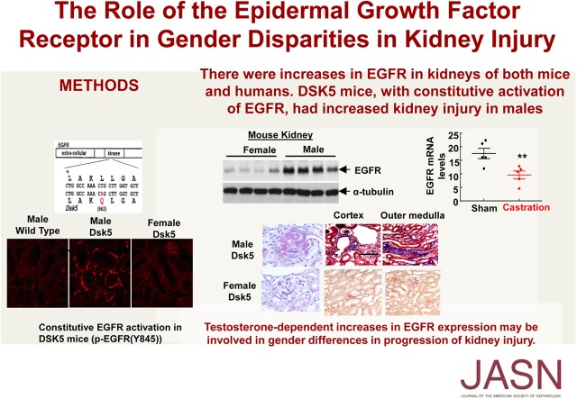
Background: Sex differences mediating predisposition to kidney injury are well known, with evidence indicating lower CKD incidence rates and slower decline in renal function in nondiabetic CKD for premenopausal women compared with men. However, signaling pathways involved have not been elucidated to date. The EGF receptor (EGFR) is widely expressed in the kidney in glomeruli and tubules, and persistent and dysregulated EGFR activation mediates progressive renal injury.

Methods: To investigate the sex differences in response to renal injury, we examined EGFR expression in mice, in human kidney tissue, and in cultured cell lines.

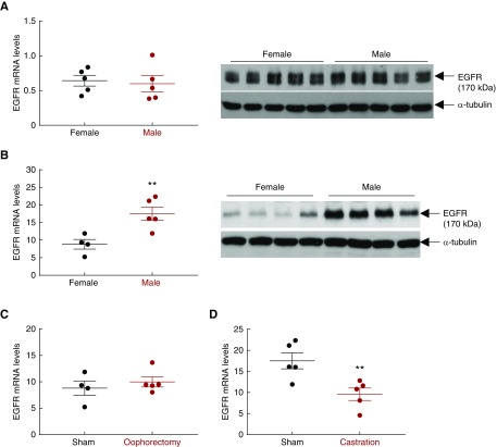
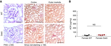
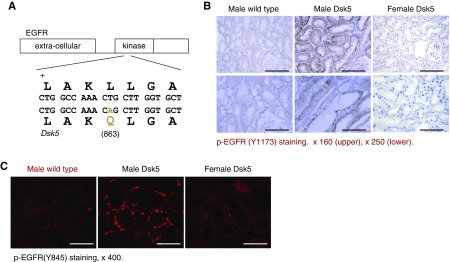
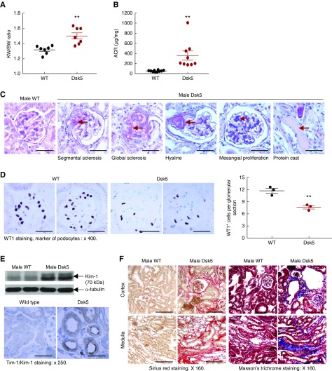
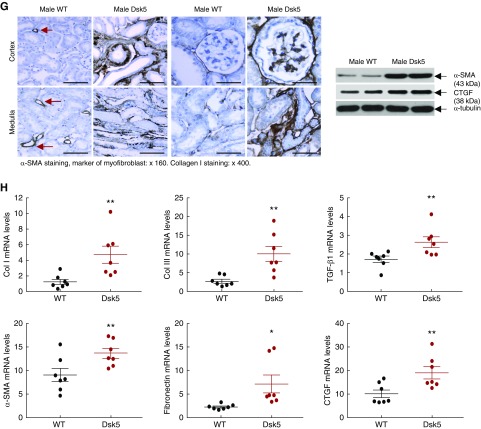
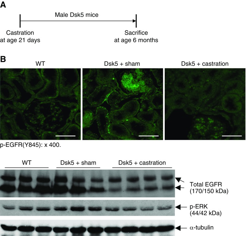
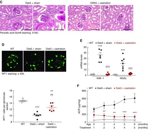
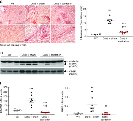
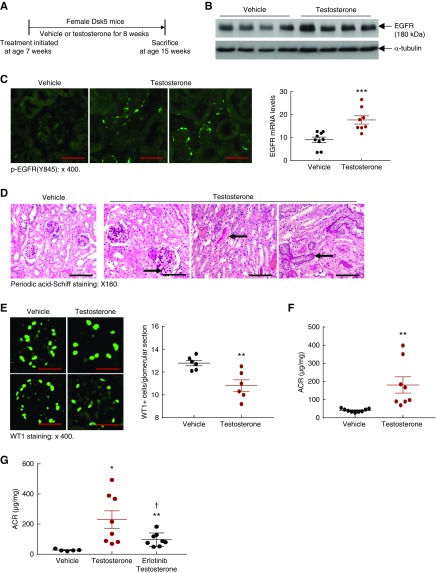
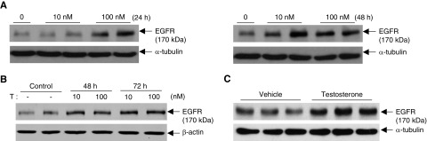
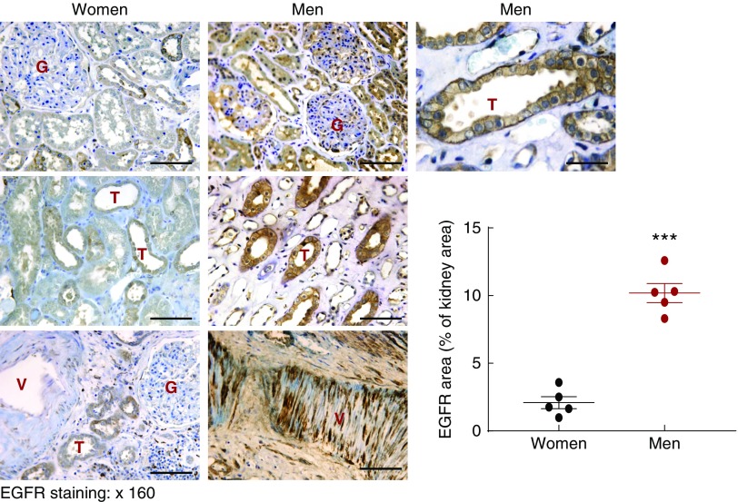
Results: In wild type mice, renal mRNA and protein EGFR levels were comparable in males and females at postnatal day 7 but were significantly lower in age-matched adult females than in adult males. Similar gender differences in renal EGFR expression were detected in normal adult human kidneys. In Dsk5 mutant mice with a gain-of-function allele that increases basal EGFR kinase activity, males had progressive glomerulopathy, albuminuria, loss of podocytes, and tubulointerstitial fibrosis, but female Dsk5 mice had minimal kidney injury. Oophorectomy had no effect on renal EGFR levels in female Dsk5 mice, while castration protected against the kidney injury in male Dsk5 mice, in association with a reduction in EGFR expression to levels seen in females. Conversely, testosterone increased EGFR expression and renal injury in female Dsk5 mice. Testosterone directly stimulated EGFR expression in cultured kidney cells.

Conclusions: These studies indicate that differential renal EGFR expression plays a role in the sex differences in susceptibility to progressive kidney injury that may be mediated at least in part by testosterone.

Keywords: DSK5; EGFR; glomerulosclerosis; testosterone; tubulointerstitial fibrosis.


Figures

None
Graphical abstract
Figure 1.
Figure 1.
Mouse kidney EGFR expression is androgen-dependent. (A) Renal EGFR mRNA and protein levels were comparable between wild-type male and female mice at prepubertal 7 days of age. n=5. (B) Renal EGFR mRNA and protein levels were significantly higher in wild-type male than female mice at postpubertal 3 months of age (**P<0.01; n=4 in female group and n=5 in male group). (C) Oophorectomy (bilateral removal of ovaries) of wild-type female mice at age 21 days had no effect on renal EGFR expression 6 weeks later. n=4 in sham group and n=5 in ovariectomized group. (D) Castration of wild-type male mice at age 21 days led to decreased renal EGFR expression 6 weeks later (**P<0.01; n=5 in each group).
Figure 2.
Figure 2.
Female Dsk5 mice had minimal kidney injury at 30 weeks of age. Female wild-type and Dsk5 mice were euthanized at 30 weeks of age. (A) Female Dsk5 mice had minimal glomerular injury and tubulointerstitial fibrosis. Original magnification, ×400 for periodic acid–Schiff (PAS) staining (scale bar, 25 µm), ×160 for Sirius Red staining (scale bar, 62.5 µm). (B) Albuminuria was comparable between female wild-type and Dsk5 mice. n=4 in each group.
Figure 3.
Figure 3.
Confirmation of basal activation of renal EGFR in Dsk5 mice. (A) Dsk5 mice have a Leu863Gln mutation within the kinase domain that stabilizes the receptor activation loop, producing a gain-of-function mutation of basal EGFR kinase activity. (B) Immunohistochemical staining determined increased renal basal EGFR activation in male but not female Dsk5 mice at 15 weeks of age as indicated by increased phospho-EGFR (Y1173) staining in male Dsk5 mice. Original magnification, ×160 for upper panel (scale bar, 62.5 6 µm) and ×250 (scale bar, 40 µm) for lower panel. (C) Immunofluorescence staining with another phospho-EGFR (Y845) antibody also confirmed increases in renal EGFR activation in male Dsk5 mice, although only minimal renal EGFR activation was found in age-matched male wild-type mice and female Dsk5 mice. Original magnification, ×400 (scale bar, 25 µm).
Figure 4.
Figure 4.
Male Dsk5 mice developed spontaneous kidney injury. Male wild-type and Dsk5 mice were euthanized at 15 weeks of age. (A) Male Dsk5 mice had kidney hypertrophy as indicated by increased kidney weight versus body weight (KW/BW) ratios (**P<0.01; n=7). (B) Male Dsk5 mice had increased albuminuria as indicated by increased urinary albumin-to-creatinine ratio (ACR) at 15 weeks of age (**P<0.01; n=10 in wild-type group and n=9 in Dsk5 group). (C) Period acid–Schiff staining showed that kidneys of male Dsk5 mice exhibited segmental and global glomerular sclerosis, mesangial expansion, hyaline, and protein casts. Original magnification, ×400 (scale bar, 25 µm). (D) Male Dsk5 mice had loss of podocytes as indicated by two representative photomicrographs from wild-type and Dsk5 mice (left panel) and quantitatively decreased WT1-positive cells (right panel; **P<0.01; n=3). Original magnification, ×400 (scale bar, 25 µm). (E) Male Dsk5 mice had epithelial cell injury as indicated by increased renal expression levels of KIM-1, a marker of proximal tubule epithelial injury. Original magnification, ×250 (scale bar, 40 µm). (F) Male Dsk5 mice exhibited tubulointerstitial fibrosis as indicated by Sirius Red and Masson trichrome staining. Original magnification, ×160 (scale bar, 62.5 µm). (G) Immunostaining and immunoblotting determined that male Dsk5 mice had increased renal expression levels of α-SMA, a marker of myofibroblasts, CTGF, and collagen I. Original magnification, ×160 for α-SMA (scale bar, 62.5 µm) and ×400 for collagen I (scale bar, 25 µm). Arrows indicate blood vessels. (H) Kidneys of male Dsk5 mice had increased mRNA levels of profibrotic and fibrotic components, including α-SMA, CTGF, TGF-β, collagen I and III (Col I and Col III), and fibronectin. *P<0.05; **P<0.01. n=7 in each group.
Figure 4.
Figure 4.
Male Dsk5 mice developed spontaneous kidney injury. Male wild-type and Dsk5 mice were euthanized at 15 weeks of age. (A) Male Dsk5 mice had kidney hypertrophy as indicated by increased kidney weight versus body weight (KW/BW) ratios (**P<0.01; n=7). (B) Male Dsk5 mice had increased albuminuria as indicated by increased urinary albumin-to-creatinine ratio (ACR) at 15 weeks of age (**P<0.01; n=10 in wild-type group and n=9 in Dsk5 group). (C) Period acid–Schiff staining showed that kidneys of male Dsk5 mice exhibited segmental and global glomerular sclerosis, mesangial expansion, hyaline, and protein casts. Original magnification, ×400 (scale bar, 25 µm). (D) Male Dsk5 mice had loss of podocytes as indicated by two representative photomicrographs from wild-type and Dsk5 mice (left panel) and quantitatively decreased WT1-positive cells (right panel; **P<0.01; n=3). Original magnification, ×400 (scale bar, 25 µm). (E) Male Dsk5 mice had epithelial cell injury as indicated by increased renal expression levels of KIM-1, a marker of proximal tubule epithelial injury. Original magnification, ×250 (scale bar, 40 µm). (F) Male Dsk5 mice exhibited tubulointerstitial fibrosis as indicated by Sirius Red and Masson trichrome staining. Original magnification, ×160 (scale bar, 62.5 µm). (G) Immunostaining and immunoblotting determined that male Dsk5 mice had increased renal expression levels of α-SMA, a marker of myofibroblasts, CTGF, and collagen I. Original magnification, ×160 for α-SMA (scale bar, 62.5 µm) and ×400 for collagen I (scale bar, 25 µm). Arrows indicate blood vessels. (H) Kidneys of male Dsk5 mice had increased mRNA levels of profibrotic and fibrotic components, including α-SMA, CTGF, TGF-β, collagen I and III (Col I and Col III), and fibronectin. *P<0.05; **P<0.01. n=7 in each group.
Figure 5.
Figure 5.
Castration led to decreases in renal EGFR expression and injury in male Dsk5 mice. (A) Male Dsk5 mice were castrated at 21 days of age and euthanized at 6 months of age. (B) Castration led to decreases in renal EGFR activation as indicated by phospho-EGFR (Y845) immunofluorescence staining, total renal EGFR expression, and p-ERK levels, a downstream signaling of EGFR activation. Original magnification, ×400 (scale bar, 20 µm). (C) Glomerular sclerosis, tubular dilation, and protein casts were apparent in sham male Dsk5 mice, whereas only mesangial expansion was seen in castrated male Dsk5 mice. Original magnification, ×160 (scale bar, 62.5 µm). (D) Loss of WT1 staining, a marker of podocytes, seen in male Dsk5 mice was significantly attenuated but was not completely prevented by castration (**P<0.01; ***P<0.001 versus wild-type group; ††P<0.01 versus sham Dsk5 group; n=6 in each group). Original magnification, ×400 (scale bar, 25 µm). (E) Tubular injury seen in male Dsk5 mice was dramatically attenuated by castration as indicated by significant reduction of renal expression levels of KIM-1 and NGAL, two markers of tubular injury (***P<0.001 versus wild-type group; †††P<0.001 versus sham Dsk5 group; n=8 in wild-type group and n=7 in other groups). (F) Castration prevented progressive albuminuria seen in male Dsk5 mice (*P<0.05; **P<0.01; ***P<0.001 versus sham Dsk5 mice; n=8 in wild-type group and n=7 in other groups). (G) Castration significantly reduced tubulointerstitial fibrosis in male Dsk5 mice as indicated by Sirius Red staining (***P<0.001 versus wild-type group; †††P<0.001 versus sham Dsk5 group; n=6). Original magnification, ×160 (scale bar, 80 µm). (H) Castration decreased renal expression levels of α-SMA and CTGF in male Dsk5 mice. (I) Male Dsk5 mice had significant increases in renal expression levels of HB-EGF and amphiregulin (AREG), two ligands for EGFR, which were significantly attenuated by castration (**P<0.01, ***P<0.001 versus wild-type group; ††P<0.01, †††P<0.001 versus sham Dsk5 group; n=8 in wild-type group and n=7 in other groups).
Figure 5.
Figure 5.
Castration led to decreases in renal EGFR expression and injury in male Dsk5 mice. (A) Male Dsk5 mice were castrated at 21 days of age and euthanized at 6 months of age. (B) Castration led to decreases in renal EGFR activation as indicated by phospho-EGFR (Y845) immunofluorescence staining, total renal EGFR expression, and p-ERK levels, a downstream signaling of EGFR activation. Original magnification, ×400 (scale bar, 20 µm). (C) Glomerular sclerosis, tubular dilation, and protein casts were apparent in sham male Dsk5 mice, whereas only mesangial expansion was seen in castrated male Dsk5 mice. Original magnification, ×160 (scale bar, 62.5 µm). (D) Loss of WT1 staining, a marker of podocytes, seen in male Dsk5 mice was significantly attenuated but was not completely prevented by castration (**P<0.01; ***P<0.001 versus wild-type group; ††P<0.01 versus sham Dsk5 group; n=6 in each group). Original magnification, ×400 (scale bar, 25 µm). (E) Tubular injury seen in male Dsk5 mice was dramatically attenuated by castration as indicated by significant reduction of renal expression levels of KIM-1 and NGAL, two markers of tubular injury (***P<0.001 versus wild-type group; †††P<0.001 versus sham Dsk5 group; n=8 in wild-type group and n=7 in other groups). (F) Castration prevented progressive albuminuria seen in male Dsk5 mice (*P<0.05; **P<0.01; ***P<0.001 versus sham Dsk5 mice; n=8 in wild-type group and n=7 in other groups). (G) Castration significantly reduced tubulointerstitial fibrosis in male Dsk5 mice as indicated by Sirius Red staining (***P<0.001 versus wild-type group; †††P<0.001 versus sham Dsk5 group; n=6). Original magnification, ×160 (scale bar, 80 µm). (H) Castration decreased renal expression levels of α-SMA and CTGF in male Dsk5 mice. (I) Male Dsk5 mice had significant increases in renal expression levels of HB-EGF and amphiregulin (AREG), two ligands for EGFR, which were significantly attenuated by castration (**P<0.01, ***P<0.001 versus wild-type group; ††P<0.01, †††P<0.001 versus sham Dsk5 group; n=8 in wild-type group and n=7 in other groups).
Figure 6.
Figure 6.
Testosterone supplementation led to increases in renal EGFR expression and injury in female Dsk5 mice. (A) Seven-week-old female Dsk5 mice were given testosterone at a dose of 10 mg/kg per day for 8 weeks and were euthanized at 15 weeks of age. (B and C) Testosterone administration led to increases in renal EGFR activation, mRNA, and protein levels (***P<0.001 versus vehicle; n=9 in vehicle group and n=8 in testosterone group). Original magnification, ×400 (scale bar, 25 µm). (D) Testosterone treatment led to mesangial expansion, protein casts, and tubular epithelial cell atrophy in female Dsk5 mice (arrows). Original magnification, ×160 (scale bar, 50 µm). (E) Testosterone administration caused loss WT1 staining, a marker of podocytes, in female Dsk5 mice (**P<0.01; n=6). Original magnification, ×400 (scale bar, 25 µm). (F) Testosterone supplementation led to increased albuminuria in female Dsk5 mice (**P<0.01; n=9 in vehicle group and n=8 in testosterone group). (G) Testosterone-induced albuminuria in female Dsk5 mice was prevented by erlotinib, an inhibitor of EGFR tyrosine kinase activity (*P<0.05, **P<0.01 versus vehicle group; P<0.05 versus testosterone group; n=5 in vehicle group and n=8 in testosterone and testosterone plus erlotinib groups).
Figure 7.
Figure 7.
Testosterone stimulated EGFR expression in cultured cells. (A) Human renal cortex proximal tubule epithelial cells were treated with testosterone for 24 and 48 hours. Testosterone stimulated EGFR expression at both time points. (B) Testosterone at 10 or 100 nM stimulated EGFR expression in differentiated podocytes at 48 and 72 hours. (C) Testosterone at 10 nM stimulated EGFR expression in differentiated mesangial cells at 48 hours.
Figure 8.
Figure 8.
Renal EGFR expression levels were higher in man than in women. Relatively normal human kidney sections from nephrectomy were stained with EGFR antibody. EGFR immunostaining was found in tubular epithelial cells (T), glomerulus (G), and arterials (V), and renal EGFR expression was higher in men and in women (***P<0.001; n=5 in each group). Original magnification, ×160 (scale bar, 50 µm).

Similar articles

Cited by

References

    1. Neugarten J, Acharya A, Silbiger SR: Effect of gender on the progression of nondiabetic renal disease: A meta-analysis. J Am Soc Nephrol 11: 319–329, 2000 - PubMed
    1. Yu M, Ryu DR, Kim SJ, Choi KB, Kang DH: Clinical implication of metabolic syndrome on chronic kidney disease depends on gender and menopausal status: Results from the Korean National Health and Nutrition Examination survey. Nephrol Dial Transplant 25: 469–477, 2010 - PubMed
    1. Benjamin EJ, Blaha MJ, Chiuve SE, Cushman M, Das SR, Deo R, et al. : Heart disease and stroke statistics-2017 update: A report from the American Heart Association. Circulation 135: e146–e603, 2017 - PMC - PubMed
    1. Maric-Bilkan C: Sex differences in micro- and macro-vascular complications of diabetes mellitus. Clin Sci (Lond) 131: 833–846, 2017 - PubMed
    1. Ricardo AC, Yang W, Sha D, Appel LJ, Chen J, Krousel-Wood M, et al. .: Sex-related disparities in CKD progression. J Am Soc Nephrol 30: 137–146, 2019 - PMC - PubMed

Publication types

MeSH terms