. 2019 Sep 12;93(19):e00202-19.
doi: 10.1128/JVI.00202-19. Print 2019 Oct 1.

Single-Dose Vaccination with a Hepatotropic Adeno-associated Virus Efficiently Localizes T Cell Immunity in the Liver with the Potential To Confer Rapid Protection against Hepatitis C Virus

Affiliations

Single-Dose Vaccination with a Hepatotropic Adeno-associated Virus Efficiently Localizes T Cell Immunity in the Liver with the Potential To Confer Rapid Protection against Hepatitis C Virus

Zelalem A Mekonnen et al. J Virol. .

Abstract

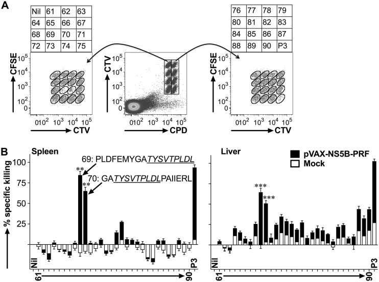
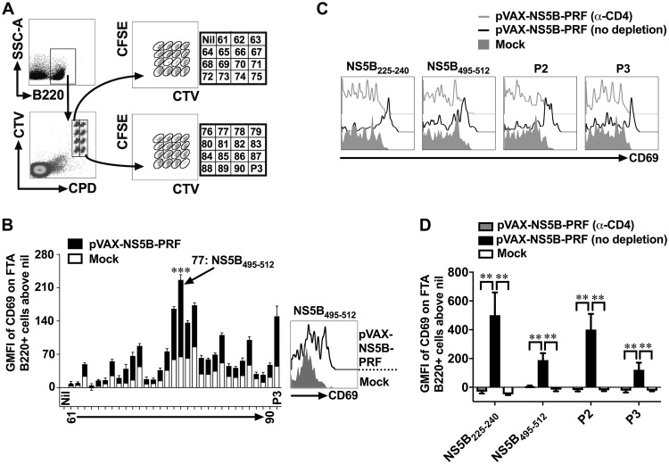
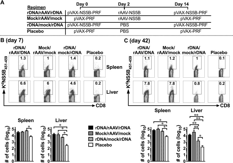
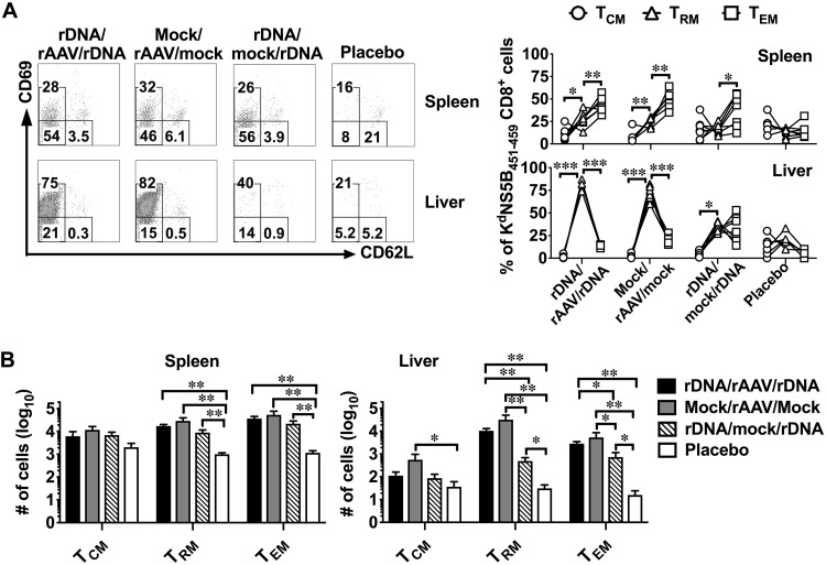
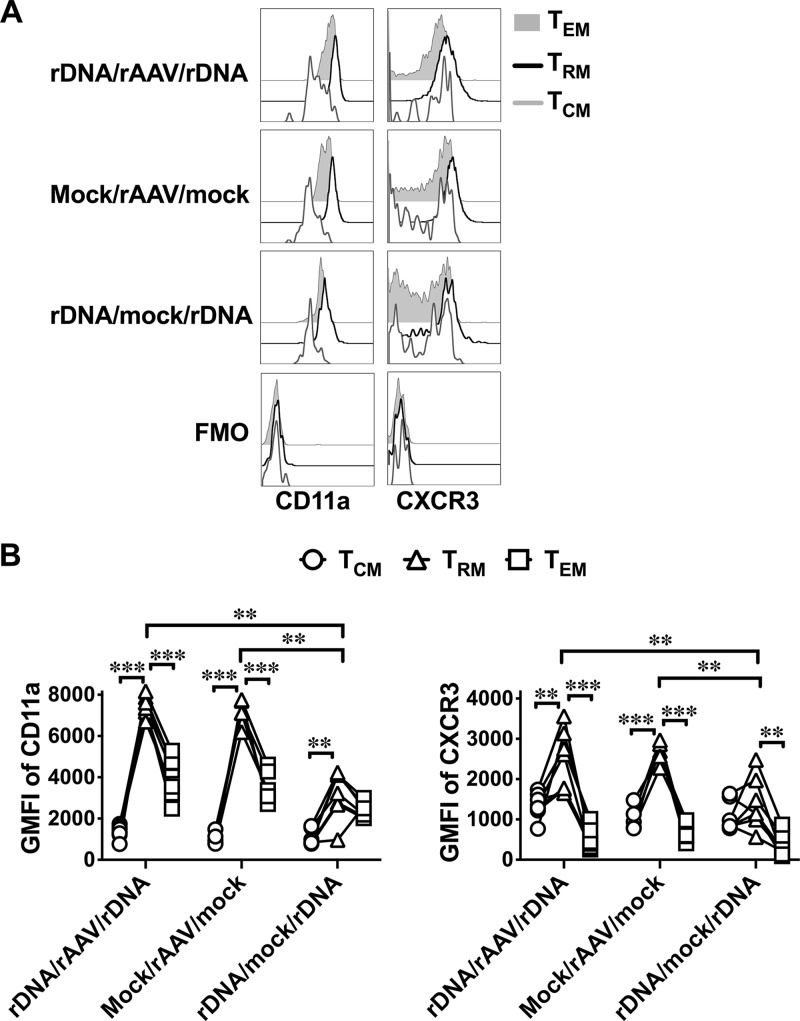
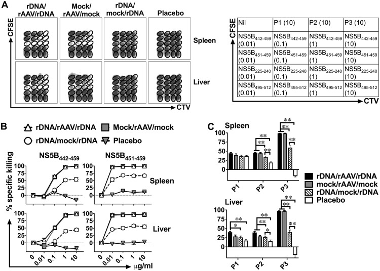
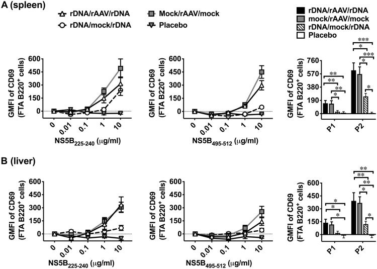
Hepatitis C virus (HCV) is a significant contributor to the global disease burden, and development of an effective vaccine is required to eliminate HCV infections worldwide. CD4+ and CD8+ T cell immunity correlates with viral clearance in primary HCV infection, and intrahepatic CD8+ tissue-resident memory T (TRM) cells provide lifelong and rapid protection against hepatotropic pathogens. Consequently, we aimed to develop a vaccine to elicit HCV-specific CD4+ and CD8+ T cells, including CD8+ TRM cells, in the liver, given that HCV primarily infects hepatocytes. To achieve this, we vaccinated wild-type BALB/c mice with a highly immunogenic cytolytic DNA vaccine encoding a model HCV (genotype 3a) nonstructural protein (NS5B) and a mutant perforin (pVAX-NS5B-PRF), as well as a recombinant adeno-associated virus (AAV) encoding NS5B (rAAV-NS5B). A novel fluorescent target array (FTA) was used to map immunodominant CD4+ T helper (TH) cell and cytotoxic CD8+ T cell epitopes of NS5B in vivo, which were subsequently used to design a KdNS5B451-459 tetramer and analyze NS5B-specific T cell responses in vaccinated mice in vivo The data showed that intradermal prime/boost vaccination with pVAX-NS5B-PRF was effective in eliciting TH and cytotoxic CD8+ T cell responses and intrahepatic CD8+ TRM cells, but a single intravenous dose of hepatotropic rAAV-NS5B was significantly more effective. As a T-cell-based vaccine against HCV should ideally result in localized T cell responses in the liver, this study describes primary observations in the context of HCV vaccination that can be used to achieve this goal.IMPORTANCE There are currently at least 71 million individuals with chronic HCV worldwide and almost two million new infections annually. Although the advent of direct-acting antivirals (DAAs) offers highly effective therapy, considerable remaining challenges argue against reliance on DAAs for HCV elimination, including high drug cost, poorly developed health infrastructure, low screening rates, and significant reinfection rates. Accordingly, development of an effective vaccine is crucial to HCV elimination. An HCV vaccine that elicits T cell immunity in the liver will be highly protective for the following reasons: (i) T cell responses against nonstructural proteins of the virus are associated with clearance of primary infection, and (ii) long-lived liver-resident T cells alone can protect against malaria infection of hepatocytes. Thus, in this study we exploit promising vaccination platforms to highlight strategies that can be used to evoke highly functional and long-lived T cell responses in the liver for protection against HCV.

Keywords: DNA vaccine; adeno-associated virus vaccine; cytotoxic T cells; helper T cells; hepatitis C virus vaccine; liver immunity.


Figures

FIG 1
FIG 1
FTA-based mapping analysis reveals immunodominant CD8+ T cell epitopes of gt3a NS5B in vivo. Female BALB/c mice (n = 7 per group) were immunized i.d. on three occasions at fortnightly intervals with 50 μg of rDNA-PRF encoding genotype 3a HCV NS5B (pVAX-NS5B-PRF) or rDNA-PRF lacking this sequence (pVAX-PRF [mock]). Thirteen days after the final immunization, mice were challenged i.v. with naive peptide-pulsed or unpulsed autologous splenocytes labeled with cell tracking dyes (cell trace violet [CTV], carboxyfluorescein succinimidyl ester [CFSE], and cell proliferation dye efluor670 [CPD]). The experimental details regarding how the FTA was constructed are described in the Materials and Methods and Table 1. (A) The representative flow cytometry plots show the fluorescently bar-coded target cell populations that were unpulsed (Nil), pulsed with an individual peptide (peptides 61 to 90) spanning NS5B385–591 or all the peptides spanning NS5B385–591 (P3), and recovered 15 h after the FTA challenge from the spleen of a pVAX-NS5B-PRF-vaccinated mouse. (B) Mean (n = 7) + SEM of the specific killing responses (see Materials and Methods for the formula used for the calculation) detected against each target cell population in the FTA recovered from the spleen (left panel) and the liver (right panel) from immunized mice for the experiment described in panel A. The sequences for the most immunodominant peptides (69 and 70) and the 9-aa sequence (NS5B451–459 [underlined]) used to synthesize a tetramer (KdNS5B451–459) in order to analyze NS5B-specific CD8+ T cells are shown. The asterisks above each bar corresponding to the mapped immunodominant peptides indicate statistically significant data for the comparisons of the killer responses between pVAX-NS5B-PRF and mock. **, P < 0.01; ***, P < 0.001.
FIG 2
FIG 2
FTA-based mapping analysis to identify immunodominant TH cell peptides of gt3a NS5B. Female BALB/c mice were vaccinated with pVAX-NS5B-PRF or mock vaccinated and challenged with an FTA comprising target cell populations pulsed with an individual peptide (peptides 61 to 90) or a peptide pool spanning NS5B385–591 (P3) as described in the legend to Fig. 1. (A) Representative dot plots showing how B220+ FTA cells recovered from the spleen of a pVAX-NS5B-PRF-vaccinated mouse were gated for analysis. (B) The bar graph shows the mean (n = 7) + SEM of the specific (beyond nil) expression (GMFI) of CD69 on gated B220+ cells within the FTA (as shown in panel A) recovered from the spleen of vaccinated mice. The representative histogram plot shows the expression of CD69 on gated B220+ FTA cells pulsed with the immunodominant NS5B495–512 peptide. The asterisks above the bar corresponding to the mapped immunodominant peptide indicate statistically significant data for the comparisons of the TH cell responses between pVAX-NS5B-PRF and mock vaccinated. ***, P < 0.001. (C and D) BALB/c mice were vaccinated with pVAX-NS5B-PRF or mock vaccinated as described for panel A. Thirteen days after the final (3rd dose) vaccination, 100 μg of anti-mouse CD4 depletion antibody was injected via the intraperitoneal (i.p.) route into each mouse from a cohort (n = 7 to 9) of pVAX-NS5B-PRF-vaccinated mice 3 days prior to FTA challenge with target cells that were unpulsed or pulsed with NS5B225–240, NS5B495–512, P2, or P3. The representative histogram plots (C) show the expression of CD69 on peptide-pulsed B220+ FTA cells, and the bar graph (D) shows the mean (n = 7 to 9) specific expression (GMFI) of CD69 on B220+ cells depicted in panel C. **, P < 0.01.
FIG 3
FIG 3
rAAV‐NS5B immunization promotes durable, NS5B‐specific intrahepatic CD8+ T cell responses. Female BALB/c mice were immunized with pVAX-NS5B-PRF (“rDNA” on the figure) or rAAV-NS5B (“rAAV” in the figure) and/or mock immunized using the vaccination regimens depicted in panel A. Seven (B) and 42 (C) days after the final vaccination, NS5B-specific CD8+ T cells derived from the spleen and the liver were analyzed using KdNS5B451–459 tetramer staining and flow cytometry. The numbers in the representative dot plots show the percentage of live CD8+ cells in the spleen and the liver that stained positive for the KdNS5B451–459 tetramer. The bar graphs show the mean (n = 3 to 7) absolute number of splenic and intrahepatic NS5B451–459-specific CD8+ T cells. Only comparisons between vaccinated groups that had a statistically significant difference are denoted: *, P < 0.05; and **, P < 0.01.
FIG 4
FIG 4
Evaluation of NS5B-specific memory CD8+ T cells reveals that rAAV-NS5B vaccination is necessary to elicit the highest numbers of intrahepatic TRM cells. Female BALB/c mice were vaccinated as described in the legend to Fig. 3A, and 42 days after the final vaccination, NS5B451–459-specific CD8+ T cells were analyzed for the formation of CD69 CD62L+ (TCM), CD69+ CD62L (TRM), or CD69 CD62L (TEM) cells using flow cytometry. (A) Representative dot plots showing the expression of CD69 and CD62L on gated live CD8+ cells that stained positive for the KdNS5B451–459 tetramer in the spleen and the liver. The numbers in each gate denote the percentage of NS5B451–459-specific CD8+ T cells that are TCM, TRM, or TEM based on the expression of CD69 and CD62L as described above. The line graphs depict the percentage of NS5B451–459-specific CD8+ cells that are TCM, TRM, or TEM cells as determined using flow cytometry, with each line representing the analysis of an individual animal used in each vaccination group. (B) Mean (n = 5 to 7) absolute number of NS5B451-459-specific CD8+ TCM, TRM, and TEM cells that formed in the spleen and the liver of vaccinated mice described in panel A. Only comparisons between vaccinated groups that had a statistically significant difference are denoted: *, P < 0.05; **, P < 0.01; and ***, P < 0.001.
FIG 5
FIG 5
Among the different memory subsets of intrahepatic NS5B-specific CD8+ T cells, TRM cells express the highest levels of CD11a and CXCR3 in vaccinated mice. Female BALB/c mice vaccinated with pVAX-NS5B-PRF and/or rAAV-NS5B (Fig. 3A) were euthanized 42 days after the final vaccination, and the expression of CD11a and CXCR3 was analyzed on intrahepatic CD69 CD62L+ CD8+ (TCM), CD69+ CD62L CD8+ (TRM), or CD69 CD62L CD8+ (TEM) cells that stained positive for KdNS5B451–459 tetramer. (A) Representative histogram plots show the expression of CD11a or CXCR3 on NS5B-specific CD8+ TCM, TRM, or TEM cells. Fluorescent minus one (FMO) plots represent the background staining for CD11a and CXCR3. (B) GMFI of CD11a or CXCR3 in each vaccinated mouse from rDNA/rAAV/rDNA-, mock/rAAV/mock-, and rDNA/mock/rDNA-vaccinated groups. Only comparisons between vaccinated groups that had a statistically significant difference are denoted: **, P < 0.01; and ***, P < 0.001.
FIG 6
FIG 6
pVAX-NS5B-PRF- and rAAV-NS5B-based vaccination regimens elicit robust NS5B-specific killer CD8+ T cell responses in vivo. Female BALB/c mice vaccinated as described in the legend to Fig. 3A were challenged i.v. 42 days after the final vaccination with peptide-pulsed autologous target cells labeled in an FTA for flow cytometry analysis similar to that described in the legend to Fig. 1. The experimental details regarding how the FTA was constructed are described in Materials and Methods and Table 2. (A) The representative density plots and the table show that the FTA is comprised of target cell populations that were unpulsed (Nil) or pulsed with titrated concentrations of the mapped immunodominant T cell peptides of NS5B or 10 μg/ml of peptide pools spanning NS5B1–207 (P1), NS5B197–395 (P2), or NS5B385–591 (P3). (B and C) Mean (n = 7) specific (above nil targets) killing responses against target cells pulsed with immunodominant CD8+ T cell peptides (NS5B442–459 and NS5B451–459) or peptide pools of NS5B recovered from the spleen (B) and the liver (C) 15 h after the FTA challenge are shown. Only comparisons between vaccinated groups that had a statistically significant difference are denoted: *, P < 0.05; and **, P < 0.01. The P values for the data depicted in the line graphs are shown in Table 3.
FIG 7
FIG 7
rAAV-NS5B-based vaccination regimens also elicit superior NS5B-specific TH cell responses in vivo. Female BALB/c mice were vaccinated as described in the legend to Fig. 3A and challenged with the peptide-pulsed FTA described in the legend to Fig. 6. Mean (n = 7) specific GMFI of CD69 on B220+ cells in the FTA pulsed with immunodominant TH cell peptides (NS5B225–240 and NS5B495–512) or peptide pools of NS5B recovered from the spleen (A) and the liver (B) 15 h after the FTA challenge are shown. Only comparisons between vaccinated groups that had a statistically significant difference are denoted: *, P < 0.05; **, P < 0.01; and ***, P < 0.001. The P values for the data depicted in the line graphs are shown in Table 4.

References

    1. WHO. 2017. Global Hepatitis Report, 2017. https://www.who.int/hepatitis/publications/global-hepatitis-report2017/en/.
    1. WHO. 2016. Global health sector strategies for viral hepatitis 2016–2021. https://www.who.int/hepatitis/strategy2016-2021/ghss-hep/en/.
    1. Borgia SM, Hedskog C, Parhy B, Hyland RH, Stamm LM, Brainard DM, Subramanian MG, McHutchison JG, Mo H, Svarovskaia E, Shafran SD. 2018. Identification of a novel hepatitis C virus genotype from Punjab, India: expanding classification of hepatitis C virus into 8 genotypes. J Infect Dis 218:1722–1729. doi: 10.1093/infdis/jiy401. - DOI - PubMed
    1. Sacks-Davis R, Grebely J, Dore GJ, Osburn W, Cox AL, Rice TM, Spelman T, Bruneau J, Prins M, Kim AY, McGovern BH, Shoukry NH, Schinkel J, Allen TM, Morris M, Hajarizadeh B, Maher L, Lloyd AR, Page K, Hellard M, InC3 Study Group. 2015. Hepatitis C virus reinfection and spontaneous clearance of reinfection—the InC3 Study. J Infect Dis 212:1407–1419. doi: 10.1093/infdis/jiv220. - DOI - PMC - PubMed
    1. Baumert TF, Fauvelle C, Chen DY, Lauer GM. 2014. A prophylactic hepatitis C virus vaccine: a distant peak still worth climbing. J Hepatol 61:S34–S44. doi: 10.1016/j.jhep.2014.09.009. - DOI - PubMed

Publication types