. 2019 Jul 23;116(30):15007-15012.
doi: 10.1073/pnas.1907214116. Epub 2019 Jul 10.

Microfluidic protein isolation and sample preparation for high-resolution cryo-EM

Affiliations

Microfluidic protein isolation and sample preparation for high-resolution cryo-EM

Claudio Schmidli et al. Proc Natl Acad Sci U S A. .

Abstract

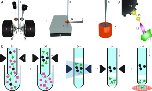
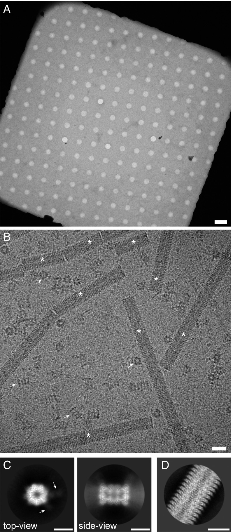
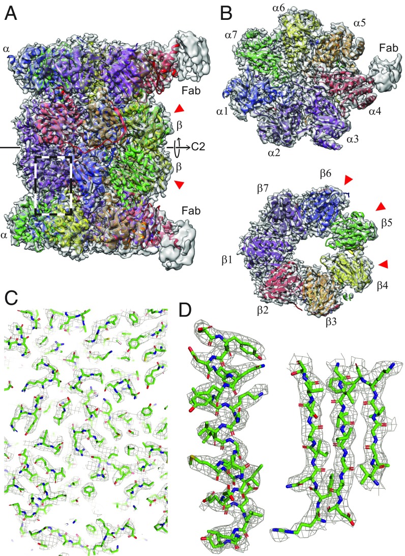
High-resolution structural information is essential to understand protein function. Protein-structure determination needs a considerable amount of protein, which can be challenging to produce, often involving harsh and lengthy procedures. In contrast, the several thousand to a few million protein particles required for structure determination by cryogenic electron microscopy (cryo-EM) can be provided by miniaturized systems. Here, we present a microfluidic method for the rapid isolation of a target protein and its direct preparation for cryo-EM. Less than 1 μL of cell lysate is required as starting material to solve the atomic structure of the untagged, endogenous human 20S proteasome. Our work paves the way for high-throughput structure determination of proteins from minimal amounts of cell lysate and opens more opportunities for the isolation of sensitive, endogenous protein complexes.

Keywords: cryo-EM; endogenous; microfluidics; protein purification; sample preparation.


Conflict of interest statement

Conflict of interest statement: H.S. and T.B. declare the following competing financial interest: The cryoWriter concept is part of patent application PCT/EP2015/065398. A former student will found a spin-off for the commercialization of the cryoWriter (March 2019).

Figures

Fig. 1.
Fig. 1.
Schematic workflow for microfluidic protein isolation and cryo-EM grid preparation. (A) Hardware for protein isolation and cryo-EM grid preparation. The electromagnetic trap consists of 2 electromagnets (1) that produce a strong magnetic field gradient via their water-cooled iron tips (2). Sample processing in the capillary (3) is monitored by a camera (4), and a UV LED (5) allows photo-cleavage (B) of the sample, both via mirrors. After protein isolation, the capillary nozzle is moved above a cryo-EM grid covered with a holey carbon film (6). The cryo-EM grid is positioned on a stage (7) that is temperature controlled by a Peltier element (8) and held with a Peltier-cooled tweezer (9). The isolated protein is directly written onto the grid and plunge-frozen in liquid ethane (10). (B) Composite material for “protein fishing.” The target protein (11) is recognized by a Fab (12) that is covalently modified by a photo-cleavable cross-linker (13). The linker molecule ends with a biotin moiety, which strongly binds to the streptavidin-coated bead (14). (C) Protein isolation workflow. (i) Magnetic beads are incubated with biotinylated Fabs and cell lysate to capture the target structures (green). Less than 900 nL of sample is aspirated into the microcapillary for the protein isolation. (ii) The magnetic beads are immobilized in the magnetic trap (1). Nonbound lysate components (red) are flushed out. (iii) Illumination with UV light breaks the cross-linker. Before photoelution, 2 air bubbles are introduced and serve as boundaries to avoid dilution of the released proteins by diffusion (see SI Appendix, Figs. S1 and S3 for details). (iv) Separation of the capturing magnetic beads and the eluted proteins. (v) The isolated target proteins are directly deposited on a cryo-EM grid for vitrification. The blue vertical arrows indicate the pump direction.
Fig. 2.
Fig. 2.
Sample quality and data collection. (A) Overview image of a grid square and the holey carbon film, showing a thin film of vitreous ice. (Scale bar: 4 μm.) (B) High-magnification image of the isolated 20S proteasome sample. Arrows indicate 20S proteasome as top and side views; asterisks denote TMV. Furthermore, small particles are visible in the background, most probably unbound Fabs. (Scale bar: 20 nm.) TMV was added to the elution buffer as a positive control for the cryo-EM grid quality (SI Appendix, Fig. S4). The contrast of the image was increased using a Gaussian blur and subsequent histogram adjustment. (C) Selected projection averages of the 20S proteasome. Arrows indicate 2 bound Fabs recognizing the α4 subunit. (Scale bar: 10 nm.) (D) Typical projection average of TMV from the same cryo-EM grid as the 20S proteasome. (Scale bar: 10 nm.)
Fig. 3.
Fig. 3.
The 3D reconstruction of the human 20S proteasome. The red arrowheads indicate the catalytically active subunits. (A) Side view showing the 2 α and 2 β rings. All 14 subunits are fitted into the mass densities. Parts of the 2 bound Fabs are visible at lower resolution, due to the high flexibility of the attached Fabs. The C2 symmetry axis is indicated. (B) Top view of an α and a β ring. Both rings have a pseudo-7-fold symmetry. Different subunits are indicated by different colors. (C) A zoom-in into the side-view region indicated by the dashed box in A, documenting the quality of the data and model fitting. An enlarged view of 1 α-helix and 3 strands of a β-sheet is shown in D as an example.

References

    1. Dubochet J., et al. , Cryo-electron microscopy of vitrified specimens. Q. Rev. Biophys. 21, 129–228 (1988). - PubMed
    1. Kühlbrandt W., Biochemistry. The resolution revolution. Science 343, 1443–1444 (2014). - PubMed
    1. Cheng Y., Single-particle cryo-EM-how did it get here and where will it go. Science 361, 876–880 (2018). - PMC - PubMed
    1. Frank J., Averaging of low exposure electron micrographs of non-periodic objects. Ultramicroscopy 1, 159–162 (1975). - PubMed
    1. van Heel M., Frank J., Use of multivariate statistics in analysing the images of biological macromolecules. Ultramicroscopy 6, 187–194 (1981). - PubMed

Publication types

MeSH terms

Substances