. 2019 Jul 10;51(7):1-14.
doi: 10.1038/s12276-019-0272-9.

Inhibition of acetylation of histones 3 and 4 attenuates aortic valve calcification

Affiliations

Inhibition of acetylation of histones 3 and 4 attenuates aortic valve calcification

Jia Gu et al. Exp Mol Med. .

Erratum in

Abstract

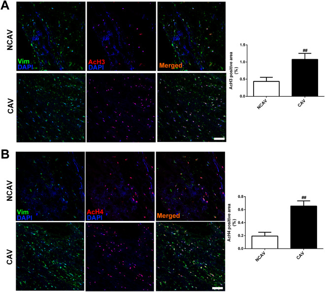
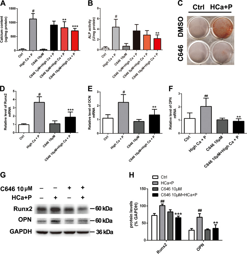
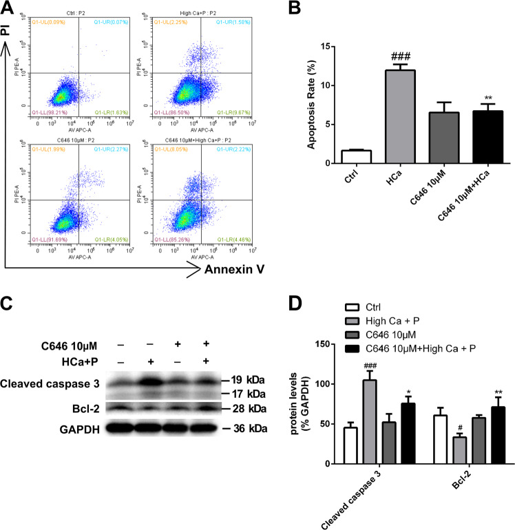
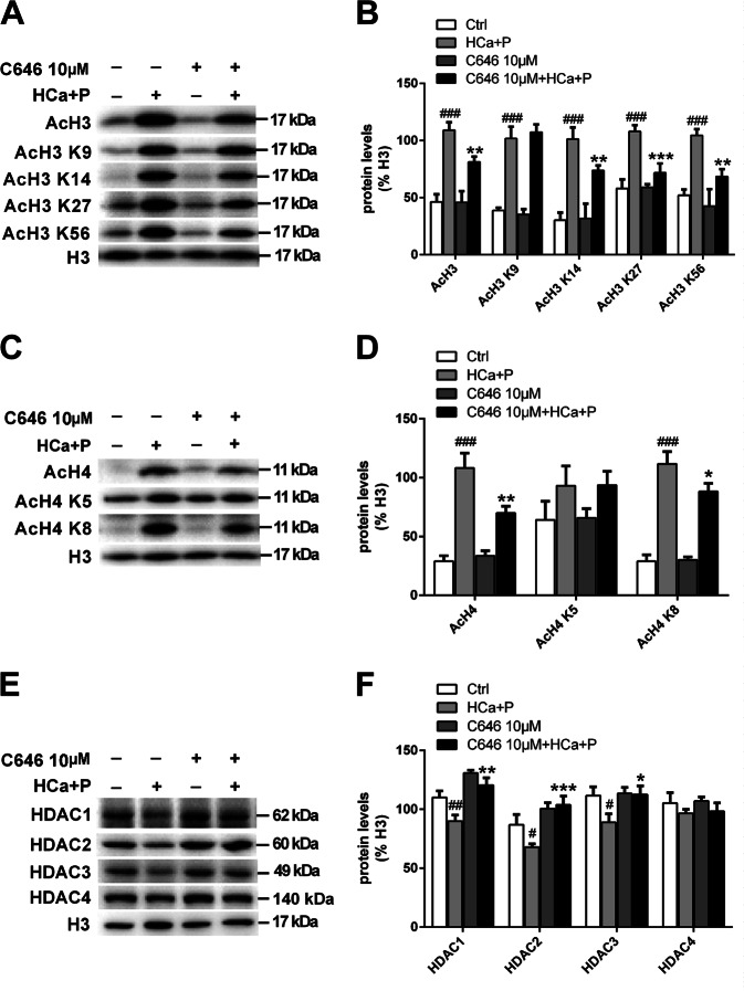
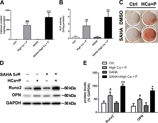
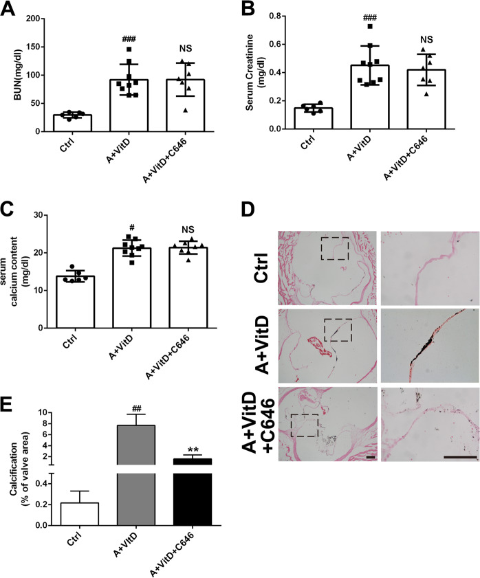
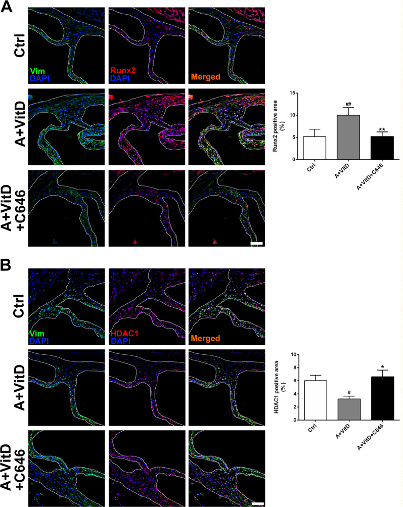
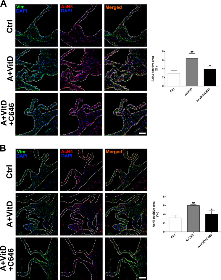
Aortic valve calcification develops in patients with chronic kidney disease who have calcium and phosphate metabolic disorders and poor prognoses. There is no effective treatment except valve replacement. However, metabolic disorders put patients at high risk for surgery. Increased acetylation of histones 3 and 4 is present in interstitial cells from human calcific aortic valves, but whether it is involved in aortic valve calcification has not been studied. In this study, we found that treating cultured porcine aortic valve interstitial cells with a high-calcium/high-phosphate medium induced calcium deposition, apoptosis, and expression of osteogenic marker genes, producing a phenotype resembling valve calcification in vivo. These phenotypic changes were attenuated by the histone acetyltransferase inhibitor C646. C646 treatment increased the levels of class I histone deacetylase members and decreased the acetylation of histones 3 and 4 induced by the high-calcium/high-phosphate treatment. Conversely, the histone deacetylase inhibitor suberoylanilide hydroxamic acid promoted valve interstitial cell calcification. In a mouse model of aortic valve calcification induced by adenine and vitamin D treatment, the levels of acetylated histones 3 and 4 were increased in the calcified aortic valves. Treatment of the models with C646 attenuated aortic valve calcification by restoring the levels of acetylated histones 3 and 4. These observations suggest that increased acetylation of histones 3 and 4 is part of the pathogenesis of aortic valve calcification associated with calcium and phosphate metabolic disorders. Targeting acetylated histones 3 and 4 may be a potential therapy for inoperable aortic valve calcification in chronic kidney disease patients.


Conflict of interest statement

The authors declare that they have no conflict of interest.

Figures

Fig. 1
Fig. 1. Levels of histone acetylation in human calcific and noncalcific aortic valves.
a Representative images of immunofluorescence staining of human aortic valves for AcH3. The expression of AcH3 was slightly higher in CAVs than in NCAVs. b Representative images of immunofluorescence staining of human aortic valves for AcH4. The expression of AcH4 was slightly higher in CAVs than in NCAVs. Scale bar = 50 μm. n = 3 for each group. ##p < 0.01 compared with NCAVs
Fig. 2
Fig. 2. The effect of C646 on pAVIC calcification induced by high-calcium/high-phosphate treatment.
a Calcium deposition was measured with a QuantiChromTM calcium assay kit. b ALP activity was measured to assess the degree of osteoblastic differentiation and calcification. c Calcium deposition was assessed by alizarin red staining. df The expression of Runx2, OPN, and OCN was assessed by qPCR. (G-H) The expression of Runx2 and OPN was assessed by western blotting. The relative protein levels were normalized to GAPDH levels. The data are shown as the means ± standard errors of the means of triplicates and are representative of three independent experiments performed. #p < 0.05, ##p < 0.01 compared with control. **p < 0.01, ***p < 0.001 compared with high Ca+P
Fig. 3
Fig. 3. The effect of C646 on apoptosis and proliferation of pAVICs after calcification.
a, b Flow cytometry analysis of apoptosis revealed that C646 inhibited apoptosis induced by high-calcium/high-phosphate treatment. The right bar graph shows the results of statistical analysis of the apoptosis ratios. c, d The expression of cleaved caspase 3 and Bcl-2 was assessed by western blotting. The relative protein levels were normalized to GAPDH levels. The data are shown as the means ± standard errors of the means of triplicates and are representative of three independent experiments performed (#p < 0.05, ###p < 0.001 compared with control; *p < 0.05, **p < 0.01 compared with high Ca+P)
Fig. 4
Fig. 4. HAT activity in pAVICs treated with high calcium/high phosphate.
a, b The expression of AcH3, AcH3 K9, AcH3 K14, AcH3 K27, and AcH3 K56 was assessed by western blotting. The relative protein levels were normalized to H3. c, d The expression of AcH4, AcH4 K5, and AcH4 K8 was assessed by western blotting. The relative protein levels were normalized to H3 levels. e, f The expression of HDAC1, HDAC2, HDAC3, and HDAC4 was assessed by western blotting. The relative protein levels were normalized to H3 levels. The data are shown as the means ± standard errors of the means of triplicates and are representative of three independent experiments performed. (#p < 0.05, ##p < 0.01, ###p < 0.001 compared with control; *p < 0.1, **p < 0.01, ***p < 0.001 compared with high Ca+P)
Fig. 5
Fig. 5. The effect of HDAC inhibition on pAVIC calcification induced by high-calcium/high-phosphate treatment.
a Calcium deposition was measured with a QuantiChromTM calcium assay kit. b ALP activity was measured to assess the degree of osteoblastic differentiation and calcification. c Calcium deposition was assessed by alizarin red staining. d, e The expression of Runx2 and OPN was assessed by western blotting. The relative protein levels were normalized to GAPDH levels. The data are shown as the means ± standard errors of the means of triplicates and are representative of three independent experiments performed (#p < 0.05, ##p < 0.01, ###p < 0.001 compared with control; *p < 0.05, **p < 0.01, ***p < 0.001 compared with high Ca+P)
Fig. 6
Fig. 6. The effect of C646 on calcification induced by adenine-containing chow and VitD injection.
a, b The effect of adenine treatment on BUN and serum creatinine concentrations. c Adenine and vitamin D treatment on serum calcium levels. d Histological von Kossa staining. e Quantification of calcification in the aortic valves. (Scale bar = 100 μm; n = 3 for each group; #p < 0.05, ###p < 0.001 compared with control)
Fig. 7
Fig. 7. The effects of HAT inhibition on osteogenesis and HDAC1.
a Representative images of immunofluorescence staining of mouse aortic valves for Runx2. b Representative images of immunofluorescence staining of mouse aortic valves for HDAC1 (scale bar = 50 μm; n = 3 for each group; #p < 0.05, ##p < 0.01 compared with control; *p < 0.05, **p < 0.01 compared with adenine+vitamin D)
Fig. 8
Fig. 8. The effect of HAT inhibition on histone acetylation.
a Representative images of immunofluorescence staining of mouse aortic valves for AcH3. b Representative images of immunofluorescence staining of mouse aortic valves for AcH4 (scale bar = 50 μm; n = 3 for each group; ##p < 0.01 compared with control; *p < 0.05 compared with adenine+vitamin D)

References

    1. Kim IY, et al. Cardiac valve calcification is associated with presence and severity of coronary artery disease in patients with pre-dialysis chronic kidney disease. Clin. Exp. Nephrol. 2015;19:1090–1097. doi: 10.1007/s10157-015-1104-4. - DOI - PubMed
    1. Raggi P, et al. All-cause mortality in hemodialysis patients with heart valve calcification. Clin. J. Am. Soc. Nephrol. 2011;6:1990–1995. doi: 10.2215/CJN.01140211. - DOI - PMC - PubMed
    1. Rattazzi M, et al. Aortic valve calcification in chronic kidney disease. Nephrol. Dial. Transplant. 2013;28:2968–2976. doi: 10.1093/ndt/gft310. - DOI - PubMed
    1. Sharma R, et al. Mitral annular calcification predicts mortality and coronary artery disease in end stage renal disease. Atherosclerosis. 2007;191:348–354. doi: 10.1016/j.atherosclerosis.2006.03.033. - DOI - PubMed
    1. Wang AY, et al. Cardiac valve calcification as an important predictor for all-cause mortality and cardiovascular mortality in long-term peritoneal dialysis patients: A prospective study. J. Am. Soc. Nephrol. 2003;14:159–168. doi: 10.1097/01.ASN.0000038685.95946.83. - DOI - PubMed

MeSH terms

Supplementary concepts