

Myosin binding protein H-like (MYBPHL): a promising biomarker to predict atrial damage
- PMID: 31292467
- PMCID: PMC6620353
- DOI: 10.1038/s41598-019-46123-w
Myosin binding protein H-like (MYBPHL): a promising biomarker to predict atrial damage
Abstract
Myosin binding protein H-like (MYBPHL) is a protein associated with myofilament structures in atrial tissue. The protein exists in two isoforms that share an identical amino acid sequence except for a deletion of 23 amino acids in isoform 2. In this study, MYBPHL was found to be expressed preferentially in atrial tissue. The expression of isoform 2 was almost exclusively restricted to the atria and barely detectable in the ventricle, arteria mammaria interna, and skeletal muscle. After atrial damage induced by cryo- or radiofrequency ablation, MYBPHL was rapidly and specifically released into the peripheral circulation in a time-dependent manner. The plasma MYBPHL concentration remained substantially elevated up to 24 hours after the arrival of patients at the intensive care unit. In addition, the recorded MYBPHL values were strongly correlated with those of the established biomarker CK-MB. In contrast, an increase in MYBPHL levels was not evident in patients undergoing aortic valve replacement or transcatheter aortic valve implantation. In these patients, the values remained virtually constant and never exceeded the concentration in the plasma of healthy controls. Our findings suggest that MYBPHL can be used as a precise and reliable biomarker to specifically predict atrial myocardial damage.
Conflict of interest statement
H.L., M.D., St.D., R.L. and M.K. are holders of a patent (No. 18 164 958.3) for MYBPHL as a biomarker for heart muscle damage. All other authors declare no competing interests.
Figures
 non-coding regions,
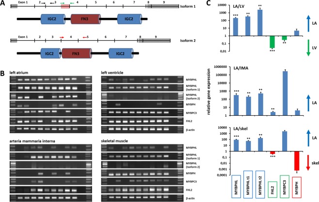
  deleted in isoform 2. Arrows indicate the location of primers used to amplify MYBPHL (black arrows), isoform 1 (green arrows) or isoform 2 (red arrows). (B) Gene expression in human LA, LV, arteria mammaria interna and skeletal muscle tissue. In each slot of the gel 20 µL of amplified fragments were loaded. (C) Quantification of relative gene expression in LA vs. LV, arteria mammaria interna and skeletal muscle tissue. Values are expressed as the mean ± SE. *p < 0.05, **p < 0.01, ***p < 0.001.
References
Publication types
MeSH terms
Substances
LinkOut - more resources
Full Text Sources
Other Literature Sources
Medical
Molecular Biology Databases
Research Materials
Miscellaneous
