. 2019 Jul 10;9(1):9973.
doi: 10.1038/s41598-019-45799-4.

New therapeutic target for pediatric anaplastic ependymoma control: study of anti-tumor activity by a Kunitz-type molecule, Amblyomin-X

Affiliations

New therapeutic target for pediatric anaplastic ependymoma control: study of anti-tumor activity by a Kunitz-type molecule, Amblyomin-X

Lorena Favaro Pavon et al. Sci Rep. .

Abstract

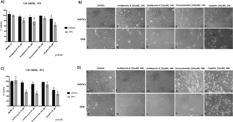
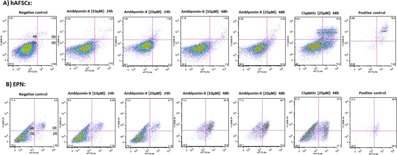
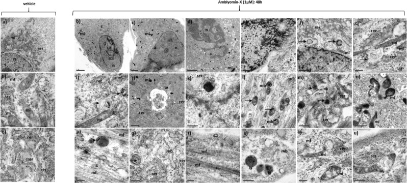
EPNs comprise a heterogeneous group of neuroepithelial tumors, accounting for about 10% of all intracranial tumors in children and up to 30% of brain tumors in those younger than 3 years. Actually, the pattern therapy for low-grade EPNs includes complete surgical resection followed by radiation therapy. Total surgical excision is often not possible due to tumor location. The aim of this study was to evaluate, for the first time, the anti-tumor activity of Amblyomin-X in 4 primary cultures derived from pediatric anaplastic posterior fossa EPN, Group A (anaplastic, WHO grade III) and one primary culture of a high grade neuroepithelial tumor with MN1 alteration, which was initially misdiagnosed as EPN: i) by in vitro assays: comparisons of temozolomide and cisplatin; ii) by intracranial xenograft model. Amblyomin-X was able to induce cell death in EPN cells in a more significant percentage compared to cisplatin. The cytotoxic effects of Amblyomin-X were not detected on hFSCs used as control, as opposed to cisplatin-treatment, which promoted a substantial effect in the hAFSCs viability. TEM analysis showed ultrastructural alterations related to the process of cell death: mitochondrial degeneration, autophagosomes and aggregate-like structures. MRI and histopathological analyzes demonstrated significant tumor mass regression. Our results suggest that Amblyomin-X has a selective effect on tumor cells by inducing apoptotic cell death and may be a therapeutic option for Group AEPNs.


Conflict of interest statement

The authors declare no competing interests.

Figures

Figure 1
Figure 1
(A) Cell viability assay of hFSCs and EPN cells treated with DMEM-LG (vehicle), Amblyomin-X (10 and 20 µM), Temozolomide (100 µM) and Cisplatin (20 µM) for 24 h. (B) Cytological aspects of hFSCs (b-e) and EPN cells (g-j) treated with Amblyomin-X (10 and 20 µM), Temozolomide (100 µM) and Cisplatin (20 µM) for 24 h of treatment. (C) Cell viability assay of hFSCs and EPN cells treated with DMEM-LG (control), Amblyomin-X (10 and 20 µM), Temozolomide (100 µM) and Cisplatin (20 µM) for 48 h. (D) Cytological aspects of hFSCs (l-o) and EPN cells (q-t) treated with Amblyomin-X (10 and 20 µM), Temozolomide (100 µM) and Cisplatin (20 µM) for 48 h of treatment. Cytological aspects of hFSCs (B.a;D.k) and EPN cells (B.f;D.p) no treated. Scale: 100 µm.
Figure 2
Figure 2
Induction of apoptosis by Amblyomin-X in (A) hFSCs and (A) EPN cells. The cells were incubated with 10 and 20 µM Amblyomin-X in DMEM-LG for 24 and 48 h, Cisplatin (20 µM) for 48 h and analyzed by flow cytometry using 7AAD and FITC-conjugated Annexin-V. It is worth mentioning that similar results were observed when we used cells derived from a neuroepithelial tumor. Quadrants 1–4: viable cells; early apoptotic cells; late apoptotic or necrotic cells; dead cells, respectively. Negative control: treatment with DMEM-LG; positive control to apoptosis: treatment with DMEM-LG and H2O2 [30%].
Figure 3
Figure 3
TEM of a primary cell culture of human EPNs treated with DMEM-LG (vehicle) (a, h, o) and primary cell culture of human EPNs treated (b-g, i-n, p-u) with Amblyomin-X (1.0 µM) in DMEM-LG for 48 h. n = nucleus; c = cytoplasm, nu = nucleoli; mi = mitochondria; rer = rough endoplasmic reticulum; crer = cisternae of rough endoplasmic reticulum; mt = microtubules; mf = microfilaments; mb = multivesicular bodies; ly = secondary lysosomes; black arrow = autophagosomes; white arrow 1 = aggregate-like structures; white arrow 2 = putative aggresomes. Scale: (a) 5.0μm; (b, c, d) 2.0μm; (e, f) 1.0μm; (g-u) 0,5 µm.
Figure 4
Figure 4
(A) a) Detection of MION-Rh labeled EPN cells by fluorescence assay. Magnification: 100×. (A) b) Stereotaxic implantation of MION-Rh labeled EPN cells in brain tumor experimental models. (A) c) Combined fluorescence and X-Ray tomography for in vivo detection of tumor (vehicle - saline) generate by infusion of MION-Rh labeled EPN cell. (A) d) Combined fluorescence and X-Ray tomography for in vivodetection of tumor treated with Amblyomin-X. (A) e) Radiofrequency bobine of animal positioning of the MRI neuroimaging equipment (2 Tesla): superconducting magnet 85310HR. (B) MRI monitoring of in vivo EPNs with no treatment (vehicle) and EPN with treatment (Amblyomin-X) of the case study 1 and 2 of the animals. Controls used are without any EPN cell transplantation, only sterile saline solution. Cases studies 1 and 2 represent fifteen independent experiments were performed. It is worth mentioning that similar results were observed when we used cells derived from a neuroepithelial tumor.
Figure 5
Figure 5
Immunopathological tumor analysis. a-i) EPN treated with vehicle. a’- I’) EPN with treatment of Amblyomin-X. a, a’) Histochemical analysis with Prussian Blue for MION-Rh detection in EPN cells.b-d; b’-d’) Hematoxylin and eosin staining.e,f;e’, f’) Immunohistochemical assay of tumor for glial fibrillary acidic protein (GFAP). g,h; g’-h’) Immunohistochemical assay of tumor for Ki67 for proliferation cellular detection. i, i’) Immunohistochemical assay of tumor for VEGF. White arrow: hemosiderin granules. Scale: 50 μm.

References

    1. Souweidane MM, Bouffet E, Finlay J. The role of chemotherapy in newly diagnosed ependymoma of childhood. PediatrNeurosurg. 1988;28:273–278. doi: 10.1159/000028664. - DOI - PubMed
    1. Bouffet E, Tabori U, Huang A, Bartels U. Ependymoma: lessons from the past, prospects for the future. Childs Nerv Syst. 2009;25:1383–1384. doi: 10.1007/s00381-009-0915-6. - DOI - PubMed
    1. Ostrom Quinn T., Gittleman Haley, Fulop Jordonna, Liu Max, Blanda Rachel, Kromer Courtney, Wolinsky Yingli, Kruchko Carol, Barnholtz-Sloan Jill S. CBTRUS Statistical Report: Primary Brain and Central Nervous System Tumors Diagnosed in the United States in 2008-2012. Neuro-Oncology. 2015;17(suppl 4):iv1–iv62. doi: 10.1093/neuonc/nov189. - DOI - PMC - PubMed
    1. Louis DN, et al. The 2016 World Health Organization Classification of Tumors of the Central Nervous System: a summary. ActaNeuropathol. 2016;131(6):803–820. doi: 10.1007/s00401-016-1545-1. - DOI - PubMed
    1. Mack SC, Taylor MD. The genetic and epigenetic basis of ependymoma. Childs Nerv Syst. 2009;25:1195–1201. doi: 10.1007/s00381-009-0928-1. - DOI - PubMed

Publication types

Substances

Supplementary concepts