. 2019 Jul 10;9(1):10021.
doi: 10.1038/s41598-019-46493-1.

Generation and characterization of a stable cell line persistently replicating and secreting the human hepatitis delta virus

Affiliations

Generation and characterization of a stable cell line persistently replicating and secreting the human hepatitis delta virus

Yi Ni et al. Sci Rep. .

Abstract

Human hepatitis delta virus (HDV) causes the most severe form of viral hepatitis. Approximately 15-25 million people are chronically infected with HDV. As a satellite virus of the human hepatitis B virus (HBV), HDV uses the HBV-encoded envelope proteins for egress from and de novo entry into hepatocytes. So far, in vitro production of HDV particles is restricted to co-transfection of cells with HDV/HBV encoding cDNAs. This approach has several limitations. In this study, we established HuH7-END cells, which continuously secrete infectious HDV virions. The cell line was generated through stepwise stable integration of the cDNA of the HDV antigenome, the genes for the HBV envelope proteins and the HBV/HDV receptor NTCP. We found that HuH7-END cells release infectious HDV particles up to 400 million copies/milliliter and support virus spread to co-cultured cells. Due to the expression of NTCP, HuH7-END cells are also susceptible to de novo HDV entry. Virus production is stable for >16 passages and can be scaled up for preparation of large HDV virus stocks. Finally, HuH7-END cells are suitable for screening of antiviral drugs targeting HDV replication. In summary, the HuH7-END cell line provides a novel tool to study HDV replication in vitro.


Conflict of interest statement

Stephan Urban is co-applicant and co-inventor on patents protecting HBV preS-derived lipopeptides (Myrcludex B) for the use of HBV/HDV entry inhibitors. The other authors in this study declare nothing to disclose.

Figures
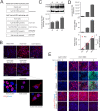
Figure 1
Figure 1
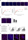
Establishment and characterization of HDV replicating cell lines. (A) Generation scheme of a HepG2-derived cell line stably expressing HDV (HepG2-HDV) and HuH7 cells expressing HDV (HuH7-HDV), HDV and the HBV envelope proteins (HuH7-HDV-Env) and HDV, HBV envelope proteins plus human NTCP (HuH7-HDV-Env-NTCP). Finally, a single cell clone of HuH7-HDV-Env-NTCP cells was named as HuH7-END. (B) Characterization of the HDV-replicating cell lines described above: HepG2-HDV, HuH7-HDV, HuH7-HDV-Env cells (top row) and HuH7-HDV-Env-NTCP (pool) and clone B1 (middle row) were seeded and stained for HDAg at day 1 post seeding (HDAg in red). Confocal image analyses of HDAg-stained single cells from the HuH7-END cells (lower row) revealed three distinguishable patterns of the subcellular location of HDAg. The numbers given in the pictures indicate the percentage of HDAg-positive cells. (C) Western blot analysis of HDAg (top) and quantification of intracellular HDV RNA (below) of HuH7-END cells at d3, d6 and d9 post seeding. (D) Analyses of particle secretion of HuH7-END cells. Cell culture medium of the indicated time frames were harvested and quantified for secreted HDV RNA (top), HBsAg (middle). The secreted infectious HDV is determined by the number of HDAg-positive HuH7-NTCP cell post-infection (bottom). For the latter the calculated MOI is shown as the red line. (E) Characterization of the HuH7-END cell line with respect to HDAg, HBV L-protein and expression of NTCP. HuH7-NTCP cells (d3 post seeding) and HuH7-END cells at d3, d6 and d9 post seeding were analyzed by IF for HDAg (row 1), HBV L-protein (row 2) and the merged pictures (row 3, HDAg in red, L protein in green). Nuclei were counterstained with Hoechst. Surface NTCP is labelled using 100 nM Atto565-labelled MyrB (row 4). Pretreatment of cells with 2 µM unlabelled MyrB (row 5) was used to ensure specific NTCP labelling.
Figure 2
Figure 2
Secretion of infectious HDV particles by HuH7-END cells. (A) HDAg-specific IF of HuH7-NTCP cells inoculated with different dilutions (1:40 to 1:2.5) of cell culture supernatants collected from HuH7-END cells between day 6–9 post seeding. HDAg-positive cells after infection were counted and depicted on the left corner of pictures. (B) HuH7-NTCP were infected with the culture supernatant of HuH7-END cells or a different source of HDV (heparin-column purified) at different MOI of HDV genome equivalents. The HDAg positive cells were counted (left) and the intracellular HDV RNA (right) were measured at 5 days post-infection. (C) Comparison of infectivity of HuH7-END derived HDV with virus obtained from co-transfection of HuH7 cells with pSVLD3 and pT7HB2.7: IF of HDAg in HuH7-NTCP cells after infection with cell culture supernatant of HuH7-END cells (collected between d6-9 post seeding and diluted 1:10) and SN (collected day 10–12 post transfection and diluted at 1:10) Infection rates were quantified by counting HDAg positive HuH7-NTCP cells at day 5 p.i. as shown on the right. (D) Scheme of large scale production and concentration of virus stocks produced from HuH7-END cells. (E) Characterization of concentrated virus stocks (according to the scheme depicted in C) collected at different time periods post seeding. Secreted HBsAg in IU/ml (left), HDV RNA in genome copies/ml (middle) and the infectivity of 1 µl concentrated virus (0.2% of inoculum) in HuH7-NTCP cells in 24-well plate were determined (right). (F) Comparative analysis of the infectivity of HuH7-END derived HDV in four HDV susceptible cell lines (HuH7-NTCP, HepG2-NTCP, differentiated HepaRG and differentiated HepaRG-NTCP). The number given in the pictures indicates the percentage of infected cells. The closing dashed lines indicate hepatic islands.
Figure 3
Figure 3
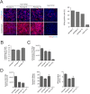
Stability of HDV replication and HDV secretion upon passaging of HuH7-END cells. (A) IF staining of HDAg in HuH7-END cells after different passages (passage 2, 8 and 16) in comparison to HDV replicating cell line HuH7-D12 (upper row). In addition, surface NTCP expression (lower row) was quantified using an Atto565-labelled MyrB derivative. The percentage of HDAg-expressing cells is depicted as bars at the right. (B) The copy number of intracellular HDV DNA from different passages were quantified by ddPCR in comparison to a single-cope gene RNase P. (C) Comparative quantification of intracellular HDV RNA of passage 2, 8 and 16 of HuH7-END versus HuH7-D12 cells. (D) Comparative analyses of secreted HDV RNA, HBsAg and infectious HDV in HuH7-END cells at passage 2, 8 and 16 in comparison to HuH7-D12 cells. Note that due to the lack of HBsAg (middle graph) HuH7-D12 cells are negative for secreted HDV RNA (left) and infectious virions (right).
Figure 4
Figure 4
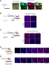
Spread of HDV from HuH7-END to susceptible co-cultured cell lines. (A) Co-cultivation of HuH7-END and HepG2-NTCP-GFP cells. HuH7-END cells were temporarily labelled via binding of Atto565-MyrB with NTCP and subsequently co-seeded with HepG2-NTCP-GFP cells (ratio 1:6). 6 hours post seeding, the two cell populations were visualized by GFP fluorescence (green identifying HepG2-NTCP-GFP cells) and Atto565-MyrB fluorescence (red identifying HuH7-END cells). 11 days post co-culture, cells were fixed and stained for HDAg (red) and GFP (green). Cells positive for HDAg (red) identify HuH7-END producer cells (white arrow) while cells positive for both HDAg and GFP (yellow arrows) are infected HepG2-NTCP-GFP cells (zoon-in picture on upper left), indicating that spread occurred. (B) Co-cultivation of HuH7-END cells with HuH7-NTCP in Transwell plate. HuH7-END cells grown in a Transwell insert for 6 days were co-cultured with HuH7-NTCP cells seeded on the bottom of well. Medium supplemented with entry inhibitor MyrB were used as a control. Eight days after co-culture, cells were stained for HDAg (red) and nuclei is counterstained by Hoechst (blue). (C) Co-cultivation of HuH7-END cells with HuH7-NTCP or HepG2-NTCP cells in coverslips. HuH7-END cells (seeded on coverslips for 6 days) and HuH7-NTCP or HepG2-NTCP cells (seeded on coverslips for 1 day) were co-cultured (left panels) in the presence or absence of the entry inhibitor MyrB. Eight days after co-culture, cells in coverslips were stained for HDAg (red) and nuclei is counterstained by Hoechst (blue) (right panels). (D) Kinetics of HDV spread determined by HDAg expression in HuH7-NTCP recipient cells co-cultured with HuH7-END cells. Left, two cell lines are pre-seeded in coverslips as shown in Fig. 4C and co-cultured for 11 days; Right, HDAg staining at different time points after co-seeding as shown by IF of HDAg (red) and nuclei (blue) in the recipient cell line HuH7-NTCP (the number given in the pictures indicates the percentage of infected cells). (E) Inhibition of HDV spread by MyrB in time course. Left, MyrB administration scheme (6 h, 1d, 2d, and 6d post co-culture 1 µM MyrB was added and replenished whenever medium was changed; Right, HDAg staining at different time points after co-seeding as shown by IF of HDAg (red) and nuclei (blue) in the recipient cell line HuH7-NTCP (the number given in the pictures indicates the percentage of infected cells).
Figure 5
Figure 5
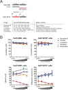
Infection of HuH7-END cells with genotype 3 HDV. (A) Alignment of genotype 1 and 3 HDV with respect to the primers used for quantification. The letter shown in the alignment is the binding site of three primer pairs (gt1, gt3 and universal) and the commonly used probe. (B) Scheme of HuH7-END cells infected with gt3 HDV (MOI of 300 genome equivalents/cell). HuH7-END cell or HuH7-NTCP cell were inoculated with gt3 HDV overnight. The cell at d7 p.i. were analyzed for intracellular HDV RNA using three primers pairs (C) and the culture supernatant between d4-7 post infection were quantified for gt1 or gt3 HDV (D).
Figure 6
Figure 6
Evaluation of drug efficacy using HuH7-END cells. (A) Schematic presentation of a combined drug screening approach using HuH7-END cells in 96-well plate. 5 different substances (MyrB, RG7834, Lonafarnib, IFN-alpha and IFN-lambda) with different modes-of-action (right) were administered for 6 days in HuH7-END cells. Following removal of the drug, cells were cultivated for another 2 days and the culture supernatant was analyzed for HBsAg and used to infect HuH7-NTCP cells. (B) Cell viability tested by WST-1 assay (upper panels) and quantification of HBsAg secretion in HuH7-END (lower left) and infectivity assay determined in HuH7-NTCP cells (lower right). NT, non-treated control.

Similar articles

Cited by

References

    1. Rizzetto M, et al. Immunofluorescence detection of new antigen-antibody system (delta/anti-delta) associated to hepatitis B virus in liver and in serum of HBsAg carriers. Gut. 1977;18:997–1003. doi: 10.1136/gut.18.12.997. - DOI - PMC - PubMed
    1. Hetzel, U. et al. Identification of a Novel Deltavirus in Boa Constrictors. MBio10, 10.1128/mBio.00014-19 (2019). - PMC - PubMed
    1. Wille Michelle, Netter Hans, Littlejohn Margaret, Yuen Lilly, Shi Mang, Eden John-Sebastian, Klaassen Marcel, Holmes Edward, Hurt Aeron. A Divergent Hepatitis D-Like Agent in Birds. Viruses. 2018;10(12):720. doi: 10.3390/v10120720. - DOI - PMC - PubMed
    1. Smedile A, et al. Influence of delta infection on severity of hepatitis B. Lancet. 1982;2:945–947. doi: 10.1016/S0140-6736(82)90156-8. - DOI - PubMed
    1. Bartenschlager R, Urban S, Protzer U. Towards curative therapy of chronic viral hepatitis. Z Gastroenterol. 2019;57:61–73. doi: 10.1055/a-0824-1576. - DOI - PubMed

Publication types