. 2019 Jul;33(7):665-675.
doi: 10.1007/s10822-019-00209-9. Epub 2019 Jul 10.

Structural analysis of the Aβ(11-42) amyloid fibril based on hydrophobicity distribution

Affiliations

Structural analysis of the Aβ(11-42) amyloid fibril based on hydrophobicity distribution

Irena Roterman et al. J Comput Aided Mol Des. 2019 Jul.

Abstract

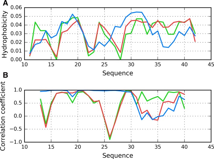
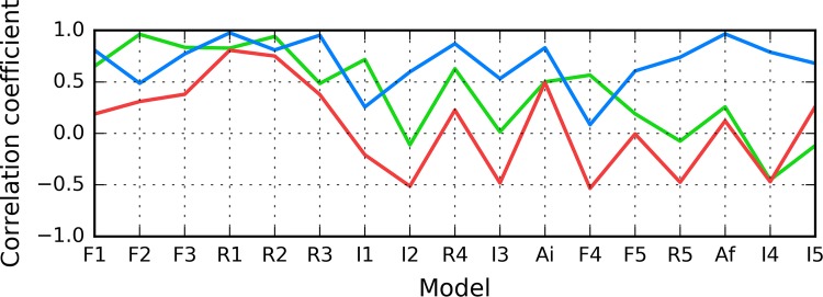
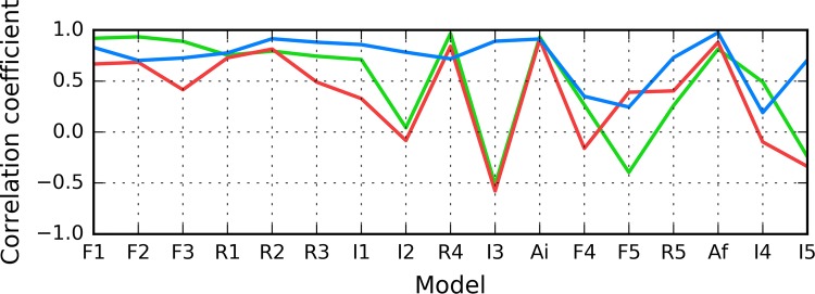
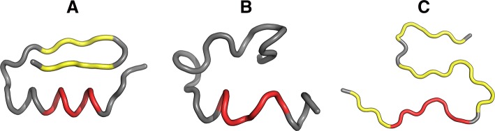
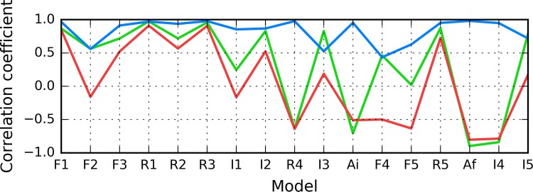
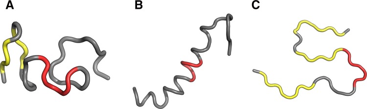
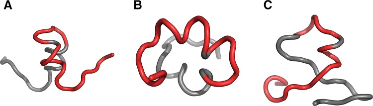
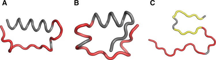
The structure of the Aβ(11-42) amyloid available in PDB makes possible the molecular analysis of the specificity of amyloid formation. This molecule (PDB ID 2MVX) is the object of analysis. This work presents the outcome of in silico experiments involving various alternative conformations of the Aβ(11-42) sequence, providing clues as to the amylodogenecity of its constituent fragments. The reference structure (PDB) has been compared with folds generated using I-Tasser and Robetta-the strongest contenders in the CASP challenge. Additionally, a polypeptide which matches the Aβ(11-42) sequence has been subjected to folding simulations based on the fuzzy oil drop model, which favors the production of a monocentric hydrophobic core. Computer simulations yielded 15 distinct structural forma (five per software package), which, when compared to the experimentally determined structure, allow us to study the role of structural elements which cause an otherwise globular protein to transform into an amyloid. The unusual positions of hydrophilic residues disrupting the expected hydrophobic core and propagating linearly along the long axis of fibril is recognized as the seed for amyloidogenic transformation in this polypeptide. This paper discusses the structure of the Aβ(11-42) amyloid fibril, listed in PDB under ID 2MXU (fragment od Aβ(1-42) amyloid).

Keywords: Amyloidosis; Aβ(11–42); Aβ(1–42); Fibrillation.


Figures

Fig. 1
Fig. 1
Theoretical (T: blue) and observed (O: red) hydrophobicity distributions in the Aβ(11–42) fibril
Fig. 2
Fig. 2
Theoretical (T: blue) and observed (O: red) hydrophobicity distributions in the Aβ(11–42) fibril presented in an overlapped mode (with all chains sharing the X axis): A all chains present in the fibril; B without one outlying chain from each end of the structure
Fig. 3
Fig. 3
Chain F analyzed as a component of the fibril: A T (blue), O (red) and H (green) hydrophobicity distributions; B correlation coefficients (HvO: blue, HvT: red, TvO: green) calculated for a 5 aa moving frame (in overlapped system). The indicated position on X axis represents the central residue in a given frame (i. e. 20 corresponds to residue 20 in the 18–19–20–21–22 frame)
Fig. 4
Fig. 4
Chain F analyzed as an individual molecule: A T (blue), O (red) and H (green) hydrophobicity distributions; B correlation coefficients (HvO: blue, HvT: red, TvO: green) calculated for a 5 aa moving frame (in overlapped system). The indicated position on X axis represents the central residue in a given frame (i. e. 20 corresponds to residue 20 in the 18–19–20–21–22 frame)
Fig. 5
Fig. 5
HvO (blue), HvT (red), TvO (green) calculated for residues 11–16 in successive structures as listed in Table 1
Fig. 6
Fig. 6
Presentation of structure I4: A theoretical (T: blue) and observed (O: red) hydrophobicity profiles; B 3D view. Red highlight in A and red fragment in B correspond to 11–16 residue range
Fig. 7
Fig. 7
HvO (blue), HvT (red), TvO (green) calculated for residues 16–22 in successive structures as listed in Table 1
Fig. 8
Fig. 8
3D view of structures R3 (A), I3 (B) and chain F (C) with 16–22 fragment colored red. Yellow fragments mark the locations of beta sheets (partially covered with red in C)
Fig. 9
Fig. 9
HvO (blue), HvT (red), TvO (green) calculated for residues 24–28 in successive structures as listed in Table 1
Fig. 10
Fig. 10
3D view of structures R4 (A), I4 (B) and chain F (C) with 24–28 fragment colored red. Yellow fragments mark the locations of beta sheets
Fig. 11
Fig. 11
Presentation of structure F1: A theoretical (T: blue) and observed (O: red) hydrophobicity profiles; B 3D view. Red highlights in A and red fragments in B correspond to 11–16, 16–22 and 24–28 residue ranges
Fig. 12
Fig. 12
Presentation of structure I5: A theoretical (T: blue) and observed (O: red) hydrophobicity profiles; B 3D view. Red highlights in A and red fragments in B correspond to 11–16, 16–22 and 24–28 residue ranges
Fig. 13
Fig. 13
3D presentation of selected structures exhibiting RD(T-O-R) values calculated for the whole chain below 0.4 (exhibiting good accordance with FOD model): F2 (A), F3 (B) and I1 (C) with fragments 11–16, 16–22 and 24–28 in red
Fig. 14
Fig. 14
3D presentation of structures accordant with the FOD model: R1 (A), R2 (B) contrasted with the amyloid structure–chain F (C)—with fragments 11–16, 16–22 and 24–28 in red. Yellow fragments mark the locations of beta sheets

References

    1. Chiti F, Dobson CM. Protein misfolding, amyloid formation, and human disease: a summary of progress over the last decade. Annu Rev Biochem. 2017;86(1):27–68. doi: 10.1146/annurev-biochem-061516-045115. - DOI - PubMed
    1. Eisenberg DS, Sawaya MR. Structural studies of amyloid proteins at the molecular level. Annu Rev Biochem. 2017;86(1):69–95. doi: 10.1146/annurev-biochem-061516-045104. - DOI - PubMed
    1. Joseph AP, de Brevern AG. From local structure to a global framework: recognition of protein folds. J R Soc Interface. 2014;11(95):20131147. doi: 10.1098/rsif.2013.1147. - DOI - PMC - PubMed
    1. Stirnemann G, Giganti D, Fernandez JM, Berne BJ. Elasticity, structure, and relaxation of extended proteins under force. Proc Natl Acad Sci U S A. 2013;110(10):3847–3852. doi: 10.1073/pnas.1300596110. - DOI - PMC - PubMed
    1. Khoury GA, Liwo A, Khatib F, Zhou H, Chopra G, Bacardit J, Bortot LO, Faccioli RA, Deng X, He Y, Krupa P, Li J, Mozolewska MA, Sieradzan AK, Smadbeck J, Wirecki T, Cooper S, Flatten J, Xu K, Baker D, Cheng J, Delbem AC, Floudas CA, Keasar C, Levitt M, Popović Z, Scheraga HA, Skolnick J, Crivelli SN, Foldit Players WeFold: a coopetition for protein structure prediction. Proteins. 2014;82(9):1850–1868. doi: 10.1002/prot.24538. - DOI - PMC - PubMed

Publication types

LinkOut - more resources