

Tc-99m HDP Single Photon Emission Computed Tomography/Computed Tomography in Stress Fracture of Base of Metatarsal Bone
- PMID: 31293314
- PMCID: PMC6593954
- DOI: 10.4103/ijnm.IJNM_68_19
Tc-99m HDP Single Photon Emission Computed Tomography/Computed Tomography in Stress Fracture of Base of Metatarsal Bone
Abstract
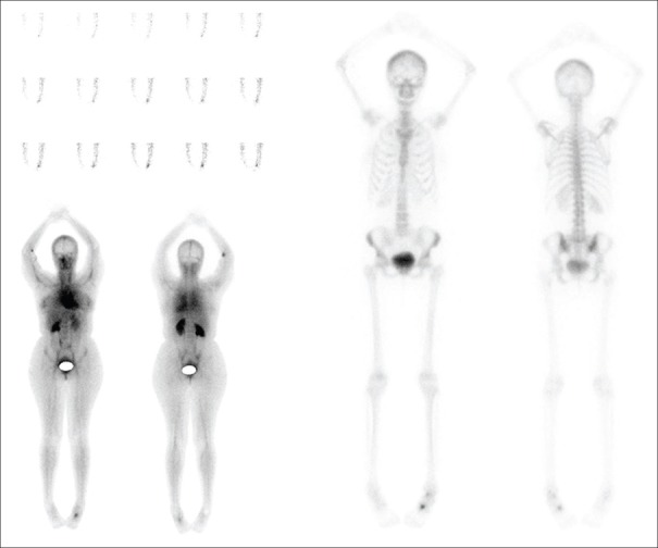
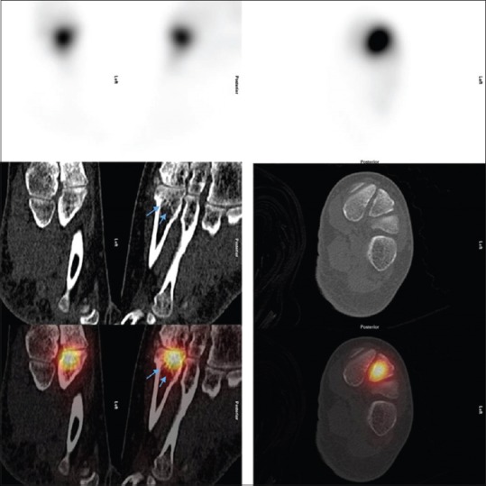
Proximal metatarsal stress fractures are common at base of the second metatarsal, typically seen in repetitive impact athletes, dancers, and those with cavus feet. It is caused by increased bone resorption rather than formation in a state of abrupt increased physical activity or intensity, leading to repetitive microfractures and eventually stress fracture. It is characterized by swelling and tenderness of a prolonged duration. We report a case of a 40-year-old female having left foot pain diagnosed with stress fracture on Tc-99m HDP single-photon emission computed tomography (SPECT)-CT. We emphasize the use of SPECT/CT in allocating active source of pain in the feet.
Keywords: Foot pain; Tc-99m HDP single-photon emission computed tomography-computed tomography; stress fracture.
Conflict of interest statement
There are no conflicts of interest.
Figures
Similar articles
-
The Utility of Tc-99m Hydroxymethylene Diphosphonate Single-photon Emission Computed Tomography/Computed Tomography in Symptomatic Os Trigonum.Indian J Nucl Med. 2018 Apr-Jun;33(2):177-179. doi: 10.4103/ijnm.IJNM_165_17. Indian J Nucl Med. 2018. PMID: 29643689 Free PMC article.
-
The Utility of Tc-99m Hydroxydiphosphonate Single-Photon Emission Computed Tomography/Computed Tomography in Symptomatic Kohler's Disease.Indian J Nucl Med. 2023 Apr-Jun;38(2):185-187. doi: 10.4103/ijnm.ijnm_187_22. Epub 2023 Jun 8. Indian J Nucl Med. 2023. PMID: 37456193 Free PMC article.
-
Prospective evaluation of planar bone scintigraphy, SPECT, SPECT/CT, 18F-NaF PET/CT and whole body 1.5T MRI, including DWI, for the detection of bone metastases in high risk breast and prostate cancer patients: SKELETA clinical trial.Acta Oncol. 2016;55(1):59-67. doi: 10.3109/0284186X.2015.1027411. Epub 2015 Apr 2. Acta Oncol. 2016. PMID: 25833330
-
Clinical Applications of Technetium-99m Quantitative Single-Photon Emission Computed Tomography/Computed Tomography.Nucl Med Mol Imaging. 2019 Jun;53(3):172-181. doi: 10.1007/s13139-019-00588-9. Epub 2019 Mar 15. Nucl Med Mol Imaging. 2019. PMID: 31231437 Free PMC article. Review.
-
Technetium-99m Methylene Diphosphonate Single-photon Emission Computed Tomography/Computed Tomography of the Foot and Ankle.World J Nucl Med. 2017 Apr-Jun;16(2):88-100. doi: 10.4103/1450-1147.203077. World J Nucl Med. 2017. PMID: 28553174 Free PMC article. Review.
Cited by
-
Fifth metatarsal fractures: an update on management, complications, and outcomes.EFORT Open Rev. 2022 Jan 11;7(1):13-25. doi: 10.1530/EOR-21-0025. EFORT Open Rev. 2022. PMID: 35073515 Free PMC article. Review.
References
-
- Simons SM. Foot injuries in the runner. In: O'Connor FG, Wilder RP, editors. Textbook of Running Medicine. New York: McGraw-Hill; 2001. pp. 213–26.
-
- Brukner P, Bennell K. Stress fractures in female athletes. Diagnosis, management and rehabilitation. Sports Med. 1997;24:419–29. - PubMed
