

Assessing Cerebral White Matter Microstructure in Children With Congenital Sensorineural Hearing Loss: A Tract-Based Spatial Statistics Study
- PMID: 31293368
- PMCID: PMC6598398
- DOI: 10.3389/fnins.2019.00597
Assessing Cerebral White Matter Microstructure in Children With Congenital Sensorineural Hearing Loss: A Tract-Based Spatial Statistics Study
Abstract
Objectives: To assess the microstructural properties of cerebral white matter in children with congenital sensorineural hearing loss (CSNHL).
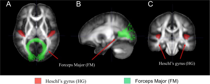
Methods: Children (>4 years of age) with profound CSNHL and healthy controls with normal hearing (the control group) were enrolled and underwent brain magnetic resonance imaging (MRI) scans with diffusion tensor imaging (DTI). DTI parameters including fractional anisotropy, mean diffusivity, axial diffusivity, and radial diffusivity were obtained from a whole-brain tract-based spatial statistics analysis and were compared between the two groups. In addition, a region of interest (ROI) approach focusing on auditory cortex, i.e., Heschl's gyrus, using visual cortex, i.e., forceps major as an internal control, was performed. Correlations between mean DTI values and age were obtained with the ROI method.
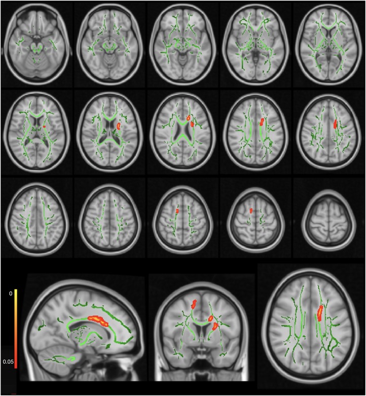
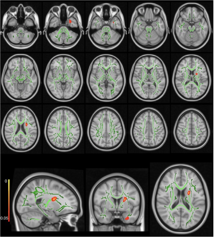
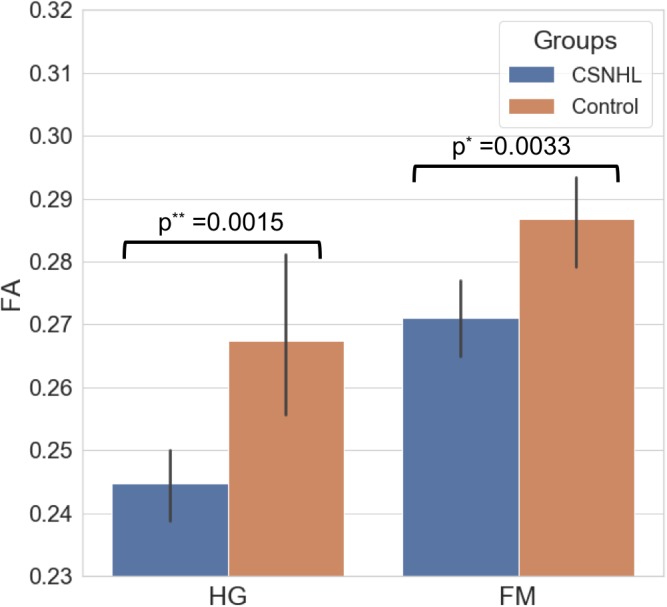
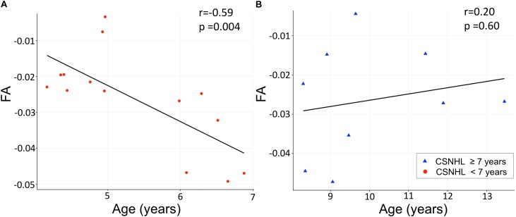
Results: The study cohort consisted of 23 children with CSHNL (11 boys and 12 girls; mean age ± SD: 7.21 ± 2.67 years; range: 4.1-13.5 years) and 18 children in the control group (11 boys and 7 girls; mean age ± SD: 10.86 ± 3.56 years; range: 4.5-15.3 years). We found the axial diffusivity values being significantly greater in the left anterior thalamic radiation, right corticospinal tract, and corpus callosum in the CSHNL group than in the control group (p < 0.05). Significantly higher radial diffusivity values in the white matter tracts were noted in the CSHNL group as compared to the control group (p < 0.05). The fractional anisotropy values in the Heschl's gyrus in the CSNHL group were lower compared to the control group (p = 0.0015). There was significant negative correlation between the mean fractional anisotropy values in Heschl's gyrus and age in the CSNHL group < 7 years of age (r = -0.59, p = 0.004).
Conclusion: Our study showed higher axial and radial diffusivities in the children affected by CNHNL as compared to the hearing children. We also found lower fractional anisotropy values in the Heschl's gyrus in the CSNHL group. Furthermore, we identified negative correlation between the fractional anisotropy values and age up to 7 years in the children born deaf. Our study findings suggest that myelination and axonal structure may be affected due to acoustic deprivation. This information may help to monitor hearing rehabilitation in the deaf children.
Keywords: congenital sensorineural hearing loss; diffusion tensor imaging; diffusivity; magnetic resonance imaging; tract-based spatial statistics; white matter.
Figures
References
-
- Andersson J. L., Jenkinson M., Smith S. (2007). Non-Linear Registration, Aka Spatial Normalisation. FMRIB technical report TR07JA2 Oxford: FMRIB Centre.
-
- Basser P. J., Mattiello J., LeBihan D. (1994). Estimation of the effective self-diffusion tensor from the NMR spin echo. J. Magn. Reson. B 103 247–254. - PubMed
LinkOut - more resources
Full Text Sources