. 2019 Jun 25:13:645.
doi: 10.3389/fnins.2019.00645. eCollection 2019.

Silencing of SNHG12 Enhanced the Effectiveness of MSCs in Alleviating Ischemia/Reperfusion Injuries via the PI3K/AKT/mTOR Signaling Pathway

Affiliations

Silencing of SNHG12 Enhanced the Effectiveness of MSCs in Alleviating Ischemia/Reperfusion Injuries via the PI3K/AKT/mTOR Signaling Pathway

Yuanzhi Li et al. Front Neurosci. .

Abstract

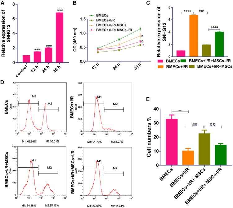
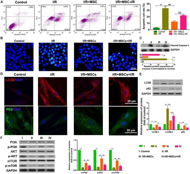
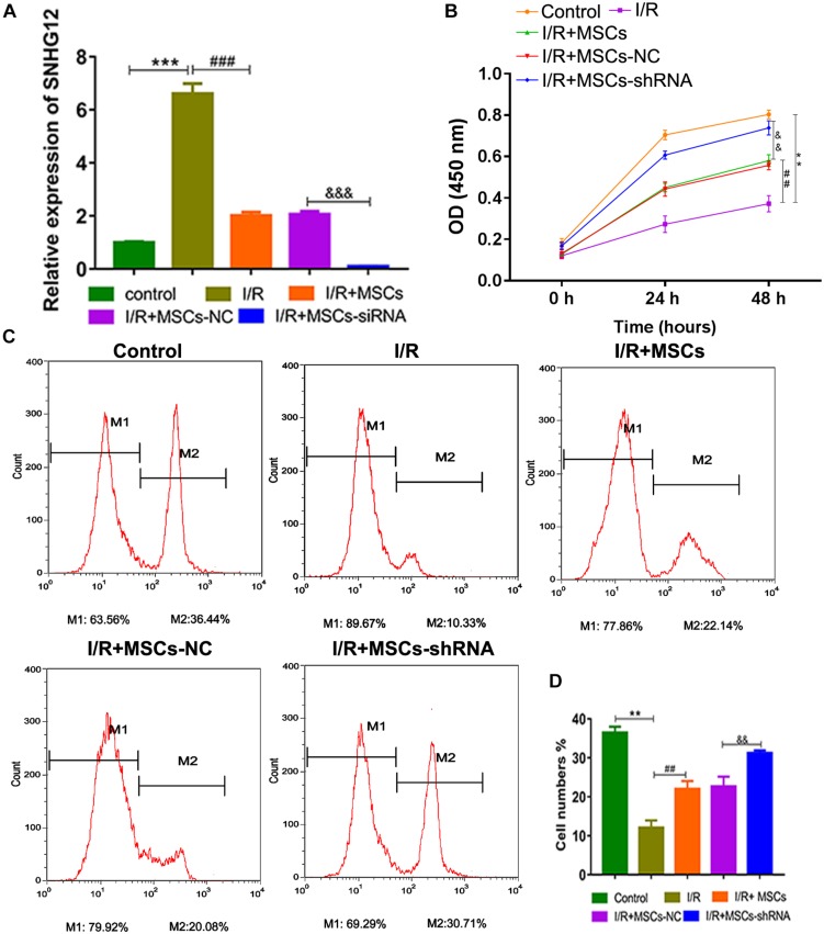
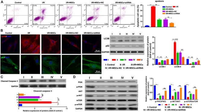
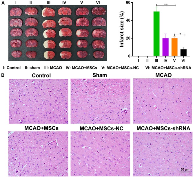
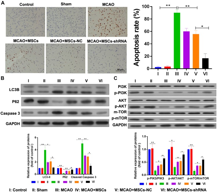
Previous studies have reported that the long non-coding RNA SNHG12 (lncRNA SNHG12) plays a critical role in regulating the function of mesenchymal stem cells (MSCs); however, the effect of lncRNA SNHG12 on MSCs in injured brain tissue has rarely been reported. We studied the effect and mechanism of lncRNA SNHG12-modified mesenchymal stem cells (MSCs) in treating brain injuries caused by ischemia/reperfusion (I/R). I/R treated rat brain microvascular endothelial cells (BMECs) were co-cultured with MSCs or I/R pretreated MSCs. Next, BMEC proliferation was detected by using CCK-8 and EdU assays, and cell apoptosis was determined by using flow cytometry and the Hoechst staining method. Autophagy of BMECs was determined using immunofluorescence and expression of associated pathway proteins were measured by western blotting. Moreover, BMEC proliferation, apoptosis, and autophagy were also determined after the BMECs had been co-cultured with shSNHG12-MSCs. In addition, a rat model of middle cerebral artery occlusion (MCAO) was used to further confirm the findings obtained with cells. I/R treatment significantly decreased the proliferation of BMECs, but increased their levels of SNHG12 expression, apoptosis, and autophagy. However, co-culturing of BMECs with MSCs markedly alleviated the reduction in BMEC proliferation and the increases in BMEC apoptosis and autophagy, as well as the phosphorylation of PI3K, AKT, and mTOR proteins in BMECs that had been induced by I/R. Furthermore, shSNHG12 remarkably enhanced the effects of MSCs. In addition, an injection MSCs reduced the infarct areas and rates of cell apoptosis in MACO rats, and reduced the phosphorylation of PI3K, AKT, and mTOR proteins. Moreover, shSNHG12 enhanced the ameliorative effect of MSCs in treating brain injuries in the MACO rats. In conclusion, silencing of SNHG12 enhanced the effects of MSCs in reducing apoptosis and autophagy of BMECs by activating the PI3K/AKT/mTOR signaling pathway.

Keywords: SNHG12; apoptosis; autophagy; ischemia/reperfusion injury; mesenchymal stem cell.


Figures

FIGURE 1
FIGURE 1
Co-culturing with MSCs promoted the proliferation of BMECs after I/R treatment. (A) Relative expression of SNHG12 in BMECs as determined by qRT-PCR. (B) Proliferation of BMECs as determined by the CCK-8 assay. (C) Relative expression of SNHG12 in BMECs co-cultured with MSCs as determined by qRT-PCR. (D) Cell proliferation as determined by the EdU assay. (E) Cell proliferation as determined by the EdU assay. The percentage of the M2 peak was analyzed as the percentage of EdU-positive cells. MSCs, mesenchymal stem cells; BMECs, brain microvascular endothelial cells; I/R, ischemia/reperfusion. Comparisons among multiple groups were assessed using ANOVA followed by Turkey’s multiple comparisons test. Compared with the control group, ∗∗P < 0.01, ∗∗∗P < 0.001, and ****P < 0.0001; compared with the I/R group, #P < 0.05, ##P < 0.01, and ###P < 0.001; compared with the I/R+MSCs group, &P < 0.05, &&P < 0.01, and &&&&P < 0.0001.
FIGURE 2
FIGURE 2
Mesenchymal stem cells reduced BMEC apoptosis and autophagy via the PI3K/AKT/mTOR pathway. (A) Apoptosis of BMECs as determined by flow cytometry. (B) Apoptosis of BMECs as determined by Hoechst staining. (C) Expression of caspase 3 as determined by western blotting. (D) Expression of LC3B (red) and P62 (green) in BMECs as determined by immunofluorescence. (E) Expression of LC3B and p62 as determined by western blotting. (F) Expression and phosphorylation of PI3K, AKT, and mTOR in BMECs as determined by western blotting. MSCs, mesenchymal stem cells; BMECs, brain microvascular endothelial cells; I/R, ischemia/reperfusion. Comparisons among multiple groups were assessed using ANOVA followed by Turkey’s multiple comparisons test. Compared with the control group, ∗∗P < 0.01 and ∗∗∗P < 0.001; compared with the I/R group, ##P < 0.01 and ###P < 0.001; compared with the I/R+MSC group, &P < 0.05, &&P < 0.01, &&&P < 0.001.
FIGURE 3
FIGURE 3
Silencing of SNHG12 enhanced the effect of MSCs in promoting the proliferation of BMECs after I/R. (A) Relative expression of SNHG12 in BMECs after being co-cultured with different MSCs, as determined by qRT-PCR. (B) Proliferation of BMECs after being cultured with different MSCs, as detected by the CCK-8 assay. (C) Cell proliferation as determined by the EdU assay. (D) Cell proliferation as determined by the EdU assay. MSCs, mesenchymal stem cells; BMECs, brain microvascular endothelial cells; I/R, ischemia/reperfusion; NC, negative control. Comparisons among multiple groups were assessed using ANOVA followed by Turkey’s multiple comparisons test. Compared with the control group, ∗∗P < 0.01 and ∗∗∗P < 0.001; compared with the I/R group, ##P < 0.01 and ###P < 0.001; compared with the I/R+MSC-NC group, &&P < 0.01 and &&&P < 0.001.
FIGURE 4
FIGURE 4
Silencing of SNHG12 enhanced the effect of MSCs in reducing apoptosis and autophagy of BMECs after I/R. (A) Apoptosis of BMECs after being co-cultured with different MSCs, as determined by flow cytometry. (B) Expression of LC3B (red) and P62 (green) in BMECs after being co-cultured with different MSCs, as determined by immunofluorescence at 400× and western blotting. (C) Expression and quantitative expression of caspase 3 as determined by western blotting. (D) Phosphorylation and quantitative phosphorylation of PI3K, AKT, and mTOR in BMECs, as determined by western blotting. MSCs, mesenchymal stem cells; BMECs, brain microvascular endothelial cells; I/R, ischemia/reperfusion. Comparisons among multiple groups were assessed using ANOVA followed by Turkey’s multiple comparisons test. Compared with the control group, ∗∗P < 0.01 and ∗∗∗P < 0.001; compared with the I/R group, ##P < 0.01 and ###P < 0.001; compared with the I/R+MSC-NC group, &P < 0.05 and &&P < 0.01.
FIGURE 5
FIGURE 5
Silencing of SNHG12 enhanced the effect of MSCs in reducing the infarct area of MACO rat brain tissue. (A) Infarct areas in MACO rats as determined by the TTC method. (B) H&E staining of brain tissues from the MACO rats. Rats in the control group are treated without any treatment, in the sham group are exposed the right CCA, ICA, and ECA followed by sutured, in the MCAO group were artery occlusion and reperfusion to construct the I/R injury mode, and in rats in the MSC treatment groups were stereotactically injected with MSCs (MSC-shRNA-NC or MSC-shRNA-SNHG12) prior to MCAO model construction. CCA, common carotid artery; ICA, internal carotid artery; ECA, external carotid artery; MSCs, mesenchymal stem cells; BMECs, brain microvascular endothelial cells; I/R, ischemia/reperfusion; MCAO, middle cerebral artery occlusion; NC, negative control. Comparisons among multiple groups were assessed using ANOVA followed by Turkey’s multiple comparisons test. *P < 0.05, ∗∗P < 0.01.
FIGURE 6
FIGURE 6
Silencing of SNHG12 enhanced the effect of MSCs in reducing apoptosis and autophagy in MACO rat brain tissue. (A) Apoptosis in MACO rat brain tissues as determined by the TUNEL method and magnified at 200×. (B) Expression of LC3B, P62, and caspase 3 in rat brain tissues as determined by western blotting. (C) Phosphorylation of PI3K, AKT, and mTOR in rat brain tissues. CCA, common carotid artery; ICA, internal carotid artery, ECA, external carotid artery; MSCs, mesenchymal stem cells; BMECs, brain microvascular endothelial cells; I/R, ischemia/reperfusion; MCAO, middle cerebral artery occlusion; NC, negative control. Comparisons among multiple groups were assessed using ANOVA followed by Turkey’s multiple comparisons test. *P < 0.05 and ∗∗P < 0.01.

Similar articles

Cited by

References

    1. Bederson J. B., Pitts L. H., Germano S. M., Nishimura M. C., Davis R. L., Bartkowski H. M. (1986). Evaluation of 2,3,5-triphenyltetrazolium chloride as a stain for detection and quantification of experimental cerebral infarction in rats. Stroke 17 1304–1308. 10.1161/01.str.17.6.1304 - DOI - PubMed
    1. Bliss T. M., Andres R. H., Steinberg G. K. (2010). Optimizing the success of cell transplantation therapy for stroke. Neurobiol. Dis. 37 275–283. 10.1016/j.nbd.2009.10.003 - DOI - PMC - PubMed
    1. Bruno S., Grange C., Collino F., Deregibus M. C., Cantaluppi V., Biancone L., et al. (2012). Microvesicles derived from mesenchymal stem cells enhance survival in a lethal model of acute kidney injury. PLoS One 7:e33115. 10.1371/journal.pone.0033115 - DOI - PMC - PubMed
    1. Chen A., Xiong L. J., Tong Y., Mao M. (2013). Neuroprotective effect of brain-derived neurotrophic factor mediated by autophagy through the PI3K/Akt/mTOR pathway. Mol. Med. Rep. 8 1011–1016. 10.3892/mmr.2013.1628 - DOI - PubMed
    1. Chen J., Hou R., Zhang X., Ye Y., Wang Y., Tian J. (2014). Calycosin suppresses breast cancer cell growth via ERβ-dependent regulation of IGF-1R, p38 MAPK and PI3K/Akt pathways. PLoS One 9:e91245. 10.1371/journal.pone.0091245 - DOI - PMC - PubMed