Review
. 2019 Jun 25:13:213.
doi: 10.3389/fnhum.2019.00213. eCollection 2019.

Analysis of Perceptual Expertise in Radiology - Current Knowledge and a New Perspective

Affiliations
Review

Analysis of Perceptual Expertise in Radiology - Current Knowledge and a New Perspective

Stephen Waite et al. Front Hum Neurosci. .

Erratum in

Abstract

Radiologists rely principally on visual inspection to detect, describe, and classify findings in medical images. As most interpretive errors in radiology are perceptual in nature, understanding the path to radiologic expertise during image analysis is essential to educate future generations of radiologists. We review the perceptual tasks and challenges in radiologic diagnosis, discuss models of radiologic image perception, consider the application of perceptual learning methods in medical training, and suggest a new approach to understanding perceptional expertise. Specific principled enhancements to educational practices in radiology promise to deepen perceptual expertise among radiologists with the goal of improving training and reducing medical error.

Keywords: attention; expertise; gist; holistic processing; perceptual learning; radiology; visual perception; visual search.


Figures

FIGURE 1
FIGURE 1
Reprinted from Goodman and Felson (2007) with permission. (A,B) Proposed lung search pattern. (A) This commonly taught search pattern for examination of the lungs during chest radiograph (CXR) interpretation involves starting at the right base () (the costophrenic angle) and examining the right and then left lungs. (B) A second look is then performed in order to compare the right and left lungs as bilateral symmetry is assumed to be useful in recognizing abnormalities (Carmody et al., 1984). Reprinted from Waite et al. (2017a) with permission. Typical scanpaths of a novice (C) and an expert (D) radiologist, both searching a CXR which has a nodule at the left base (arrow in D). This free search pattern (D) is typically employed by experts and differs from the formal radiologic training given in residency. Instead, it indicates the flexible use of search strategies as a function of immediate visual information. The expert radiologist (D) has more efficient scanpaths (red lines) than the novice (C), with fewer fixations (circles), less coverage of the image, fewer saccades, and faster arrival at the abnormality.
FIGURE 2
FIGURE 2
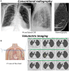
(A) Conventional radiography (2D medical imaging) such as CXR, mammography, and plain film bone X-rays, is based on the fact that tissue will absorb photons from an X-ray beam in relation to the electron density of the tissue. The number of photons passing through the region of interest will be detected by image detectors that convert the body’s direct attenuation of the photons into digital images. The resulting images are a two-dimensional projection of a three-dimensional structure. (B) In volumetric imaging such as CT and MRI, cross-sectional images of the body are obtained to represent a “slice” of the person being imaged, such as the slices in a loaf of bread. Once a number of successive slices are generated, they can be digitally “stacked” to form a three-dimensional image of the patient. On the CT scan above there is a nodule in the right upper lobe (yellow circle). As a radiologist scrolls through the stack, the nodule will present as a suddenly appearing (and then disappearing) lesion, simulating motion.
FIGURE 3
FIGURE 3
Figure modified from Wolfe et al. (2011) with permission. Two-pathway architecture for visual processing. The selective pathway can bind features and recognize objects but is capacity-limited. At its bottleneck, preference for further processing is given to items with certain basic attributes (such as color, orientation, and size) when those attributes match the appearance of a target object. However, these attributes do not fully explain the efficiency of search in the real world, where elements are arranged in a rule-governed manner—for example, people generally appear on horizontal surfaces. The regularity of scenes provides two kinds of scene-based guidance—semantic guidance, referring to the knowledge of the probability of the presence of an object in a scene and its probable location, and episodic guidance, referring to the memory of a specific previously encountered scene. In conjunction with the selective pathway, the non-selective pathway extracts statistics such as velocity, direction of motion, and size, rapidly from the entire image. Although the non-selective pathway does not support precise object recognition, it provides information used in scene-based guidance to direct attention to important locations (such as the probable locations of nodules on CXR’s). Conscious experience of the visual world is comprised of the products of both pathways (Wolfe et al., 2011).
FIGURE 4
FIGURE 4
Reprinted from Chin et al. (2018) with permission. Examples of mammogram and face stimuli in upright and inverted orientations. (A) Upright and inverted abnormal mammogram. (B) Upright and inverted smiling face. The inversion effect posits that stimuli are processed as an integrated whole rather than a sum of its parts; therefore, the inverted image is harder to recognize and process (Chin et al., 2018).
FIGURE 5
FIGURE 5
Reprinted from Litchfield and Donovan (2016) with permission. Two different experience groups—expert radiologists and psychology students—searched for lung nodules from CXR images using the flash-preview moving window (FPMW) paradigm. Participants looked at the target word for 15 s (“lung nodule”). They then saw a fixation cross for 200 ms, then either a mask preview (random array of colored pixels) or a CXR (a ‘scene’ preview) for 250 ms. Next, participants saw a mask for 50 ms and then a second fixation cross for 400 ms. Following a second presentation of the fixation cross, they conducted a windowed search, with a 2.5-degree radius window restricting the field of view (Litchfield and Donovan, 2016).
FIGURE 6
FIGURE 6
Reprinted from Ravesloot et al. (2017) with permission. Graph estimating image interpretation skill development during residency, as measured by the Dutch Radiology Progress test. Image score measures performance on image interpretation skills as opposed to factual knowledge. The score represents percentage from the maximum possible score and is calculated by subtracting the number of incorrect answers from the number of correct answers to account for guessing (making a negative value possible). The slope represents the speed of skill development and measures 16.8% during the first year of training. The slope decreases by 50% every year until it reaches 2.0% at the end of training. Note that the maximum image-score is estimated at 55.8%. Dotted lines represent the middle 95% of performances (Ravesloot et al., 2017).
FIGURE 7
FIGURE 7
Reprinted from Sasaki et al. (2010) with permission. Texture-discrimination tasks such as depicted here are frequently used in visual perceptual learning (VPL) studies. The subject is first asked to report whether a ‘T’ (as in this example) or an ‘L’ is presented in the center of the display to ensure fixation, and then whether the orientation of the target (which is comprised of the three elements with orientations that depart from those of the other elements in the display) is vertical (as in this example) or horizontal. VPL of the target orientation is examined.
FIGURE 8
FIGURE 8
Reprinted from Chen et al. (2017) with permission. (A) Example of an image shown during perceptual training of hip fracture identification. (B) Arrows represent the feedback provided in case of a wrong answer. Top novices achieved expert level accuracy in hip fracture detection in under 1 h of perceptual training (Chen et al., 2017).

References

    1. Alexander R. G., Zelinsky G. J. (2009). The frankenbear experiment: looking for part-based similarity effects on search guidance with complex objects. J. Vis. 9 1184–1184. 10.1167/9.8.1184 - DOI
    1. Alexander R. G., Zelinsky G. J. (2011). Visual similarity effects in categorical search. J. Vis. 11:9. 10.1167/11.8.9 - DOI - PMC - PubMed
    1. Alzubaidi M., Balasubramanian V., Patel A., Panchanathan S., Black J. A. (2010). “What catches a radiologist’s eye? A comprehensive comparison of feature types for saliency prediction,” in Proceedings of the Medical Imaging 2010: Computer-Aided Diagnosis, (Bellingham: SPIE; ).
    1. Andrew L. B. (2006). Expert witness testimony: the ethics of being a medical expert witness. Emerg. Med. Clin. North Am. 24 715–731. 10.1016/j.emc.2006.05.001 - DOI - PubMed
    1. Andriole K. P., Wolfe J. M., Khorasani R., Treves S. T., Getty D. J., Jacobson F. L. (2011). Optimizing analysis, visualization, and navigation of large image data sets: one 5000-section CT scan can ruin your whole day. Radiology 259 346–362. 10.1148/radiol.11091276 - DOI - PMC - PubMed