. 2019 Jun 26:11:156.
doi: 10.3389/fnagi.2019.00156. eCollection 2019.

Synaptopathy as a Mechanism for Age-Related Vestibular Dysfunction in Mice

Affiliations

Synaptopathy as a Mechanism for Age-Related Vestibular Dysfunction in Mice

Guoqiang Wan et al. Front Aging Neurosci. .

Abstract

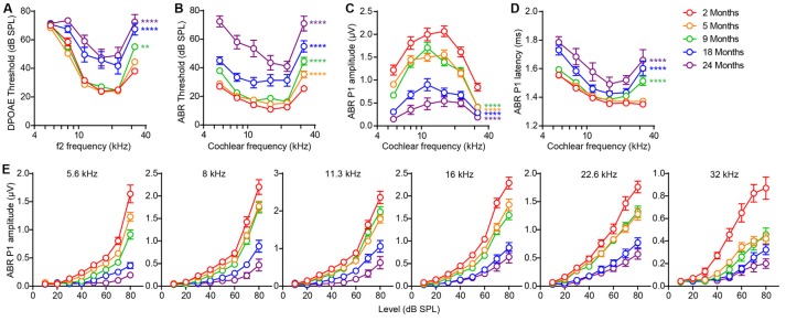
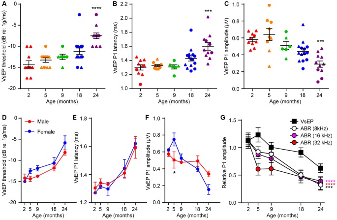
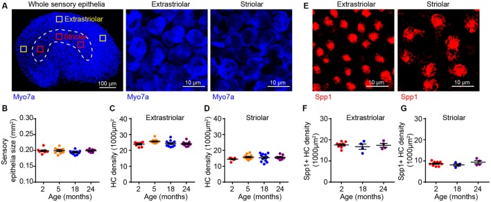
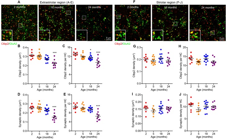
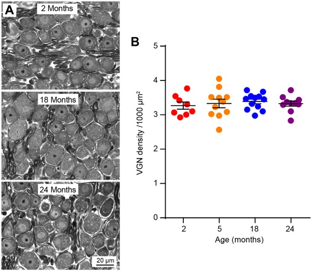
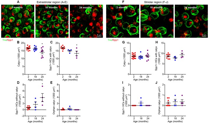
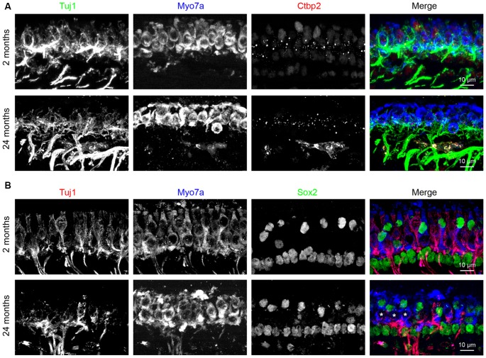
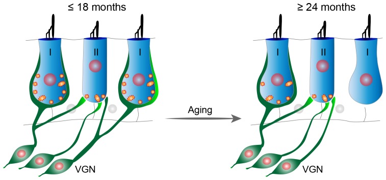
Age-related decline of inner ear function contributes to both hearing loss and balance disorders, which lead to impaired quality of life and falls that can result in injury and even death. The cellular mechanisms responsible for the ear's functional decline have been controversial, but hair cell loss has been considered the key cause for a long time. However, recent studies showed that in the cochlea, loss of inner hair cell (IHC) synapses precedes hair cell or neuronal loss, and this synaptopathy is an early step in the functional decline. Whether a similar process occurs in the vestibular organ, its timing and its relationship to organ dysfunction remained unknown. We compared the time course of age-related deterioration in vestibular and cochlear functions in mice as well as characterized the age-associated changes in their utricles at the histological level. We found that in the mouse, as in humans, age-related decline in vestibular evoked potentials (VsEPs) occurs later than hearing loss. As in the cochlea, deterioration of VsEPs correlates with the loss of utricular ribbon synapses but not hair cells or neuronal cell bodies. Furthermore, the age-related synaptic loss is restricted to calyceal innervations in the utricular extrastriolar region. Hence, our findings suggest that loss of extrastriolar calyceal synapses has a key role in age-related vestibular dysfunction (ARVD).

Keywords: calyx; hair cells; inner ear; ribbon synapses; synaptopathy; utricle; vestibular dysfunction.


Figures

Figure 1
Figure 1
Signs of age-related hearing loss in FVB/N mice are detectable at 5 months of age. (A) Distortion-product otoacoustic emission (DPOAE) threshold, (B) auditory brainstem response (ABR) threshold, (C) ABR peak 1 amplitude, and (D) latency were measured on 2 (n = 10), 5 (n = 12), 9 (n = 12), 18 (n = 12) and 24 (n = 10) month old FVB/N mice. ABR peak 1 latency was analyzed at 80 dB sound pressure level (SPL) and amplitude analyzed by averaging 70 and 80 dB SPL. **p < 0.01 and ****p < 0.0001 by two-way ANOVA comparing to 2-month-old mice. (E) ABR peak 1 amplitude was plotted against the acoustic stimulus level (10–80 dB SPL) at 5.6, 8, 11.3, 16, 22.6 and 32 kHz. Input-output curves of ABR P1 amplitudes from 5, 9, 18 and 24-month-old mice were significantly different from that of 2-month-old mice at all frequencies tested (p < 0.0001 by two-way ANOVA).
Figure 2
Figure 2
Age-related defects in vestibular evoked potentials (VsEPs) are seen only in 24 month-old-mice. (A) VsEP threshold, (B) VsEP peak 1 latency and (C) VsEP peak 1 amplitude at 2.5 dB re 1 g/ms were measured on 2 (n = 10), 5 (n = 10), 9 (n = 6), 18 (n = 15) and 24 (n = 11) month old FVB/N mice. Data from males and females are presented as triangles and circles respectively. ***p < 0.001 and ****p < 0.0001 by one-way ANOVA comparing to 2-month-old mice followed by Dunn’s post-test. (D) VsEP threshold, (E) VsEP peak 1 latency and (F) VsEP peak 1 amplitude at 2.5 dB re 1 g/ms of either male or female mice were analyzed separately. The numbers of males and females at each point are 9 and 1 (2 months), 4 and 6 (5 months), 7 and 8 (18 months), 2 and 4 (9 months), 8 and 3 (24 months) respectively. *p = 0.03 by two-way ANOVA. (G) Comparison of the time course of age-related changes in VsEP peak 1 amplitude and ABR peak 1 amplitude. VsEP and ABR (8, 16, 32 kHz) peak 1 amplitude was normalized to data from 2-month-old mice. The relative amplitude was plotted against age of the mice. ***p < 0.001 and ****p < 0.0001 by two-way ANOVA comparing to changes in VsEP amplitude.
Figure 3
Figure 3
Utricular size and density of utricular hair cells do not change with aging. (A) Myo7a immunofluorescence images showing the entire sensory epithelium of an utricle. Squares represent high magnification sampling of extrastriolar and striolar (dotted lines) areas. (B) The size of utricle sensory epithelium in 2 (n = 9), 5 (n = 9), 18 (n = 11) and 24 (n = 7) month old FVB/N mice. Density of utricular hair cells in extrastriolar area (C) and striolar area (D) in 2 (n = 8), 5 (n = 12), 18 (n = 12) and 24 (n = 9) month old FVB/N mice. (E) Representative confocal images of Spp1+ type I hair cells in extrastriolar and striolar regions. (F) Density of type I hair cell (Spp1+) in 2 (n = 9), 18 (n = 4) and 24 (n = 4) month old utricular extrastriola. (G) Density of type I hair cell (Spp1+) in 2 (n = 9), 18 (n = 4) and 24 (n = 4) month old utricular striola.
Figure 4
Figure 4
Age-related decline in utricular ribbon synapse density is restricted to the extrastriolar region and occurs only at 24 months of age. (A–E) Extrastriolar and (F–J) striolar regions of 2, 18 and 24 month old mouse utricles were analyzed separately after immunostaining with GluA2 and Ctbp2. (A) Representative confocal images of extrastriolar ribbon synapses. (B,C) Density of synaptic ribbon and (D,E) putative ribbon synapses either normalized to unit area (B,D) or each hair cell (C,E) in 2 (n = 8), 5 (n = 12), 18 (n = 12) and 24 (n = 9) month old utricular extrastriola. *p < 0.05 and ***p < 0.001 by one-way ANOVA comparing to 2-month-old group followed by Dunn’s post-test. (F) Representative confocal images of striolar ribbon synapses. (G,H) Density of synaptic ribbon and (I,J) putative ribbon synapses either normalized to unit area (G,I) or each hair cell (H,J) in 2 (n = 8), 5 (n = 6), 18 (n = 12) and 24 (n = 9) month old utricular striola.
Figure 5
Figure 5
Vestibular ganglion neuron (VGN) density does not change during aging. (A) Representative plastic section and light microscopy images of VGN in aging mice. (B) VGN density in 2 (n = 8), 5 (n = 11), 18 (n = 12) and 24 (n = 10) month old mice.
Figure 6
Figure 6
Loss of calyces in 24-month-old utricular extrastriola. (A–E) Extrastriolar and (F–J) striolar regions of 2, 18 and 24-month-old mouse utricles were analyzed separately after immunostaining with tenascin-C (Tnc) and secreted phosphoprotein 1 (Spp1). (A) Representative confocal images of extrastriolar calyces and type I hair cells. (B) Density of calyx (Tnc+) in 2 (n = 14), 18 (n = 9) and 24 (n = 8) month old utricular extrastriola. (C) Density of type I hair cell associated with calyx in 2 (n = 9), 18 (n = 4) and 24 (n = 4) month old utricular extrastriola. (D) Density of orphan type I hair cell and (E) density of orphan calyx in 2 (n = 9), 18 (n = 4) and 24 (n = 4) month old utricular extrastriola. *p < 0.05 by one-way ANOVA comparing to 2-month-old group followed by Dunn’s post-test. (F) Representative confocal images of striolar calyces and type I hair cells. (G) Density of calyx (Tnc+) in 2 (n = 15), 18 (n = 9) and 24 (n = 8) month old utricular striola. (H) Density of type I hair cell associated with calyx in 2 (n = 9), 18 (n = 4) and 24 (n = 4) month old utricular striola. (I) Density of orphan type I hair cells and (J) density of orphan calyx in 2 (n = 9), 18 (n = 4) and 24 (n = 4) month old utricular striola.
Figure 7
Figure 7
Loss of calyceal synapses but not bouton synapses in 24-month-old utricular extrastriola. (A–E) Extrastriolar and (F–J) striolar regions of 2, 18 and 24-month-old mouse utricles were analyzed separately after immunostaining with tenascin-C (Tnc) and Ctbp2. (A) Representative confocal images of extrastriolar calyces and synaptic ribbons. Density of total synaptic ribbon (B), noncalyceal ribbon (C) and calyceal ribbon (D) in 2 (n = 13), 18 (n = 8) and 24 (n = 7) month old utricular extrastriola. (E) Number of synaptic ribbons associated with each calyx in 2 (n = 13), 18 (n = 8) and 24 (n = 7) month old utricular extrastriola. *p < 0.05 and **p < 0.01 by one-way ANOVA comparing to 2-month-old group followed by Dunn’s post-test. (F) Representative confocal images of striolar calyces and synaptic ribbons. Density of total synaptic ribbon (G), noncalyceal ribbon (H) and calyceal ribbon in (I) 2 (n = 14), 18 (n = 7) and 24 (n = 9) month old utricular striola. (J) Number of synaptic ribbons associated with each calyx in 2 (n = 14), 18 (n = 7) and 24 (n = 9) month old utricular striola.
Figure 8
Figure 8
Loss of calyceal innervations and ribbons of type I hair cells in 24-month-old utricular extrastriola. (A) Co-labeling of calyceal terminals (Tuj1), hair cells (Myo7a) and synaptic ribbons (Ctbp2) in 2 and 24 months old utricular extrastriolar region. (B) Co-labeling of calyceal terminals (Tuj1), hair cells (Myo7a) and type II hair cells + supporting cells (Sox2) in 2 and 24 months old utricular extrastriolar region. Asterisks (*) indicate Sox2 type I hair cells that had lost calyceal innervations in 24-months-old utricle.
Figure 9
Figure 9
Proposed model of age-related vestibular synaptopathy in FVB/N mouse utricle. Utricular structure and function are normal until 18 months of age, but by 24 months of age there is a significant loss of calyceal nerve terminals and their associated synaptic ribbons in the extrastriolar region with a corresponding alteration in VsEPs.

References

    1. Agrawal Y., Zuniga M. G., Davalos-Bichara M., Schubert M. C., Walston J. D., Hughes J., et al. (2012). Decline in semicircular canal and otolith function with age. Otol. Neurotol. 33, 832–839. 10.1097/mao.0b013e3182545061 - DOI - PMC - PubMed
    1. Alidina A., Lyon M. J. (1990). Aging rat vestibular ganglion: I. Quantitative light microscopic evaluation. Am. J. Otolaryngol. 11, 174–181. 10.1016/0196-0709(90)90034-s - DOI - PubMed
    1. Anniko M. (1983). The aging vestibular hair cell. Am. J. Otolaryngol. 4, 151–160. 10.1016/s0196-0709(83)80037-4 - DOI - PubMed
    1. Baird R. A., Desmadryl G., Fernández C., Goldberg J. M. (1988). The vestibular nerve of the chinchilla: II. Relation between afferent response properties and peripheral innervation patterns in the semicircular canals. J. Neurophysiol. 60, 182–203. 10.1152/jn.1988.60.1.182 - DOI - PubMed
    1. Barin K., Dodson E. E. (2011). Dizziness in the elderly. Otolaryngol. Clin. North Am. 44, 437–454, x. 10.1016/j.otc.2011.01.013 - DOI - PubMed