. 2019 Jun 21:10:708.
doi: 10.3389/fphys.2019.00708. eCollection 2019.

Monitoring the Early Antiproliferative Effect of the Analgesic-Antitumor Peptide, BmK AGAP on Breast Cancer Using Intravoxel Incoherent Motion With a Reduced Distribution of Four b-Values

Affiliations

Monitoring the Early Antiproliferative Effect of the Analgesic-Antitumor Peptide, BmK AGAP on Breast Cancer Using Intravoxel Incoherent Motion With a Reduced Distribution of Four b-Values

Natacha Raissa Doudou et al. Front Physiol. .

Abstract

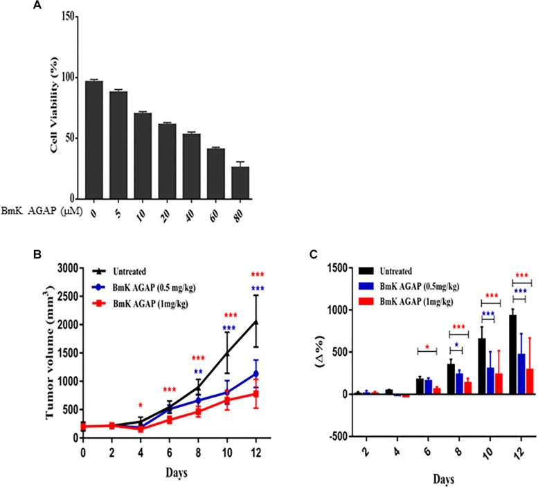
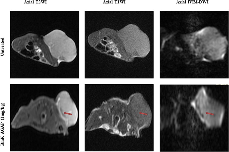
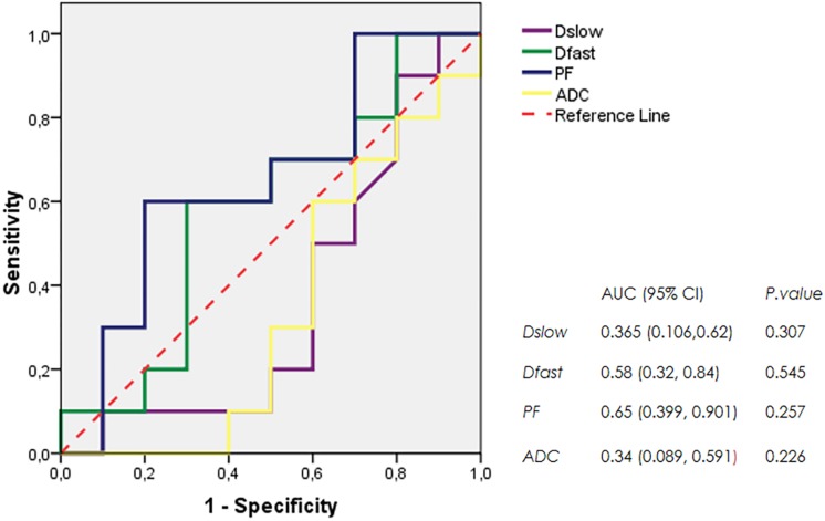
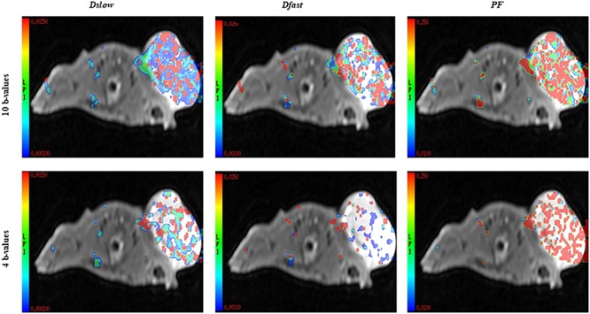
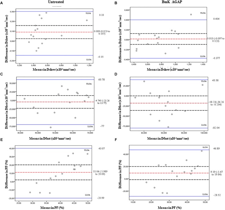
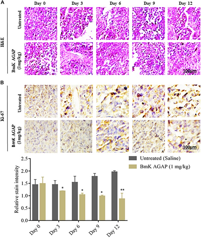
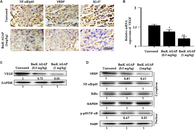
Background: The present study aimed to investigate the possibility of using intravoxel incoherent motion (IVIM) diffusion magnetic resonance imaging (MRI) to quantitatively assess the early therapeutic effect of the analgesic-antitumor peptide BmK AGAP on breast cancer and also evaluate the medical value of a reduced distribution of four b-values. Methods: IVIM diffusion MRI using 10 b-values and 4 b-values (0-1,000 s/mm2) was performed at five different time points on BALB/c mice bearing xenograft breast tumors treated with BmK AGAP. Variability in Dslow, Dfast, PF, and ADC derived from the set of 10 b-values and 4 b-values was assessed to evaluate the antitumor effect of BmK AGAP on breast tumor. Results: The data showed that PF values significantly decreased in rBmK AGAP-treated mice on day 12 (P = 0.044). PF displayed the greatest AUC but with a poor medical value (AUC = 0.65). The data showed no significant difference between IVIM measurements acquired from the two sets of b-values at different time points except in the PF on the day 3. The within-subject coefficients of variation were relatively higher in Dfast and PF. However, except for a case noticed on day 0 in PF measurements, the results indicated no statistically significant difference at various time points in the rBmK AGAP-treated or the untreated group (P < 0.05). Conclusion: IVIM showed poor medical value in the early evaluation of the antiproliferative effect of rBmK AGAP in breast cancer, suggesting sensitivity in PF. A reduced distribution of four b-values may provide remarkable measurements but with a potential loss of accuracy in the perfusion-related parameter PF.

Keywords: Buthus martensii Karsch; analgesic–antitumor peptide; breast neoplasms; intravoxel incoherent motion; magnetic resonance imaging.


Figures

FIGURE 1
FIGURE 1
BmK AGAP inhibits the growth of breast xenograft tumors. (A) IC50 values of rBmK AGAP for MDA-MB-231 cells. Different concentrations of rBmKAGAP were used to treat the cells for 48 h; cell viability was measured by MTT assay. (B) Tumor volume of tumors from rBmK AGAP-treated and untreated mice model. Breast xenograft tumor volume was calculated from measuring the length, height, and width of tumors with digital caliper following rBmK AGAP treatment. (C) Percentage changes in tumor volumes. The data were statistically significant at *P < 0.05, ∗∗P < 0.01, and ∗∗∗P < 0.001 compared with untreated group. The data represent the mean ± SEM of three independent experiments.
FIGURE 2
FIGURE 2
MR images of a tumor of a mouse from the untreated group or the BmK AGAP (1 mg/kg)-treated group, corresponding to day 12 of treatment. Axial T2WIs, axial T1WIs, and axial IVIM images for b = 800 s/mm2. An area of necrosis is noticed under the sheath (red arrow).
FIGURE 3
FIGURE 3
ROC curves generated from ADC values and IVIM parameters Dslow, Dfast, and PF.
FIGURE 4
FIGURE 4
IVIM parametric maps acquired from 10 b-values versus 4 b-values merged with a T2WI.
FIGURE 5
FIGURE 5
Bland–Altman plots of the parameters measured using the 10 b-values versus 4 b-values. The average differences (red dashed lines), its 95% confidence intervals (black dashed lines), and upper line of agreement (ULOA), and lower line of agreement (LLOA) (solid blue lines) are displayed. Dslow (A,B), Dfast (C,D), and PF (E,F).
FIGURE 6
FIGURE 6
Histologic analyses. (A) H&E staining to determine the morphologic changes that formed the basis of the breast xenograft tumor diagnosis. (B) Immunohistochemical assessment of proliferation markers in excised tumor tissues. rBmK AGAP (1 mg/kg)-treated xenograft tumor tissues were stained with antibodies against Ki-67 and examined by immunohistochemical staining at different time points (scale bars = 100 μm; magnification, 200x). The data were statistically significant at *P < 0.05 and ∗∗P < 0.01 as compared with untreated tumors. The data represent the mean ± SD of three independent experiments.
FIGURE 7
FIGURE 7
Histologic analyses. VEGF and NF-κB signaling pathway are involved in BmK AGAP inhibition of breast xenograft tumor growth. (A) Immunohistochemical assessment of proliferation markers in excised tumor tissues. Xenograft tumor tissues were stained with antibodies against NF-κB/p65, VEGF, and Ki-67 and examined by immunohistochemical staining after day 12 of rBmK AGAP (1 mg/kg) treatment (scale bars = 100 μm; magnification, 200x). (B) Relative gene expression of VEGF following rBmK AGAP treatment. Mice were treated with different concentrations of rBmK AGAP for 12 days, and the expression of VEGF and GAPDH (internal control) were analyzed by qPCR. (C) VEGF, protein expression following rBmK AGAP treatment of mice bearing breast xenograft tumors. rBmK AGAP-treated tissues were lysed and subjected to 12% SDS-PAGE and analyzed by Western blotting with antibodies against VEGF. (D) rBmK AGAP suppresses the expression of VEGF, NF-κB/p65, IκBα, and p-p65/NF-κB in breast xenograft tumors as analyzed by Western blotting. GAPDH or PARP was used as an internal control. The data were statistically significant at *P < 0.05 and ∗∗P < 0.01 as compared with untreated tumors. The data represent the mean ± SD of three independent experiments.

References

    1. Al-Asmari A. K., Islam M., Al-Zahrani A. M. (2016). In vitro analysis of the anticancer properties of scorpion venom in colorectal and breast cancer cell lines. Oncol. Lett. 11 1256–1262. 10.3892/ol.2015.4036 - DOI - PMC - PubMed
    1. Al-Asmari A. K., Riyasdeen A., Islam M. (2018). Scorpion venom causes apoptosis by increasing reactive oxygen species and cell cycle arrest in MDA-MB-231 and HCT-8 cancer cell lines. J. Evid. Based Integr. Med. 23:2156587217751796. - PMC - PubMed
    1. Al-Mahmood S., Sapiezynski J., Garbuzenko O. B., Minko T. (2018). Metastatic and triple-negative breast cancer: challenges and treatment options. Drug Deliv. Transl. Res. 5 1483–1507. 10.1007/s13346-018-0551-3 - DOI - PMC - PubMed
    1. Bains L. J., Zweifel M., Thoeny H. C. (2012). Therapy response with diffusion MRI: an update. Cancer Imaging 12 395–402. 10.1102/1470-7330.2012.9047 - DOI - PMC - PubMed
    1. Che S., Zhao X., Ou Y., Li J., Wang M., Wu B., et al. (2016). Role of the intravoxel incoherent motion diffusion weighted imaging in the pre-treatment prediction and early response monitoring to neoadjuvant chemotherapy in locally advanced breast cancer. Medicine 95:e2420. 10.1097/MD.0000000000002420 - DOI - PMC - PubMed