Review
. 2019 Jun 25:10:819.
doi: 10.3389/fpls.2019.00819. eCollection 2019.

Engineered Male Sterility by Early Anther Ablation Using the Pea Anther-Specific Promoter PsEND1

Affiliations
Review

Engineered Male Sterility by Early Anther Ablation Using the Pea Anther-Specific Promoter PsEND1

Edelín Roque et al. Front Plant Sci. .

Abstract

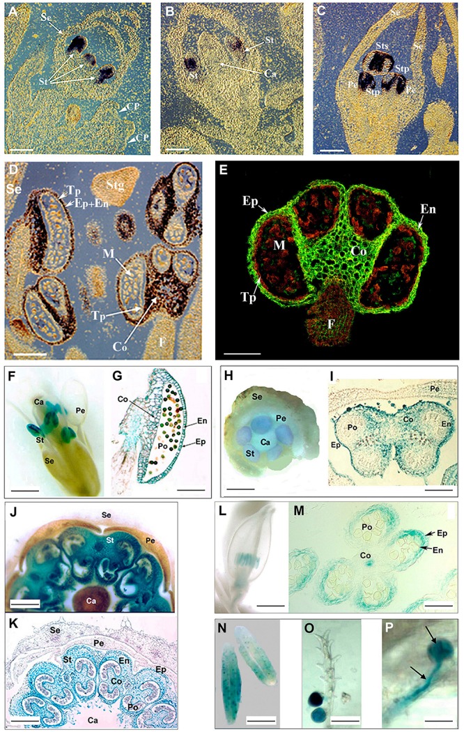
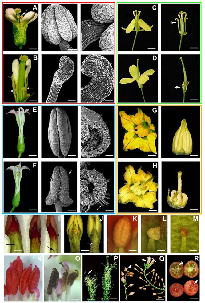
Genetic engineered male sterility has different applications, ranging from hybrid seed production to bioconfinement of transgenes in genetic modified crops. The impact of this technology is currently patent in a wide range of crops, including legumes, which has helped to deal with the challenges of global food security. Production of engineered male sterile plants by expression of a ribonuclease gene under the control of an anther- or pollen-specific promoter has proven to be an efficient way to generate pollen-free elite cultivars. In the last years, we have been studying the genetic control of flower development in legumes and several genes that are specifically expressed in a determinate floral organ were identified. Pisum sativum ENDOTHECIUM 1 (PsEND1) is a pea anther-specific gene displaying very early expression in the anther primordium cells. This expression pattern has been assessed in both model plants and crops (tomato, tobacco, oilseed rape, rice, wheat) using genetic constructs carrying the PsEND1 promoter fused to the uidA reporter gene. This promoter fused to the barnase gene produces full anther ablation at early developmental stages, preventing the production of mature pollen grains in all plant species tested. Additional effects produced by the early anther ablation in the PsEND1::barnase-barstar plants, with interesting biotechnological applications, have also been described, such as redirection of resources to increase vegetative growth, reduction of the need for deadheading to extend the flowering period, or elimination of pollen allergens in ornamental plants (Kalanchoe, Pelargonium). Moreover, early anther ablation in transgenic PsEND1::barnase-barstar tomato plants promotes the developing of the ovaries into parthenocarpic fruits due to the absence of signals generated during the fertilization process and can be considered an efficient tool to promote fruit set and to produce seedless fruits. In legumes, the production of new hybrid cultivars will contribute to enhance yield and productivity by exploiting the hybrid vigor generated. The PsEND1::barnase-barstar construct could be also useful to generate parental lines in hybrid breeding approaches to produce new cultivars in different legume species.

Keywords: Pisum sativum; PsEND1 promoter; barnase; hybrid seeds; male sterility; parthenocarpy; pollen allergens; transgene bioconfinement.


Figures

FIGURE 1
FIGURE 1
PsEND1 expression in pea and other plant species. (A) RNA in situ hybridization in sections of two pea floral buds using digoxigenin-labeled antisense PsEND1 RNA probes. Purple color indicates the localization of PsEND1 expression. No expression was detected in the common primordia (CP) to petals and stamens (white arrows). The expression of PsEND1 begins to be detected in the stamen primordia (St) of floral buds at day 12 before anthesis (d-12). (B) In flowers at d-10, the PsEND1expression is detected in the upper part of the stamen primordia where the anther locules will develop. (C) In flowers at d-8, PsEND1 expression is only detected in those tissues that will be involved in anther architecture both in antesepalous and antepetalous stamens (Sts, Stp). (D) Close-view of a flower at d-6 showing anthers with strong hybridization signal in the epidermis (Ep), endothecium (En), middle layer, and connective (Co). No expression was detected in the anther filament (F), tapetum (Tp), and microspores (M). (E) Immunolocalization (anti-IgG-FITC) of the PsEND1 protein in paraffin sections of a pea stamen. The protein is localized (green fluorescence) in the same anther tissues than the RNA. (F) PsEND1::uidA expression in transgenic Arabidopsis thaliana flowers. GUS activity (blue) was only detected in the anther but not in the filament. (G) Transgenic A. thaliana anther section showing GUS activity in the structural tissues of the pollen sacs. (H) Young PsEND1::uidA Nicotiana tabacum flower showing GUS activity only in the stamen (St) primordia. (I) Transgenic N. tabacum anther showing GUS activity in the structural tissues of the pollen sacs but not in the pollen grains or tapetum. (J) Solanum lycopersicum PsEND1::uidA flower showing specific GUS activity in the anthers. (K) Transgenic tomato flower section showing GUS activity in the tissues involved in the architecture of the pollen sacs but not in the tapetum or in the pollen grains. (L) Expression of the PsEND1::uidA construct in the anthers of an Oryza sativa floret. (M) Section of a rice floret showing GUS activity in the expected anther tissues. (N) Expression of the uidA gene in the anthers of transgenic Triticum aestivum plants carrying the PsEND1::uidA construct. (O) Mature pollen adhering to the stigma showing GUS activity in a transgenic wheat flower. (P) Close-view of a germinating pollen grain, with pollen tube (arrows) growing in the style. Ca, carpel; Co, connective; En, endothecium; Ep, epidermis; Pe, petals; Po, pollen; Se, sepals; St, stamens; Tp, tapetum. Scale bars represent 100 μm in A, B, C, D, E, G, I, K, and M; 2.0 mm in F, H, J, and L; 0.5 mm in N; and 200 μm in O and P. Adapted from Gómez et al. (2004), Roque et al. (2007), Beltrán et al. (2007), and Pistón et al. (2008).
FIGURE 2
FIGURE 2
Engineered anther ablation in model plants and crops. Red box (A. thaliana). (A) Left: wild-type (WT) A. thaliana flower showing normal anthers (arrow). Center and right: WT A. thaliana stamen observed by scanning electron microscopy (SEM). The black arrow indicates the cell types (toothed edges) present in the anther epidermis and the white one those of the filament (lengthened). (B) Left: transgenic A. thaliana PsEND1::barnase-barstar flower (two sepals and two petals were detached). Anther ablation is evident and no pollen sacs were formed (white arrows). The anther filament is short because it does not undergo the lengthening process. Center and right: PsEND1::barnase-barstar stamen observed by SEM. The hook-shaped structures (white arrows) shown are cellular types usually present in the filament but not those present in the epidermis of WT pollen sacs. Green box (B. napus). (C) Left: WT oilseed rape (Brassica napus) cv. Drakkar flower showing normal stamens. Right: Id, but with detached sepals and petals to observe the normal anthers and filaments (white arrow). (D) Left: male sterile flower of a PsEND1::barnase-barstar oilseed rape plant showing the absence of developed stamens. Right: Id, but with detached sepals and petals to see the ablated anthers and the reduction of the filament length (white arrow). Blue box (N. tabacum). (E) Left: WT tobacco (N. tabacum) cv. Petite Havana SR1 flower after anthesis showing normal anthers and full-length filaments. Center: WT tobacco anther with its characteristic four locules fully developed observed by SEM. Right: section of a WT tobacco pollen sac showing mature pollen grains. (F) Left: PsEND1::barnase-barstar tobacco flower after anthesis showing collapsed lobes and reduced filaments. Center: Tobacco PsEND1::barnase-barstar anther showing an arrowhead shape with collapsed locules and increased number of trichomes (white arrow). Right: section of a PsEND1::barnase-barstar pollen sac, no pollen grains can be observed into the collapsed locules. Orange box (S. lycopersicum). (G) Left: WT tomato (S. lycopersicum) cv. Micro-Tom flower at anthesis. Showing the staminal cone (black arrow) formed by the fully developed stamens in the center. Right: Isolated WT staminal cone covering the carpel. (H) Left: tomato PsEND1::barnase-barstar flower at anthesis. Right: anther ablation in the PsEND1::barnase-barstar flowers made visible the style and ovary of the carpel. (I) Flowers from a Kalanchoe blossfeldiana cv. “Tenorio” WT plant (center) and two male sterile lines (left and right) 1 day prior to anthesis. The WT flowers show anthers with fully developed locules, whereas the transgenic ones show collapsed structures at the end of a short filament instead of a four-lobed anther (black arrows). (J) Flowers from a K. blossfeldiana cv. “Hillary” WT plant (left) and a male sterile line (right) 1 day prior to anthesis with ablated anthers (white arrow). (K) WT anther from a “Tenorio” plant showing the normal four-lobed shape. (L) Close-view of a PsEND1::barnase-barstar “Tenorio” ablated anther with a short filament. (M) Close-view of a PsEND1::barnase-barstar “Hillary” ablated anther showing necrotic tissues and a short filament. (N) Pelargonium zonale stamens from WT flowers 1 day prior to anthesis showing fully developed locules and filaments. (O) P. zonale transgenic PsEND1::barnase-barstar stamens showing collapsed and necrotic anthers at the end of a short filament instead of a normal four-lobed anther with a fully expanded filament. (P) A. thaliana WT plant (left) showing fruits (siliques, white arrow) after flower fertilization compared with a more branched transgenic male sterile PsEND1::barnase-bastar plant showing more branches and flowers and the absence of siliques (right). (Q) Comparative panel showing how the flowering branches of WT tobacco plants were fertilized normally and produced capsules (left arrowhead), while the branches of transgenic plants do not show the formation of capsules and continue growing to produce more unfertilized flowers, which finally senesce (right arrowhead). (R) WT Micro-Tom tomato fruit showing the presence of seeds (upper arrowhead) compared with a seedless PsEND1::barnase-barstar parthenocarpic fruit (bottom arrowhead). Scale bars represent 2.0 mm in A and B; 100 μm in A and B center; 200 μm in A and B right; 0.5 cm in C, D, E, F, G, and H; 0.2 cm in I, J, N, and O; 400 μm in K, L, and M; 5.0 cm in P and Q; and 1.0 cm in R. Adapted from Roque et al. (2007), García-Sogo et al. (2010, 2012), Beltrán et al. (2007), and Medina et al. (2013).

References

    1. Beals T. P., Goldberg R. B. (1997). A novel cell ablation strategy blocks tobacco anther dehiscence. Plant Cell 9 1527–1545. 10.1105/tpc.9.9.1527 - DOI - PMC - PubMed
    1. Beltrán J. P., Cañas L. A. (2018). “Grain and forage legumes: nutritional value and agriculture sustainability,” in Functional genomics in Medicago truncatula. Methods and Protocols. Methods in Molecular Biology Series n 1822, eds Cañas L., Beltrán J. P. (New York, NY: Humana Press; ). - PubMed
    1. Beltrán J. P., Roque E., Medina M., Madueño F., Gómez M. D., Cañas L. A. (2007). Androesterilidad inducida mediante ingeniería genética. fundamentos y aplicaciones biotecnológicas. An. R. Acad. Nac. Farm. 73 1237–1264.
    1. Benlloch R., Navarro C., Beltrán J. P., Cañas L. A. (2003). Floral development of the model legume Medicago truncatula: ontogeny studies as a tool to better characterize homeotic mutations. Sex. Plant Reprod. 15 231–241.
    1. Canales C., Bhatt A. M., Scott R. (2002). EXS, a putative LRR receptor kinase, regulates male germline cell number and tapetal identity and promotes seed development in Arabidopsis. Curr. Biol. 12 1718–1727. 10.1016/s0960-9822(02)01151-x - DOI - PubMed