

Ultrastructure Morphological Characterization of Different Passages of Rat Dental Follicle Stem Cells at In vitro Culture
- PMID: 31293886
- PMCID: PMC6585478
- DOI: 10.4103/JMAU.JMAU_44_18
Ultrastructure Morphological Characterization of Different Passages of Rat Dental Follicle Stem Cells at In vitro Culture
Abstract
Introduction: Stem cells play important roles in tissue renewal and repair. Tissue-derived stem cells have been demonstrated for their applications in tissue engineering and regenerative medicine. Expansion of primary stem cells isolated from tissues to a large quantity through in vitro culture is needed for application of the stem cells. However, it is known that tissue stem cells commonly reduce or lose their stemness properties during in vitro culture. In this study, we assessed ultrastructural changes of rat dental follicle stem cells (DFSCs) during in vitro culture. It is our attempt to explain the loss of stemness properties in cultured tissue-stem cells at the ultrastructural level.
Method: DFSCs was isolated from first molars of Sprague Dawley rat pups and cultured in medium consisting of alpha-MEM plus 20% FBS. Cells were passaged at 1 to 3 ratio at 90% confluence, and collected at passages 3, 6, 7 and 9 for assessment of ultrastructure morphology by transmission electron microscopy.
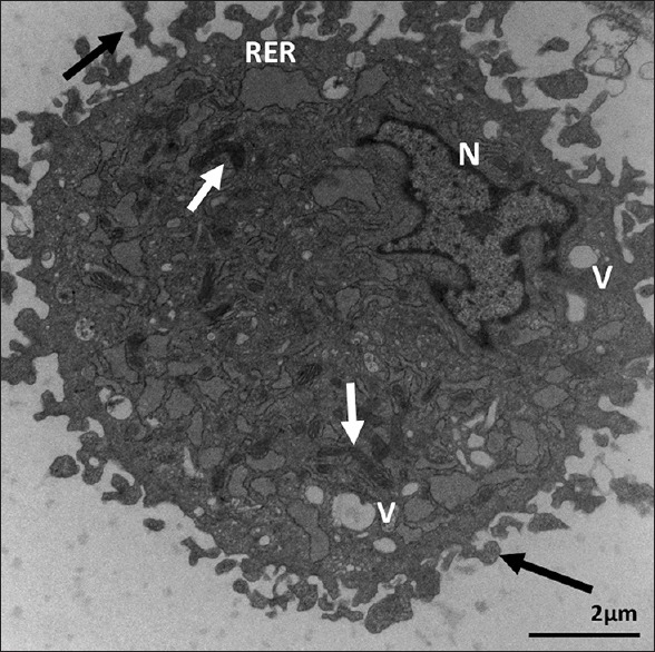
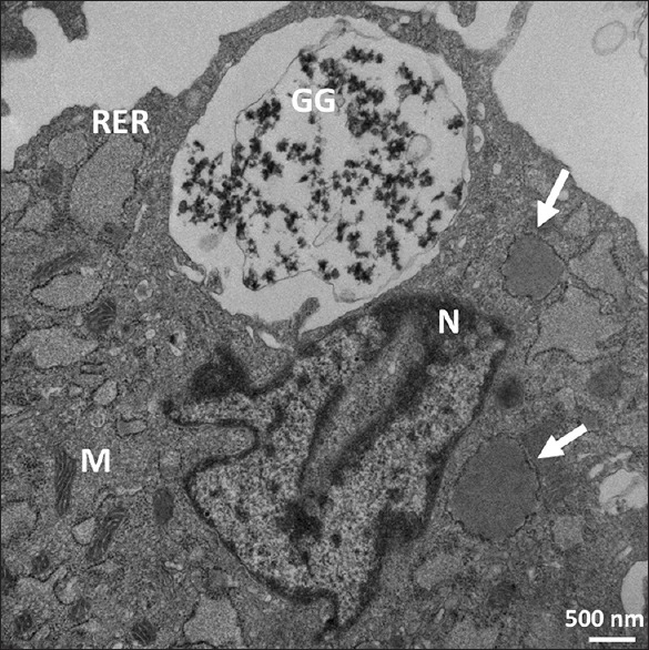
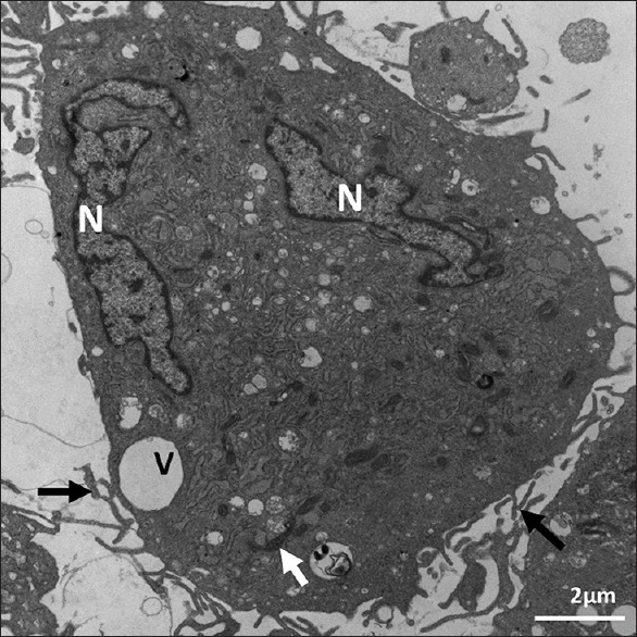
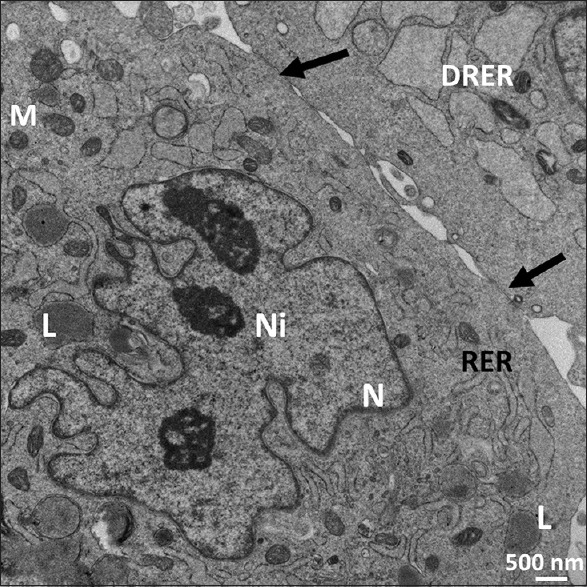
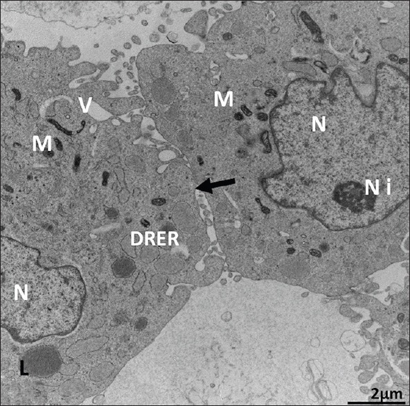
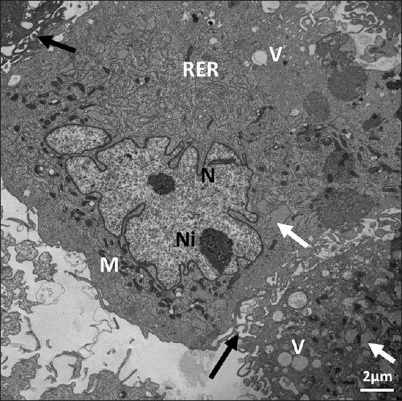
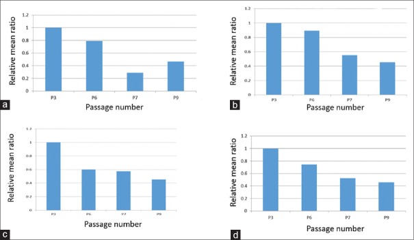
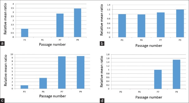
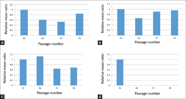
Results: Of the four passages (3, 6, 7, and 9) examined, dilated rough endoplasmic reticulum (RER) was abundant in Passage 3 but less so in Passages 6, 7, and 9. The dilated RER contained lipid in Passages 3, 7, and 9. The mono- and polyribosomes in Passages 3 and 6 were located between the mitochondria and the RER. Mono- and polyribosomes were abundant in Passage 7, although mainly monoribosomes were present in Passage 9. Membrane-bound glycogen granules were in vacuoles bulging off the cells in Passage 3. Some glycogen granules were grouped in the periphery of a stem cell in Passage 9. Nuclei shapes were irregular and mainly euchromatic in Passages 6, 7, and 9. The mitochondria were dark and scarce in Passage 9; irregular, small, and dark in Passage 7; and small and rounded in Passage 6, and they were spread in the cytoplasm away from the nucleus in Passage 3. Cell contacts were seen in Passages 6, 7, and 9. The ultrastructure morphology of the examined DFScs was not very different from the morphology criteria of the undifferentiated cells. Large vacuoles in Passage 3 were mainly at the periphery of the cell, with the small vacuoles in the cell center. Small vacuoles were scattered in the cell center of Passage 6 and the larger ones were observed at the cell's periphery.
Conclusions: We observed the following ultrastructural changes: decreases of fine cell cytoplasmic processes, dilated cytoplasmic vacuoles, cytoplasmic pinocytotic vesicles, and nuclear heterochromatin with increasing cell passage number. Conversely, mean ratios of lipid globules, nuclear euchromatin, irregular nuclear shape, and cell contact between cells were increased with passage number. The observations may suggest an increase in committed cells among the population after long-term culture of DFSCs.
Keywords: Dental follicle; mitochondria; nuclei; stem cells; transmission electron microscopy; ultrastructure.
Conflict of interest statement
There are no conflicts of interest.
Figures
Similar articles
-
Ultrastructural comparison of porcine putative embryonic stem cells derived by in vitro fertilization and somatic cell nuclear transfer.J Reprod Dev. 2016 Apr 22;62(2):177-85. doi: 10.1262/jrd.2015-124. Epub 2016 Jan 28. J Reprod Dev. 2016. PMID: 26821870 Free PMC article.
-
Morphology of in vitro expanded human muscle-derived stem cells.Biomed Pap Med Fac Univ Palacky Olomouc Czech Repub. 2008 Dec;152(2):235-8. doi: 10.5507/bp.2008.036. Biomed Pap Med Fac Univ Palacky Olomouc Czech Repub. 2008. PMID: 19219213
-
[ULTRASTRUCTURAL CHANGES OF THE STEM CELLS IN THE CYCLE MONOLAYER--SPHERES--MONOLAYER].Tsitologiia. 2016;58(1):16-22. Tsitologiia. 2016. PMID: 27220247 Russian.
-
Dental Follicle Stem Cells: Tissue Engineering and Immunomodulation.Stem Cells Dev. 2019 Aug 1;28(15):986-994. doi: 10.1089/scd.2019.0012. Epub 2019 May 7. Stem Cells Dev. 2019. PMID: 30968740 Review.
-
Clinical application prospects and transformation value of dental follicle stem cells in oral and neurological diseases.World J Stem Cells. 2023 Apr 26;15(4):136-149. doi: 10.4252/wjsc.v15.i4.136. World J Stem Cells. 2023. PMID: 37181000 Free PMC article. Review.
Cited by
-
Optimal good manufacturing practice-compliant production of dental follicle stem cell sheet and its application in Sprague-Dawley rat periodontitis.World J Stem Cells. 2025 May 26;17(5):104116. doi: 10.4252/wjsc.v17.i5.104116. World J Stem Cells. 2025. PMID: 40503360 Free PMC article.
-
Characterizing Organelles in Live Stem Cells Using Label-Free Optical Diffraction Tomography.Mol Cells. 2021 Nov 30;44(11):851-860. doi: 10.14348/molcells.2021.0190. Mol Cells. 2021. PMID: 34819398 Free PMC article.
References
-
- Shanti RM, Li WJ, Nesti LJ, Wang X, Tuan RS. Adult mesenchymal stem cells: Biological properties, characteristics, and applications in maxillofacial surgery. J Oral Maxillofac Surg. 2007;65:1640–7. - PubMed
-
- Varga V, Hollý D, Vojtaššák J, Bohmer D, Polák S, Danišovič L. Morphological characterization of in vitro expanded human dental pulp-derived stem cells. Biol Sect Zool. 2011;66:706–11.
-
- d’Aquino R, Graziano A, Sampaolesi M, Laino G, Pirozzi G, De Rosa A, et al. Human postnatal dental pulp cells co-differentiate into osteoblasts and endotheliocytes: A pivotal synergy leading to adult bone tissue formation. Cell Death Differ. 2007;14:1162–71. - PubMed
-
- Kadar K, Kiraly M, Porcsalmy B, Molnar B, Racz GZ, Blazsek J, et al. Differentiation potential of stem cells from human dental origin – Promise for tissue engineering. J Physiol Pharmacol. 2009;60(Suppl 7):167–75. - PubMed
-
- Suchanek J, Soukup T, Visek B, Ivancakova R, Kucerova L, Mokry J, et al. Dental pulp stem cells and their characterization. Biomed Pap Med Fac Univ Palacky Olomouc Czech Repub. 2009;153:31–5. - PubMed
LinkOut - more resources
Full Text Sources