

Hyaluronic Acid: Molecular Mechanisms and Therapeutic Trajectory
- PMID: 31294035
- PMCID: PMC6603175
- DOI: 10.3389/fvets.2019.00192
Hyaluronic Acid: Molecular Mechanisms and Therapeutic Trajectory
Abstract
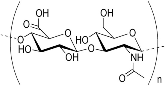
Hyaluronic acid (also known as hyaluronan or hyaluronate) is naturally found in many tissues and fluids, but more abundantly in articular cartilage and synovial fluid (SF). Hyaluronic acid (HA) content varies widely in different joints and species. HA is a non-sulfated, naturally occurring non-protein glycosaminoglycan (GAG), with distinct physico-chemical properties, produced by synoviocytes, fibroblasts, and chondrocytes. HA has an important role in the biomechanics of normal SF, where it is partially responsible for lubrication and viscoelasticity of the SF. The concentration of HA and its molecular weight (MW) decline as osteoarthritis (OA) progresses with aging. For that reason, HA has been used for more than four decades in the treatment of OA in dogs, horses and humans. HA produces anti-arthritic effects via multiple mechanisms involving receptors, enzymes and other metabolic pathways. HA is also used in the treatment of ophthalmic, dermal, burns, wound repair, and other health conditions. The MW of HA appears to play a critical role in the formulation of the products used in the treatment of diseases. This review provides a mechanism-based rationale for the use of HA in some disease conditions with special reference to OA.
Keywords: adjuvant therapy; cancer therapy; hyaluronan; hyaluronic acid; ophthalmic diseases; osteoarthritis; viscosupplementation; wound healing.
Figures
References
-
- Sugahara K, Schwartz NB, Dorfman A. Biosynthesis of hyaluronic acid by Streptococcus. J Biol Chem. (1979) 254:6252–61. - PubMed
Publication types
LinkOut - more resources
Full Text Sources
Other Literature Sources