. 2019 Sep 1;160(9):2189-2203.
doi: 10.1210/en.2019-00144.

Human Trophoblast Differentiation Is Associated With Profound Gene Regulatory and Epigenetic Changes

Affiliations

Human Trophoblast Differentiation Is Associated With Profound Gene Regulatory and Epigenetic Changes

Youn-Tae Kwak et al. Endocrinology. .

Abstract

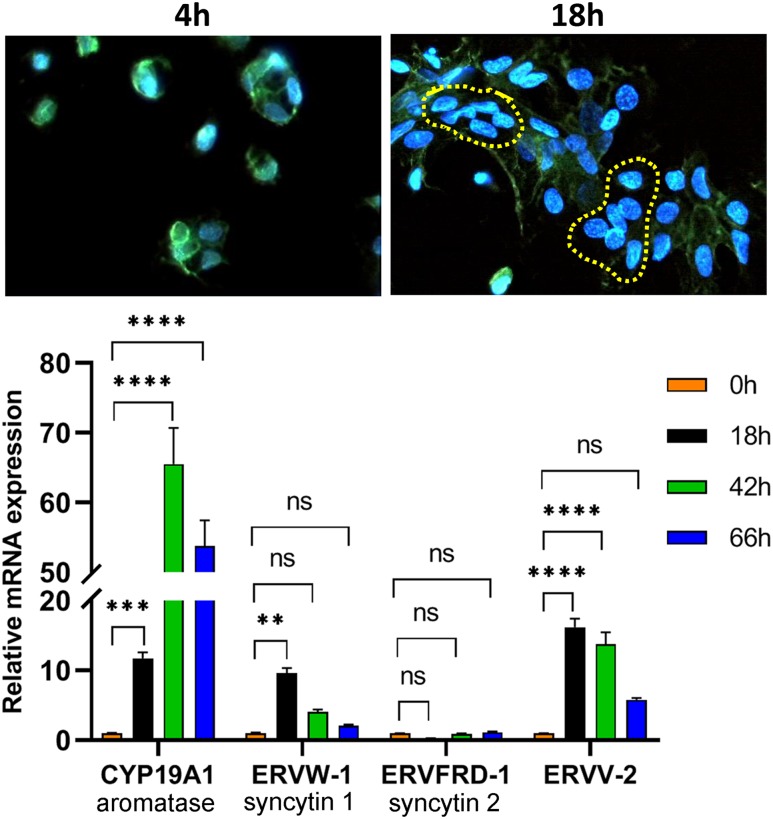
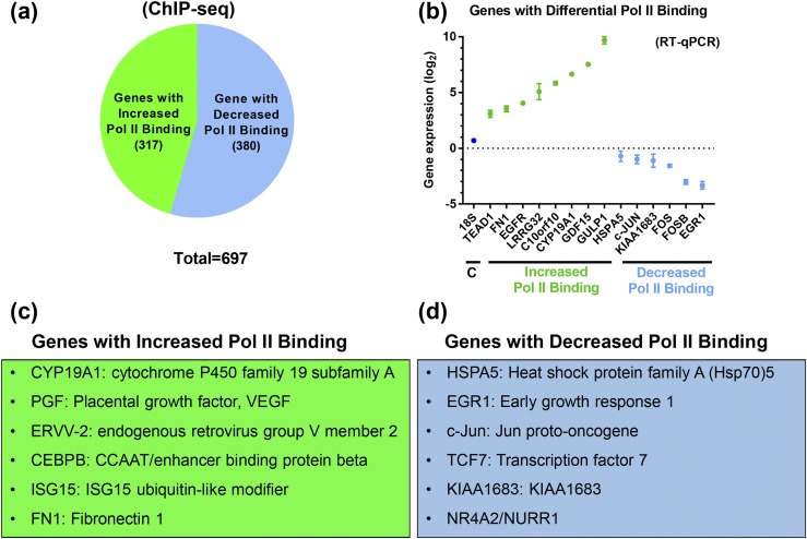
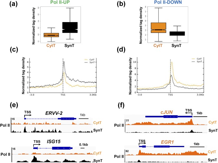
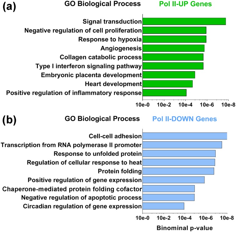
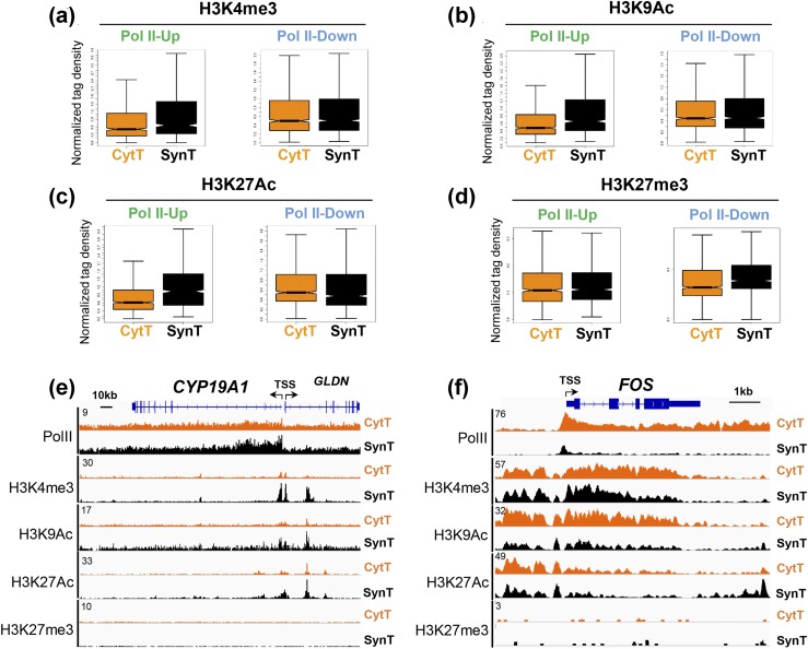
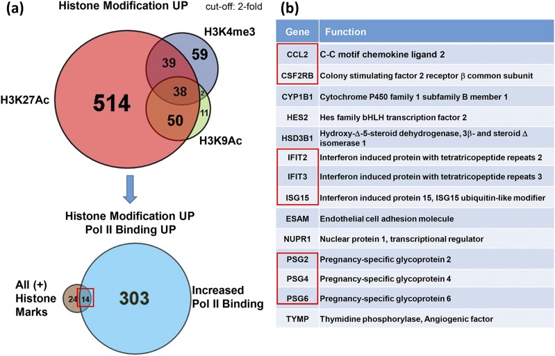
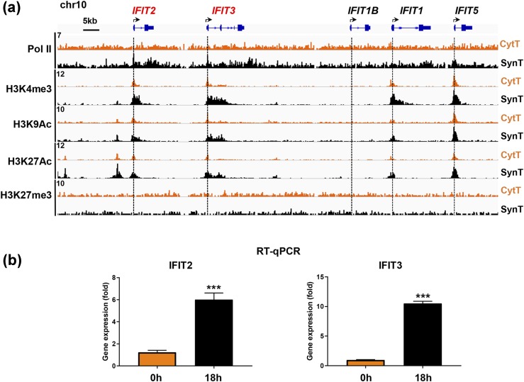
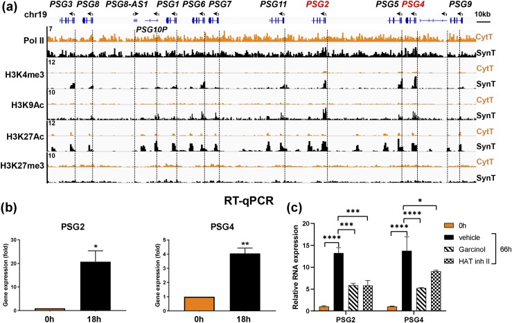
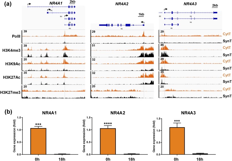
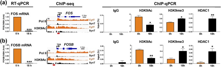
Defective placental implantation and vascularization with accompanying hypoxia contribute to preeclampsia (PE), a leading cause of maternal and neonatal morbidity and mortality. Genetic and epigenetic mechanisms underlying differentiation of proliferative cytotrophoblasts (CytTs) to multinucleated syncytiotrophoblast (SynT) are incompletely defined. The SynT performs key functions in nutrient and gas exchange, hormone production, and protection of the fetus from rejection by the maternal immune system. In this study, we used chromatin immunoprecipitation sequencing of midgestation human trophoblasts before CytT and after SynT differentiation in primary culture to analyze changes in binding of RNA polymerase II (Pol II) and of active and repressive histone marks during SynT differentiation. Our findings reveal that increased Pol II binding to promoters of a subset of genes during trophoblast differentiation was closely correlated with active histone marks. This gene set was enriched in those controlling immune response and immune modulation, including interferon-induced tetratricopeptide repeat and placenta-specific glycoprotein gene family members. By contrast, genes downregulated during SynT differentiation included proinflammatory transcription factors ERG1, cFOS, and cJUN, as well as members of the NR4A orphan nuclear receptor subfamily, NUR77, NURR1, and NOR1. Downregulation of proinflammatory transcription factors upon SynT differentiation was associated with decreased promoter enrichment of endogenous H3K27Ac and H3K9Ac and enhanced binding of H3K9me3 and histone deacetylase 1. However, promoter enrichment of H3K27me3 was low in both CytT and SynT and was not altered with changes in gene expression. These findings provide important insight into mechanisms underlying human trophoblast differentiation and may identify therapeutic targets for placental disorders, such as PE.


Conflict of interest statement

Current Affiliation: S. Muralimanoharan’s current affiliation is the Department of Molecular Medicine, Institute of Biotechnology, University of Texas Health Science Center, San Antonio, San Antonio, Texas 78229.

Disclosure Summary: The authors have nothing to disclose.

All data generated or analyzed during this study are included in this published article or in the data repositories listed in References.

Figures

Figure 1.
Figure 1.
Trophoblast fusion and induction of SynT-specific CYP19A1 and the fusogenic proteins ERVW-1 (syncytin 1) and ERVV-2 are significantly increased within 18 h of human trophoblast culture. (Top) Freshly isolated midgestation human CytT were plated on coverslips and cultured for 4 h or 18 h. The cells were subjected to immunostaining using a primary antibody to plakoglobin and a secondary antibody conjugated to Alexa Fluor 488. The yellow dashed lines encircle cell clusters in which the cell membranes have fused to form syncytia. Images were captured using a Zeiss confocal microscope at ×400 magnification. (Bottom) Midgestation CytT were analyzed before (0 h) or after 18, 42, and 66 h of culture for expression of CYP19A1, ERVW-1, ERVFRD-1, and ERVV-2 by RT-qPCR using specific primers. Data are the mean ± SEM of triplicate samples. Significantly (**P < 0.01; ***P < 0.001; ****P < 0.0001) different from 0 h. ns, not significant.
Figure 2.
Figure 2.
Pol II is differentially bound to promoters of a subset of genes during SynT differentiation. In these studies, ChIP-seq libraries were prepared from CytT isolated from midgestation human placentas before and after SynT differentiation in culture. (a) Numbers of genes that manifested a more than twofold increase (green) or more than twofold decrease (blue) in Pol II binding in SynT vs CytT. (b) RT-qPCR was used to assess expression of selected genes manifesting increased (green) and decreased (blue) binding of endogenous Pol II by ChIP-seq. (c) Representative genes that showed increased Pol II binding in SynT vs CytT. (d) Representative genes that showed decreased Pol II binding in SynT vs CytT. VEGF, vascular endothelial growth factor.
Figure 3.
Figure 3.
Genome-wide changes in Pol II binding occur at the TSS and gene body during SynT differentiation. Box plots of Pol II density at genes that manifested (a) increased Pol II binding in SynT compared with CytT (Pol II-UP) and (b) decreased Pol II binding in SynT compared with CytT (Pol II-DOWN). Metagene analysis of Pol II distribution in (c) Pol II-UP and (d) Pol II-DOWN genes in CytT vs SynT. Genome browser views of representative (e) Pol II-UP and (f) Pol II-DOWN genes in CytT vs SynT.
Figure 4.
Figure 4.
Gene ontology (GO) analyses were performed using the Database for Annotation, Visualization, and Integrated Discovery tool to assess biological processes that are represented in the Pol II-UP and Pol II-DOWN genes in SynT vs CytT. (a) Functional annotation of biological processes showed that Pol II-UP genes are enriched in functions involved in hypoxia, angiogenesis, and placental development. (b) By contrast, Pol II-DOWN gene functions included cellular adhesion and negative regulation of apoptosis.
Figure 5.
Figure 5.
Genes with increased Pol II promoter binding in SynT vs CytT show increased binding of active histone marks. (a–d) Box plot analyses of density of active and repressive histone marks in the promoter regions of Pol II-UP and Pol II-DOWN genes in SynT vs CytT. Genome browser views of binding of Pol II and postranslationally modified histones in representative (e) Pol II-UP (CYP19A1) and (f) Pol II-DOWN (cFOS) genes.
Figure 6.
Figure 6.
Genes that manifest enrichment of all three active histone marks (H3K4me3, H3K9Ac, and H3K927Ac) and increased Pol II binding in SynT vs CytT comprise a unique subset of genes involved in immune modulation. (a) Venn diagram of genes that manifest upregulated binding of one or more active histone marks in SynT compared with CytT (top). Venn diagram of genes that manifest enrichment of all three active histone marks and increased Pol II binding (bottom) comprise a unique subset of (b) genes primarily involved in immune response and immune modulation.
Figure 7.
Figure 7.
(a) Genome browser view of ChIP-seq analysis of interferon-inducible gene IFIT family members on human chromosome 10 indicates that increased binding of Pol II to the gene promoters in SynT vs CytT is correlated with increased active histone marks. (b) These changes in binding of endogenous Pol II and histone H3 modifications indicative of active chromatin are correlated with upregulated expression of the IFIT2 and IFIT3 genes in SynT (18 h of culture) vs CytT (0 h) analyzed by RT-qPCR. Data are the mean ± SEM of triplicate samples. Significantly different from 0 h (***P < 0.001).
Figure 8.
Figure 8.
Binding of Pol II and active histone marks is closely correlated with PSG gene expression during trophoblast differentiation. (a) Genome browser view of binding of Pol II and of active and repressive histone marks at the promoters of the cluster of PSG genes on the long arm of chromosome 19. (b) RT-qPCR was performed to assess expression of PSG2 and PSG4 genes during differentiation of freshly isolated CytT from midgestation placenta. (c) Freshly isolated CytT from term human placenta were analyzed for PSG2 and PSG4 mRNA expression before (0 h) or after 66 h in culture in the absence (vehicle) or presence of 5 μM garcinol or HAT inhibitor (inh) II. Data are the mean ± SEM of triplicate samples. Significantly different from 0 h or from vehicle (*P < 0.05; **P < 0.01; ***P < 0.001; ****P < 0.0001).
Figure 9.
Figure 9.
Decreased binding of Pol II and of the active histone marks H3K9Ac and H3K27Ac to the promoters and enhancers of the NR4A1 (NUR77), NR4A2 (NURR1), and NR4A3 (NOR1) genes is correlated with downregulation of their expression during SynT differentiation, whereas binding of H3K4me3 and H4K27me3 remain unchanged. (a) Genome browser views of binding of Pol II and of active and repressive histone marks to the promoters/enhancers of the NR4A1, NR4A2, and NR4A3 genes. (b) RT-qPCR of RNA isolated from midgestation placenta before and after 18 h of culture revealed marked downregulation of all three genes during SynT differentiation. Data are the mean ± SEM of triplicate samples. Significantly different from 0 h (***P < 0.001; ****P < 0.0001).
Figure 10.
Figure 10.
Decreased expression of the cFOS and FOSB genes during SynT differentiation is associated with decreased binding of Pol II and the active histone mark H3K9Ac and with increased recruitment of the repressive histone mark H3K9me3 and of HDAC1 to their promoters. RT-qPCR of (a) cFOS and (b) FOSB mRNA expression before (0 h, CytT) and after (18 h, SynT) differentiation of midgestation human trophoblasts in culture. Genome browser views of binding of Pol II and of the active histone mark H3K9Ac to the (a) cFOS and (b) FOSB gene promoters/enhancers before (0 h, CytT) and after (18 h, SynT) differentiation in culture. ChIP-qPCR of binding of H3K9Ac, H3K9me3, and HDAC1 to the (a) cFOS and (b) FOSB gene promoters/enhancers before (0 h, CytT) and after (18 h, SynT) differentiation of trophoblasts isolated from term human placenta. Antibody to nonimmune IgG was used as a control for ChIP. Data are the mean ± SEM of triplicate samples. Significantly different from 0 h (*P < 0.05; **P < 0.01; ***P < 0.001).

References

    1. Everett RB, MacDonald PC. Endocrinology of the placenta. Annu Rev Med. 1979;30(1):473–488. - PubMed
    1. Red-Horse K, Zhou Y, Genbacev O, Prakobphol A, Foulk R, McMaster M, Fisher SJ. Trophoblast differentiation during embryo implantation and formation of the maternal-fetal interface. J Clin Invest. 2004;114(6):744–754. - PMC - PubMed
    1. McMaster MT, Zhou Y, Fisher SJ. Abnormal placentation and the syndrome of preeclampsia. Semin Nephrol. 2004;24(6):540–547. - PubMed
    1. Arck PC, Hecher K. Fetomaternal immune cross-talk and its consequences for maternal and offspring’s health. Nat Med. 2013;19(5):548–556. - PubMed
    1. PrabhuDas M, Bonney E, Caron K, Dey S, Erlebacher A, Fazleabas A, Fisher S, Golos T, Matzuk M, McCune JM, Mor G, Schulz L, Soares M, Spencer T, Strominger J, Way SS, Yoshinaga K. Immune mechanisms at the maternal-fetal interface: perspectives and challenges. Nat Immunol. 2015;16(4):328–334. - PMC - PubMed

Publication types