. 2020 Jan;88(1):127-134.
doi: 10.1002/prot.25777. Epub 2019 Aug 2.

A free-energy landscape for the glucagon-like peptide 1 receptor GLP1R

Affiliations

A free-energy landscape for the glucagon-like peptide 1 receptor GLP1R

Raphael Alhadeff et al. Proteins. 2020 Jan.

Abstract

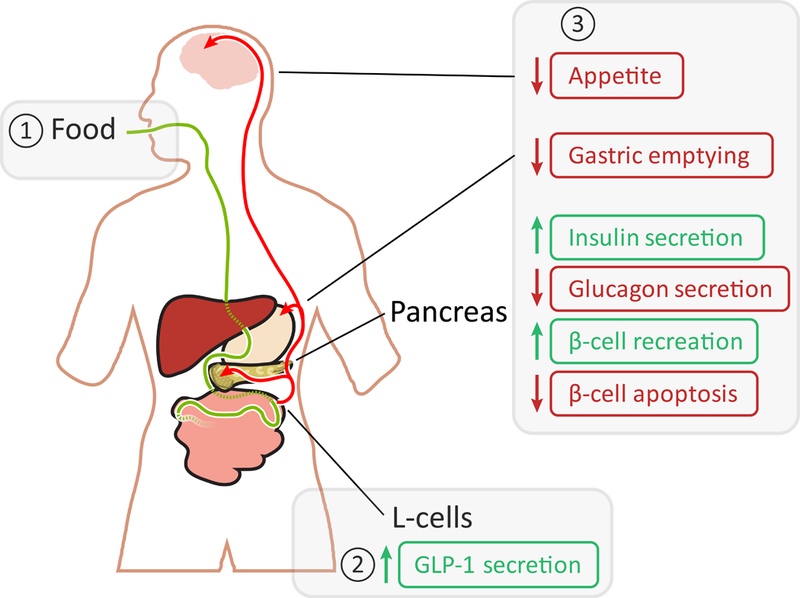
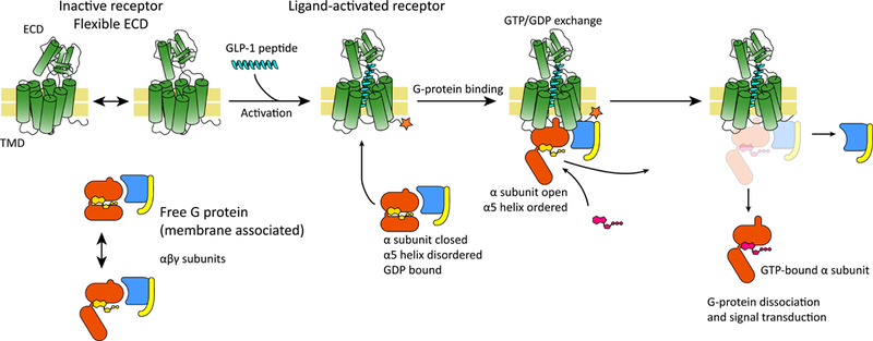
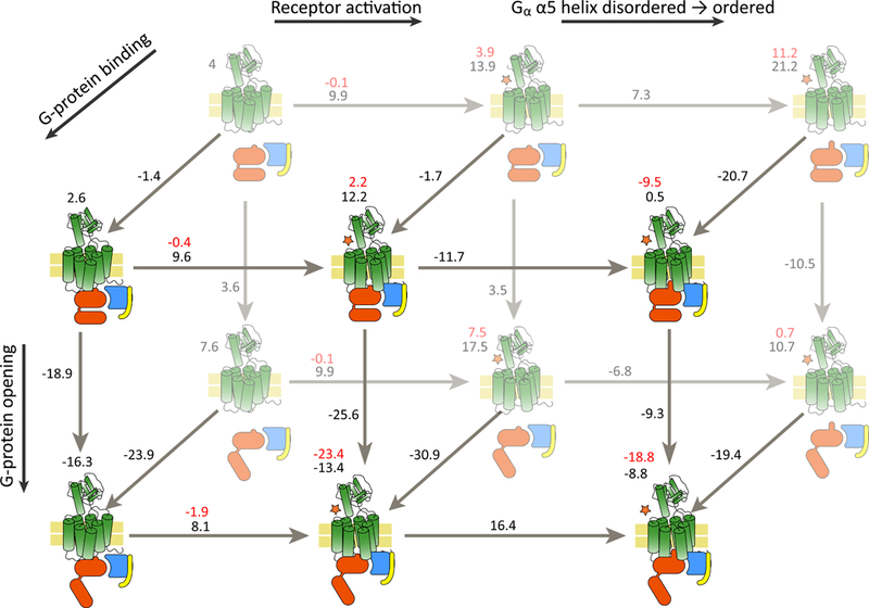
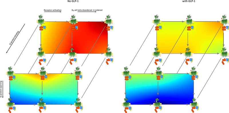
G-protein-coupled receptors (GPCRs) are among the most important receptors in human physiology and pathology. They serve as master regulators of numerous key processes and are involved in as well as cause debilitating diseases. Consequently, GPCRs are among the most attractive targets for drug design and pharmaceutical interventions (>30% of drugs on the market). The glucagon-like peptide 1 (GLP-1) hormone receptor GLP1R is closely involved in insulin secretion by pancreatic β-cells and constitutes a major druggable target for the development of anti-diabetes and obesity agents. GLP1R structure was recently solved, with ligands, allosteric modulators and as part of a complex with its cognate G protein. However, the translation of this structural data into structure/function understanding remains limited. The current study functionally characterizes GLP1R with special emphasis on ligand and cellular partner binding interactions and presents a free-energy landscape as well as a functional model of the activation cycle of GLP1R. Our results should facilitate a deeper understanding of the molecular mechanism underlying GLP1R activation, forming a basis for improved development of targeted therapeutics for diabetes and related disorders.

Keywords: GLP-1R; GPCR; diabetes; energy-landscape.


Figures

Fig. 1.
Fig. 1.
Physiological effects of GLP1R. Food consumption is marked with a green line and GLP-1 in the blood is marked with red arrows. The small green and red vertical arrows denote increase or decrease, respectively. The boxed 1, 2 and 3 show the canonical sequence of event for GLP-1, where 1 denotes the trigger, 2 the release and 3 the effects in the brain, stomach, and pancreas.
Fig. 2.
Fig. 2.
Canonical activation sequence for GLP1R. The receptor harbors the GPCR-typical 7 transmembrane helices domain (TMD) and an extracellular domain (ECD) which bears some flexibility, represented schematically in the figure with a two-headed arrow. The inactive receptor binds the ligand, GLP-1, undergoes a conformational change and becomes active (highlighted with a star in the figure). Cellular G protein bound to the activated receptor then undergoes conformational changes (namely, opening and ordering of the α5 domain; see text), allowing the exchange of GDP with GTP (shown in yellow and magenta). Finally, the GTP-bound G protein dissociates and the subunits elicit cellular response.
Fig. 3.
Fig. 3.
State total free energy. The various states as described above and their associated MCPT total free energy are shown as illustrations, with the energy labeled above it. The difference in free energy between two states is shown in black on the arrow, and the red is the energy after parametrically adding the binding energy of the ligand. The reaction coordinates are labeled with thick arrows on the edges of the scheme. The receptor is illustrated in green, with a star denoting activation, the G-protein subunits are illustrated in red, blue and yellow for α, β, and γ, respectively.
Fig. 4.
Fig. 4.
Free-energy landscapes. The energies from Fig. 3 are shown as landscapes (heat maps) in 2D, projected on the same state scheme as in Fig. 3. The barriers are unknown and were not computed, and so the figure presents a linear interpolation of the total energies, without taking barriers into account. The state scheme has been geometrically stretched to accommodate the maps more clearly. Red is high energy and blue is low energy.

Similar articles

Cited by

References

    1. Alam U, Asghar O, Azmi S, & Malik RA (2014) General aspects of diabetes mellitus. Handb Clin Neurol 126:211–222. - PubMed
    1. Caspard H, et al. (2018) Recent trends in the prevalence of type 2 diabetes and the association with abdominal obesity lead to growing health disparities in the USA: An analysis of the NHANES surveys from 1999 to 2014. Diabetes Obes Metab 20(3):667–671. - PMC - PubMed
    1. Kerru N, Singh-Pillay A, Awolade P, & Singh P (2018) Current anti-diabetic agents and their molecular targets: A review. Eur J Med Chem 152:436–488. - PubMed
    1. Baggio LL & Drucker DJ (2014) Glucagon-like peptide-1 receptors in the brain: controlling food intake and body weight. J Clin Invest 124(10):4223–4226. - PMC - PubMed
    1. Heppner KM & Perez-Tilve D (2015) GLP-1 based therapeutics: simultaneously combating T2DM and obesity. Front Neurosci 9:92. - PMC - PubMed

Publication types

LinkOut - more resources