. 2019 Sep;192(3):311-323.
doi: 10.1667/RR15266.1. Epub 2019 Jul 11.

RABiT-II-DCA: A Fully-automated Dicentric Chromosome Assay in Multiwell Plates

Affiliations

RABiT-II-DCA: A Fully-automated Dicentric Chromosome Assay in Multiwell Plates

Ekaterina Royba et al. Radiat Res. 2019 Sep.

Abstract

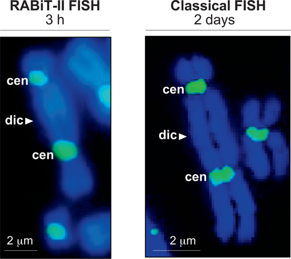
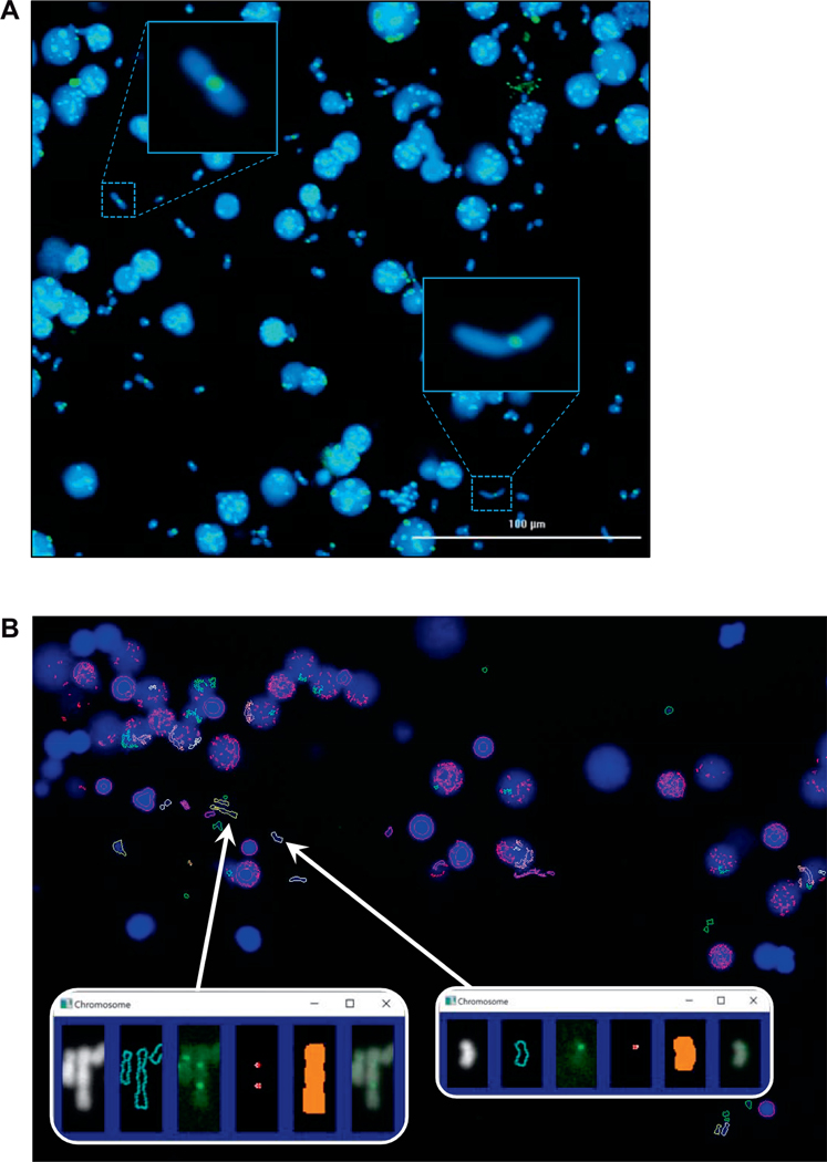
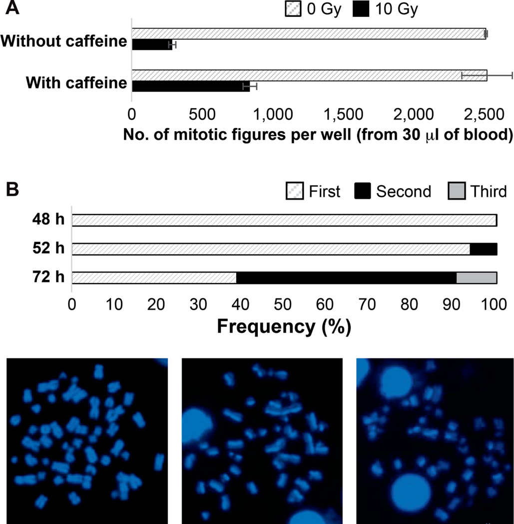
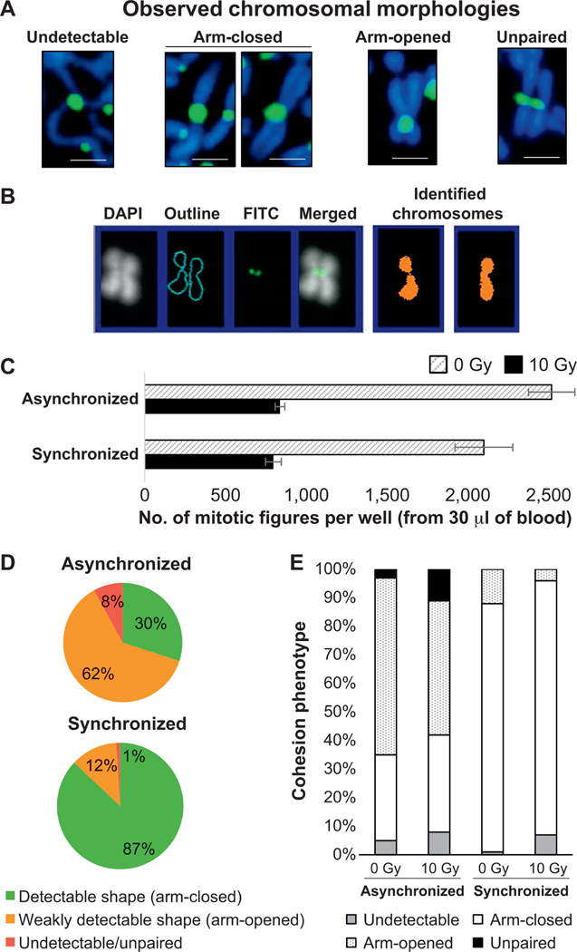
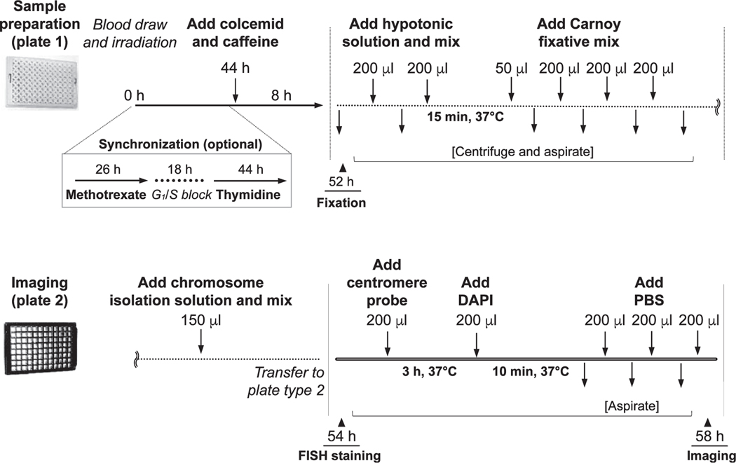
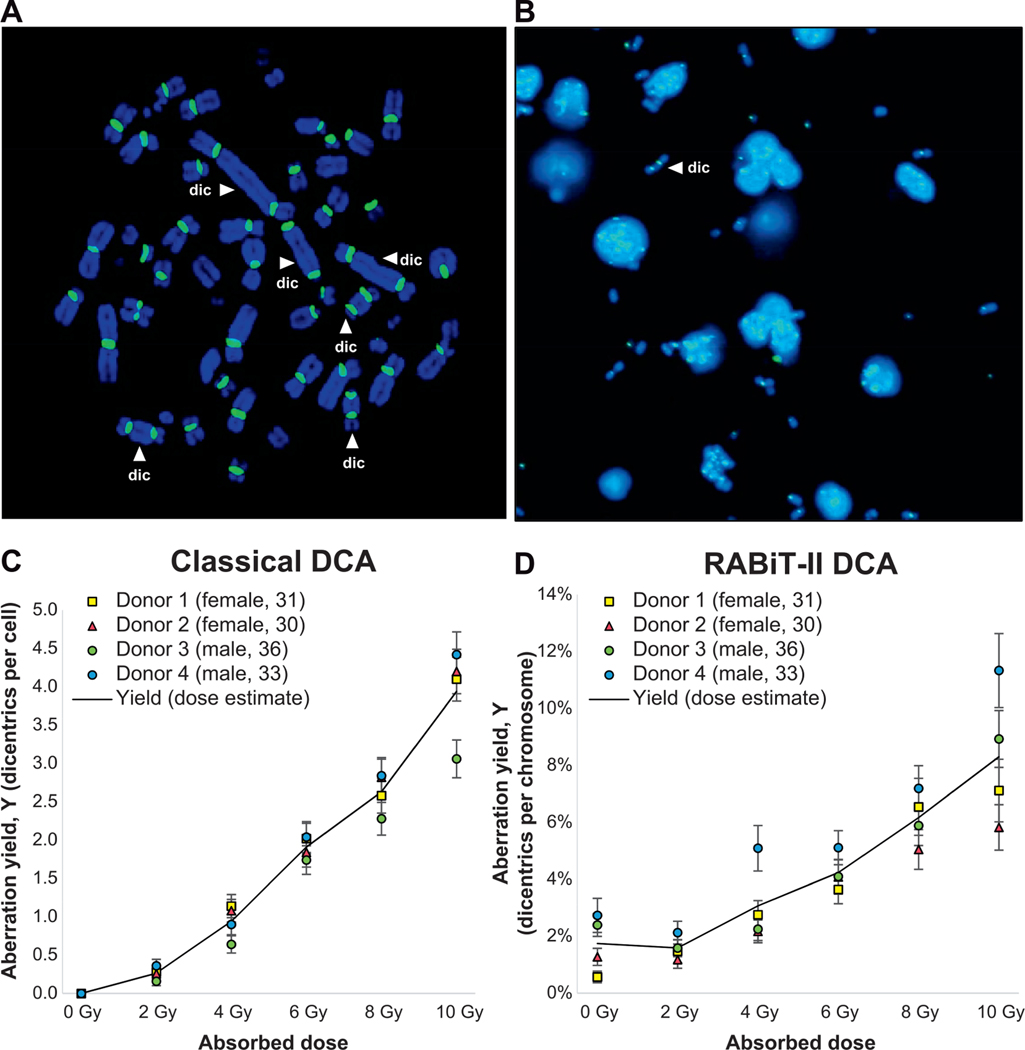
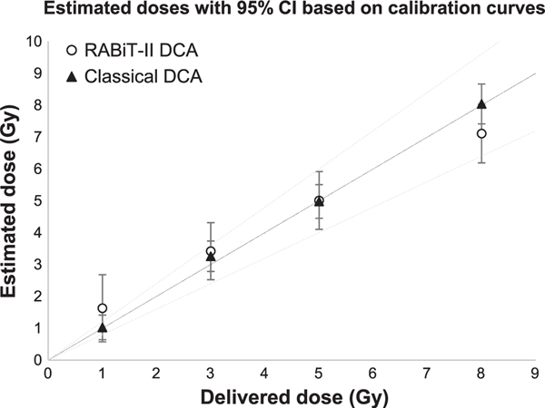
We developed a fully-automated dicentric chromosome assay (DCA) in multiwell plates. All operations, from sample loading to chromosome scoring, are performed, without human intervention, by the second-generation Rapid Automated Biodosimetry Tool II (RABiT-II) robotic system, a plate imager and custom software, FluorQuantDic. The system requires small volumes of blood (30 µl per individual) to determine radiation dose received as a result of a radiation accident or terrorist attack. To visualize dicentrics in multiwell plates, we implemented a non-classical protocol for centromere FISH staining at 37°C. The RABiT-II performs rapid analysis of chromosomes after extracting them from metaphase cells. With the use of multiwell plates, many samples can be screened at the same time. Thus, the RABiT-II DCA provides an advantage during triage when risk-based stratification and medical management are required for a large population exposed to unknown levels of ionizing radiation.


Figures

FIG. 1.
FIG. 1.
Comparison of the RABiT-II and the classical FISH staining with centromere probes. Panel A: Representative image of a dicentric stained with centromere probe at 37°C (without rehydration of samples in PBS, incubation in formaldehyde, pepsin treatment, dehydration in ethanol series and washes). Image captured at 20× magnification (BioTek Cytation Cell 1). Scale bar 2 μm. Panel B: Representative image of a dicentric after staining following classical FISH protocol at 80°C (including rehydration of samples in PBS, incubation in formaldehyde, pepsin treatment, dehydration in ethanol series and washes). Image captured at 63× magnification (Metafer 4 Master Station). Scale bar = 2 μm; dic = dicentric chromosome; cen = centromere.
FIG. 2.
FIG. 2.
Automated detection of dicentrics in the RABiT-II system. Panel A: Chromosomes released from mitotic cells (30 μl of whole blood), 0 Gy sample. Panel B: Output from the image analysis software FluorQuantDic version 3.3. Large blue objects are intact nuclei. White outlines correspond to identified monocentric chromosomes. Red outlines are objects which are either too big or too small to be scored as chromosomes. Green objects are too round to be chromosomes. Yellow objects are identified dicentric chromosomes. Inserts are identified dicentric (left) and monocentric (right) chromosomes. Panels from left to right: chromosome image (DAPI); the identified chromosome outline; centromere signal (FITC); identified centromeres; the mask for selecting which centromeres are associated with the chromosome; a merged DAPI/FITC image.
FIG. 3.
FIG. 3.
Assay development and quality control. Panel A: Caffeine allows G2/M arrested lymphocytes to enter mitosis after severe DNA damage. Quantification data represents total number of mitotic figures scored in one well after 10 Gy irradiation without (286 mitotic cell spreads per well) or with (834 mitotic cell spreads per well) caffeine. Data and standard errors were calculated based on three independent experimental trials. Panel B: Frequency of second division metaphases in the RABiT-II DCA protocol after 52 h of culture. Images represent differentiation of sister chromatids by 33258 Hoechst fluorescence after different cycles of BrdU incorporation: first (bifiliar BrdU incorporation, on the left), second (unifiliar BrdU incorporation, in the middle), and third (one fourth BrdU incorporation, on the right) cell cycle metaphase spreads.
FIG. 4.
FIG. 4.
Uniform shapes of chromosomes after synchronization. Panel A: Common chromosomal morphologies frequently observed in the same well (from drop of blood from healthy volunteer, no irradiation, asynchronously cultured, 8 h incubation with colcemid). Images representing chromosome 1 were cropped and magnified from Supplementary Fig. S1 (http://dx.doi.org/10.1667/RR15266.1.S1). Panel B: FluorQuantDic classifier misrecognized the chromosome with opened arms as two chromosomes with closed arms. Panel C: Quantification data of mitotic figures before and after synchronization. Standard errors were calculated based on three independent experimental trials. Panel D: Proportion of mitotic cells with variable shapes of chromosomes (that can or cannot be detected using FluorQuantDic software) before and after synchronization. Panel E: Quantification results for distribution of chromosomal shapes before and after synchronization calculated for 1,000 prometaphase cells (0 Gy and 10 Gy irradiated samples). White and dashed populations represent cells with closed and open arms, respectively. Dark gray and black bars refer to undetectable or unpaired populations, respectively.
FIG. 5.
FIG. 5.
Schematic of the end-to-end automated RABiT-II DCA. The operating parameters are divided into several subprograms, which allows the user to perform various tasks including routine controls of the cell culture, synchronization, harvesting, fixation, staining and sample imaging.
FIG. 6.
FIG. 6.
Dose-response calibration curves for the RABiT-II and classical DCA. Panel A: Dicentric scoring strategy in the classical DCA. Representative example of a metaphase cell (10 Gy irradiated sample) suitable for analysis (complete set of centromeres, dicentrics are accompanied by acentric fragments, etc.). For each sample, analysis was performed in 50 metaphases using Isis software. Distribution of identified dicentrics (indicated by white arrows) was recorded; multicentrics are converted to the dicentric chromosome equivalents. Panel B: Dicentric scoring strategy in the RABiT-II DCA. Representative image (4 Gy sample) used for analysis. Scoring was performed automatically by the FluorQuantDic software (450 images per sample). A dicentric is indicated by the white arrow. Panel C: Preliminary dose-response curve for 137Cs γ rays for classical DCA. The left axis shows the yield of DCs as a fraction of 50 cells scored. Panel D: Preliminary dose-response curve for 137Cs γ rays performed using the RABiT-II system and scored without human intervention. The left axis shows the yield of dicentrics as a fraction of normal chromosomes.
FIG. 7.
FIG. 7.
The reliability of individual dose estimations by RABiT-II DCA and classical DCA. Four dose estimations of samples from two different donors (male and female, 29 and 30 years old, respectively) and the corresponding 95% CI were determined at each dose. The RABiT-II DCA and classical DCA calibration curves were used for dose-prediction calculations. Reconstructed doses are based on eight replicates for each donor; black line is an ideal reconstruction (reconstructed dose = physical dose) and dashed lines are ±20%.

Similar articles

Cited by

  • Nuclear and Radiological Emergencies: Biological Effects, Countermeasures and Biodosimetry.
    Obrador E, Salvador-Palmer R, Villaescusa JI, Gallego E, Pellicer B, Estrela JM, Montoro A. Obrador E, et al. Antioxidants (Basel). 2022 May 31;11(6):1098. doi: 10.3390/antiox11061098. Antioxidants (Basel). 2022. PMID: 35739995 Free PMC article. Review.
  • Sex differences in radiation research.
    Taliaferro LP, Agarwal RK, Coleman CN, DiCarlo AL, Hofmeyer KA, Loelius SG, Molinar-Inglis O, Tedesco DC, Satyamitra MM. Taliaferro LP, et al. Int J Radiat Biol. 2024;100(3):466-485. doi: 10.1080/09553002.2023.2283089. Epub 2023 Nov 27. Int J Radiat Biol. 2024. PMID: 37991728 Free PMC article.
  • Plasma levels of serum amyloid A1 and mortality after exposure to high-dose radiation.
    Klokov D. Klokov D. Ann Transl Med. 2020 Jun;8(11):661. doi: 10.21037/atm.2020.03.172. Ann Transl Med. 2020. PMID: 32617281 Free PMC article. No abstract available.
  • The RABiT-II DCA in the Rhesus Macaque Model.
    Royba E, Repin M, Balajee AS, Shuryak I, Pampou S, Karan C, Brenner DJ, Garty G. Royba E, et al. Radiat Res. 2020 Nov 1;196(5):501-509. doi: 10.1667/RR15547.1. Radiat Res. 2020. PMID: 33022052 Free PMC article.
  • RENEB Inter-Laboratory Comparison 2021: Inter-Assay Comparison of Eight Dosimetry Assays.
    Port M, Barquinero JF, Endesfelder D, Moquet J, Oestreicher U, Terzoudi G, Trompier F, Vral A, Abe Y, Ainsbury L, Alkebsi L, Amundson SA, Badie C, Baeyens A, Balajee AS, Balázs K, Barnard S, Bassinet C, Beaton-Green LA, Beinke C, Bobyk L, Brochard P, Brzoska K, Bucher M, Ciesielski B, Cuceu C, Discher M, D Oca MC, Domínguez I, Doucha-Senf S, Dumitrescu A, Duy PN, Finot F, Garty G, Ghandhi SA, Gregoire E, Goh VST, Güçlü I, Hadjiiska L, Hargitai R, Hristova R, Ishii K, Kis E, Juniewicz M, Kriehuber R, Lacombe J, Lee Y, Lopez Riego M, Lumniczky K, Mai TT, Maltar-Strmečki N, Marrale M, Martinez JS, Marciniak A, Maznyk N, McKeever SWS, Meher PK, Milanova M, Miura T, Monteiro Gil O, Montoro A, Moreno Domene M, Mrozik A, Nakayama R, O'Brien G, Oskamp D, Ostheim P, Pajic J, Pastor N, Patrono C, Pujol-Canadell M, Prieto Rodriguez MJ, Repin M, Romanyukha A, Rößler U, Sabatier L, Sakai A, Scherthan H, Schüle S, Seong KM, Sevriukova O, Sholom S, Sommer S, Suto Y, Sypko T, Szatmári T, Takahashi-Sugai M, Takebayashi K, Testa A, Testard I, Tichy A, Triantopoulou S, Tsuyama N, Unverricht-Yeboah M, Valente M, Van Hoey O, Wilkins RC, Wojcik A, Wojewodzka M, Younghyun L, Zafiropoulos D, Abend M. Port M, et al. Radiat Res. 2023 Jun 1;199(6):535-555. doi: 10.1667/RADE-22-00207.1. Radiat Res. 2023. PMID: 37310880 Free PMC article.

References

    1. Repin M, Turner HC, Garty G, Brenner DJ. Next generation platforms for high-throughput biodosimetry. Radiat Prot Dosimetry 2014; 159:105–10. - PMC - PubMed
    1. Repin M, Pampou S, Karan C, Brenner DJ, Garty G. RABiT-II: Implementation of a high-throughput micronucleus biodosimetry assay on commercial biotech robotic systems. Radiat Res 2017; 187:492–8. - PMC - PubMed
    1. Cytogenetic dosimetry: Applications in preparedness for and response to radiation emergencies. EPR-Biodosimetry 2011; Vienna: International Atomic Energy Agency; 2011.
    1. Sullivan JM, Prasanna PGS, Grace MB, Wathen LK, Wallace RL, Koerner JF, et al. Assessment of biodosimetry methods for a mass-casualty radiological incident: medical response and management considerations. Health Phys 2013; 105:540–54. - PMC - PubMed
    1. Flood AB, Boyle HK, Du G, Demidenko E, Nicolalde RJ, Williams BB, et al. Advances in a framework to compare biodosimetry methods for triage in large-scale radiation events. Radiat Prot Dosimetry 2014; 159:77–86. - PMC - PubMed

Publication types