. 2019 Jul 11;15(7):e1008062.
doi: 10.1371/journal.pgen.1008062. eCollection 2019 Jul.

Reproduction disrupts stem cell homeostasis in testes of aged male Drosophila via an induced microenvironment

Affiliations

Reproduction disrupts stem cell homeostasis in testes of aged male Drosophila via an induced microenvironment

Yi Chieh Chang et al. PLoS Genet. .

Abstract

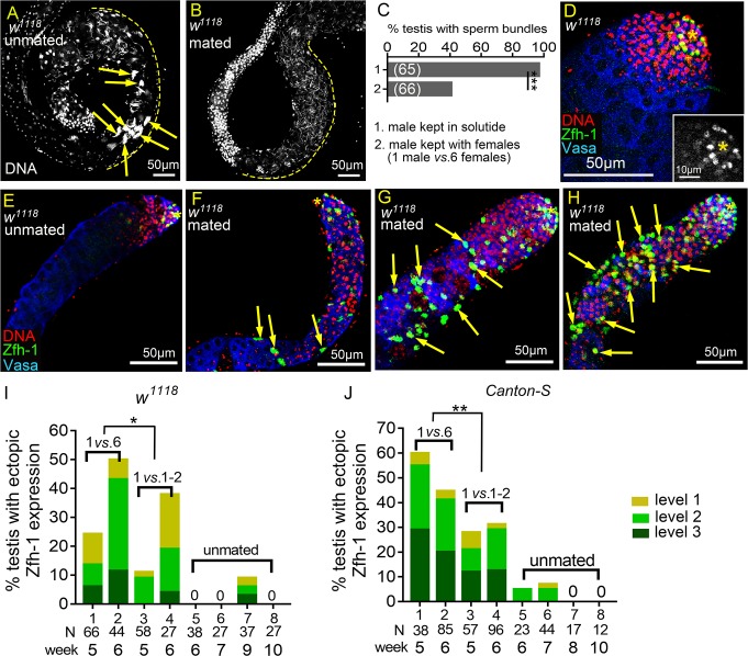
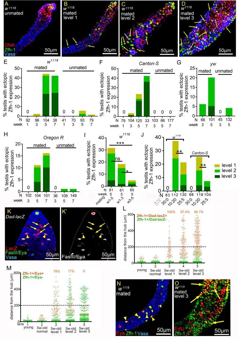
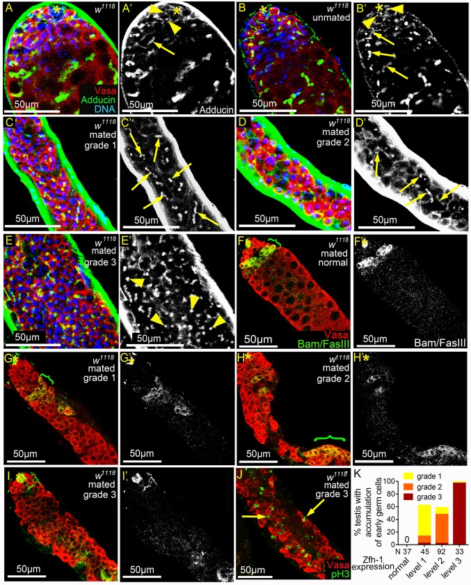
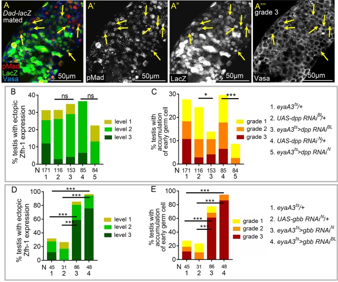
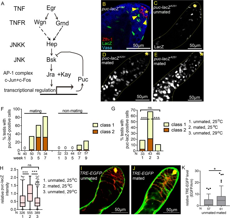
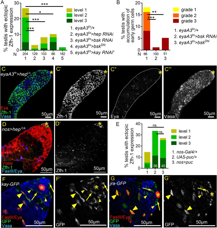
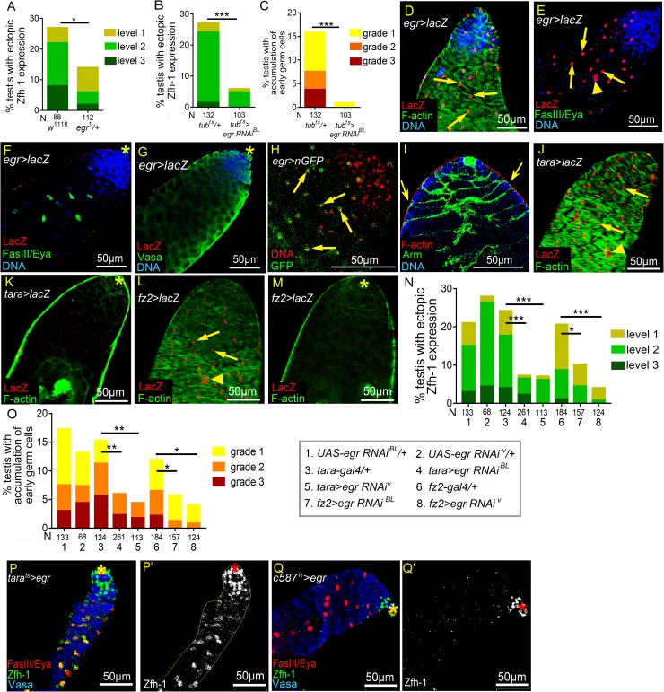
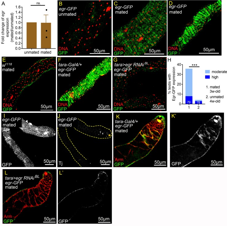
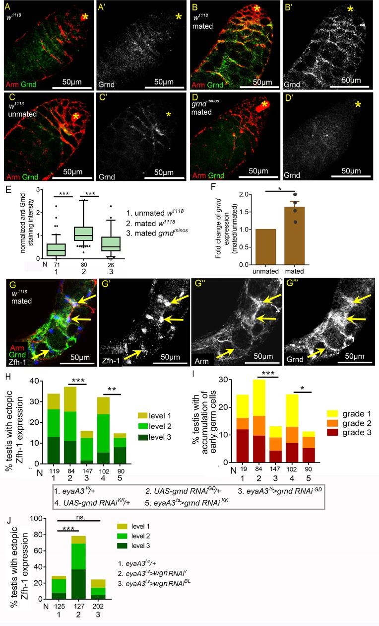
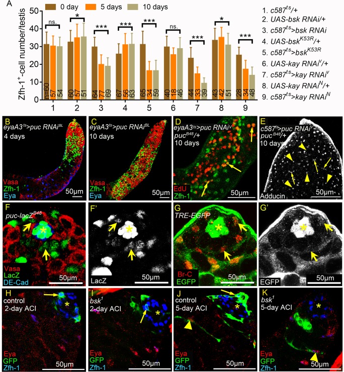
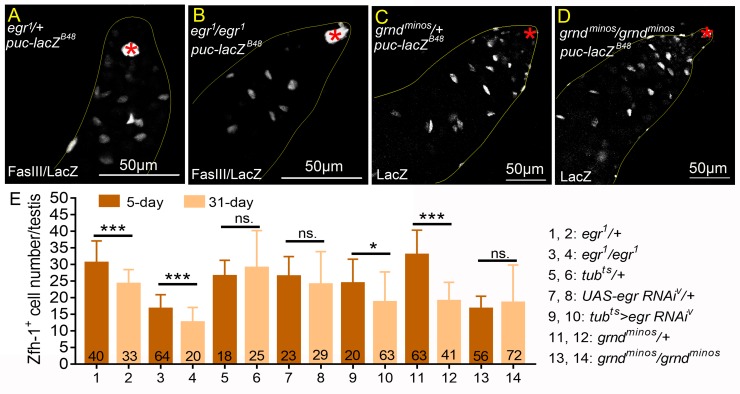
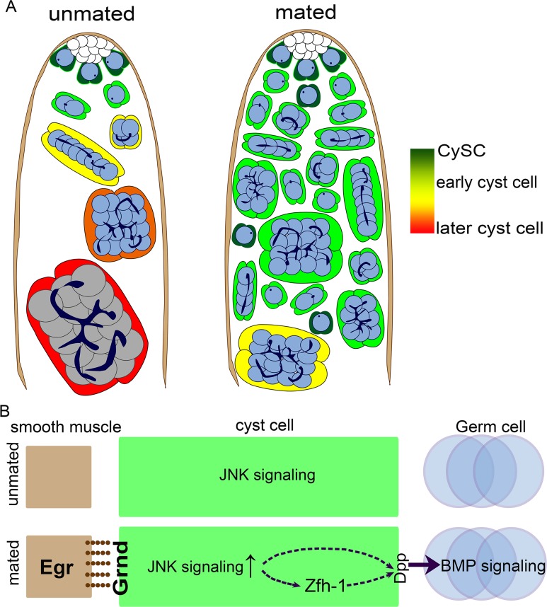
Stem cells rely on instructive cues from their environment. Alterations in microenvironments might contribute to tissue dysfunction and disease pathogenesis. Germline stem cells (GSCs) and cyst stem cells (CySC) in Drosophila testes are normally maintained in the apical area by the testicular hub. In this study, we found that reproduction leads to accumulation of early differentiating daughters of CySCs and GSCs in the testes of aged male flies, due to hyperactivation of Jun-N-terminal kinase (JNK) signaling to maintain self-renewal gene expression in the differentiating cyst cells. JNK activity is normally required to maintain CySCs in the apical niche. A muscle sheath surrounds the Drosophila testis to maintain its long coiled structure. Importantly, reproduction triggers accumulation of the tumor necrosis factor (TNF) Eiger in the testis muscle to activate JNK signaling via the TNF receptor Grindelwald in the cyst cells. Reducing Eiger activity in the testis muscle sheath suppressed reproduction-induced differentiation defects, but had little effect on testis homeostasis of unmated males. Our results reveal that reproduction in males provokes a dramatic shift in the testicular microenvironment, which impairs tissue homeostasis and spermatogenesis in the testes.


Conflict of interest statement

The authors have declared that no competing interests exist.

Figures

Fig 1
Fig 1. Reproduction induces ectopic Zfh-1 expression away from the hub.
(A and B) Basal ends of 5w-old w1118 testes stained for DNA to visualize the bundles of spermatid nuclei (Hoescht 33342). (A) Testis from males kept in solitude. Sperm bundles are indicated by arrows. (B) Testis from a mated male (1 male vs. 6 females). There was no sperm bundle at the basal end. (C) Percentages of the testes containing at least one sperm bundle. (D-H) Testes from w1118 males immunostained for Zfh-1 (CySC/early cyst cell, green), co-stained for Vasa (germ cells, blue) and DNA (nuclei, red). Asterisks indicate testis hubs. (D) Apical region of a 1-3-day-old testis. Zfh-1-positive cells and small, DNA-bright Vasa-positive early germ cells were only present in the apical region. Inset shows the Zfh-1 expression in the tip. (E-H) 5w-old testes from the males kept in solitude (unmated) (E), or from single males mated with six females (1 vs. 6) (F-H). Ectopic Zfh-1-positive cells are indicated by arrows in the testes exhibiting level 1 (F), level 2 (G) and level 3 (H) ectopic Zfh-1 expression. In level 2 (G) and level 3 (H) testes, ectopic Zfh-1-positive cells were usually associated with small germ cells. (I and J) Percentages of w1118 (I) and Canton-S (J) testes with ectopic Zfh-1 expression. Extra Zfh-1-positive cells (levels 1–3) were observed in testes from single male mated with 6 females (1 vs. 6) and 1–2 males (1 vs. 1–2) but rarely seen in the testes from unmated males. The asterisks indicate the significance comparing total testes from week-5 and -6 males in 1 vs. 6 to 1 vs. 1–2 mating ratio. N = number of testes scored. P-values shown in C, I, and J were calculated with Chi-squared test. *p<0.05, **p<0.01, and ***p<0.001.
Fig 2
Fig 2. Reproduction disrupts cyst cell differentiation.
(A-D) 5w-old w1118 testes immunostained for Zfh-1 (green), co-stained for Vasa (blue), and DNA (red). Asterisks mark the hubs. Arrows indicate ectopic Zfh-1-positive cells. (A) Normal testis from an unmated male (30 vs. 0). Zfh-1-positive cells and early germ cells were located near the hub. (B-D) Testes from mated w1118 males (10 vs.20), exhibiting ectopic Zfh-1expression at level 1 (B), level 2 (C), and level 3 (D). (E-H) Percentages of testes with ectopic Zfh-1-positive cells from the males at different ages. Numbers of the testes scored are shown in the parentheses. (E-H) Testes from mass-mated (10 vs. 20) or unmated (30 vs. 0) males of w1118 (E), Canton-S (F), yw (G), and Oregon R (H) at different ages. (I) Testes from 5w-old w1118 males mated with females (10 vs. 20) for five weeks (week 1–5), the first three weeks (week 1–3), or the last three weeks (week 3–5). (J) Testes from 4w-old w1118 and Canton-S males mated to females with the ratios of 30:0, 10:20, and 25:5. P-values in I and J were calculated with Chi-squared test. ns: p>0.05, *p<0.05, **p<0.01, and ***p<0.001. (K and K’) 3-day-old Dad-lacZ testis immunostained for β-galactosidase (red in K), co-stained for FasIII and Eya (green in K, and white in K’) and Vasa (blue in K). Arrows indicate the LacZ-positive and Eya-negative cyst cells. Arrowheads mark the cells double positive for LacZ and Eya. Asterisks indicate hubs. (L and M) Graphs showing the distance of Zfh-1-positive cells to the hub in the mated testes from Dad-lacZ (L) and w1118 males (M). Each dot indicates a single Zfh-1-positive cell. Ectopic Zfh-1-positive cells are defined as cells 200 μm away from the hub (dash line). Percentages of the double positive cells among the total ectopic Zfh-1-positive cells are shown at the top of each column. (N) A testis from mated 5w-old w1118 exhibiting ectopic Zfh-1 expression in differentiated cyst cells. Arrowheads mark the ectopically expressed Zfh-1 (green) in Eya (red)-positive cells away from the hub (large, Vasa-positive cells). (O) EdU labeling of a testis displaying level 3 ectopic Zfh-1 expression from mated 5w-old w1118 males. The arrows indicate the double-positive cells for EdU (red) and Zfh-1 (green) away from the hub. Mass-mating schemes were conducted for all experiments.
Fig 3
Fig 3. Reproduction leads to early germ cell accumulation and reduced Bam expression.
(A-E’) w1118 testes immunostained for Adducin (green, white in A’-E’), co-stained for Vasa (red) and DNA (blue). Dot spectrosomes and thin fusomes are indicated by arrowheads and arrows, respectively. Asterisks mark the hubs. (A-B’) Dot spectrosomes and thin fusomes were observed only in the apical region of the testes from 1-3-day-old (A and A’) and 5w-old unmated (B and B’) males. (C-D’) Thin fusomes were found in the middle part of the grade 1 (arrows in C and C’) and grade 2 (arrows D and D’) testes that exhibit excess early germ cells. (E and E’) Dot-like spectrosomes (arrowheads) were observed away from the hub in the grade 3 testes. (F-I’) Testes from mated 5w-old w1118 males immunostained for Bam and FasIII (green in F-I and white in F’-I’), co-stained for Vasa (red). Bam-positive regions are indicated with brackets. Asterisks mark the hubs. (G-I’) Testes with expansion of early germ cells. Bam expression was markedly reduced and the onset of Bam expression was delayed in grade 1 and 2 testes (G and H). Anti-Bam signals were not detected in grade 3 testes (I). (J) Testis from mated 5w-old w1118 male immunostained for pH3 (green), co-stained for Vasa (red). Arrows indicate the single germ cells undergoing mitosis. (K) Percentages of mated 5w-old w1118 testes exhibiting excess small germ cells. The percentages and severity of accumulation of early germ cells (Y-axis) were positively correlated with the degrees of ectopic Zfh-1 expression (X-axis). Mass-mating of 10 males and 20 females was conducted for all experiments.
Fig 4
Fig 4. dpp-dependent BMP signaling activation in germ cells by reproduction.
(A-A”‘) A grade 3 Dad-lacZ testis from mated male immunostained for pMad (red in A, and white in A’), co-stained for β-galactosidase (green in A, and white in A”), and Vasa (blue in A, and white in A’”). Arrows indicate the small germ cells positive for both pMad and LacZ. (B and C) Knockdown of dpp in cyst lineage cells decreased the percentages of germ cell accumulation (C), but did not significantly suppress ectopic Zfh-1expression (B). (D and E) Reducing gbb from cyst cell lineage failed to suppress ectopic Zfh-1 expression (D) and expansion of early germ cells (E) in mated testes. All p-values were calculated with Chi-squared test. ns: p>0.05, *p<0.05, **p<0.01, and ***p<0.001. Mass-mating was conducted for all experiments.
Fig 5
Fig 5. JNK signaling is hyperactivated in somatic cyst cells in mated testes.
(A) Diagram of JNK signaling pathway. (B) Testis from 3-day-old puc-lacZA251 male immunostained for β-galatosidase (green), co-stained for Zfh-1 (red) and Vasa (blue). Arrowheads indicate the LacZ-positive early cyst cells. (C-E) Testes from 4w-old puc-lacZA251 males immunostained for β-galactosidase, co-stained for FasIII (asterisks). Testes exhibiting class 1 and class 2 of extra puc-lacZ-positive cells are shown in D and E. (F) The percentages of testes with extra puc-lacZ-positive cells were much higher in mated males than unmated males. N: number of testes scored. (G) Percentages of 4w-old testes with extra puc-lacZ-positive cells. Overproduction of puc-lacZ-positive cells was observed in mated testes, but not in unmated testes at both 25°C and 29°C. N: number of testes scored. (H) Box-and-whisker plots showing the relative anti-β-galactosidase intensity in single early cyst cells in 4w-old puc-lacZ testes. N: number of the puc-lacZ-positive cells scored. (I-K) Testes of TRE-EGFP immunostained for GFP (green), co-stained for Arm (red). (I and J) Reduced TRE-EGFP expression in cyst cells of the unmated 4w-old males (I) compared to the mated 4w-old males (J). (K) The relative TRE-EGFP intensity in early cyst cells of unmated and mated 4w-old males. N: number of the cyst cells scored. P-values were calculated by Mann-Whitney test in H and K, and by Chi-squared test in G, and P. ns: p>0.05, *p<0.05, **p<0.01, and ***p<0.001. Asterisks indicate the hubs. Mass-mating was conducted for experiments shown in D-H, J, and K. Unmated of 30 males per vial was in C, F, G, H, I, and K.
Fig 6
Fig 6. JNK signaling acts in cyst lineage to influence Zfh-1 expression.
(A and B) Reducing JNK signaling activity in cyst cell lineage suppressed reproduction-induced ectopic Zfh-1 expression (A) and expansion of early germ cells (B). N: number of testes scored. Animals were maintained at 25°C during development. (C-C”‘) 1w-old testis immunostained for Zfh-1 (green in C and white in C’), co-stained with Eya (red in C, and white in C”) and Vasa (blue in C and white in C”‘). Constitutive activation of JNK signaling led to massive accumulation of Zfh-1-positive cells (C), loss of Eya expression (C”), and block of spermatocyte differentiation (C”‘). (D and D’) 1w-old testis immunostained for Zfh-1 (green in D; white in D’), co-stained for Vasa (red in D), and Eya and FasIII (blue in D). Zfh-1 expression and germ cell differentiation appeared normal upon overexpression of HepCA in germ cells. Animals were maintained at 25°C during development. (E) Reduction of JNK signaling in germ cells by Puc overexpression did not suppress ectopic Zfh-1 expression in testes of mated 4w-old males. N = number of the testes scored. (F-G’) 1-3-day-old testes immunostained for GFP (green in F and G, and white in F’ and G’), co-stained for Vasa (blue in F and G), and for Eya and FasIII (red in F and G). Both Kay-GFP and Jra-GFP proteins expressed from the BAC genomic clones were detected in the nucleus of cyst cells, but not in the nucleus of germ cells. Arrows mark the CySCs, and small and large arrowheads indicate early and late cyst cells, respectively. P-values in A, B, and E are obtained by Chi-squared test. ns: p>0.05, *p<0.05, **p<0.01, and ***p<0.001. Asterisks indicate the hubs. Mass-mating was conducted for experiments shown in A, B, and E.
Fig 7
Fig 7. Egr induces ectopic Zfh-1 expression from testis sheath.
(A-C) Percentages of testes with ectopic Zfh-1 expression (A and B) and accumulation of early germ cells (C) from mated 5w-old (A) and 4w-old (B and C) males. (A) One copy of egr1 allele significantly reduced the percentage of mated testes exhibiting ectopic Zfh-1 expression at 5w-old. N = number of the testes scored. (B-C) Suppression of reproduction-induced ectopic Zfh-1 expression (B) and accumulation of early germ cells (C) via ubiquitous knockdown of egr. (D-G) Testes from 1-3-day-old egr-Gal4>UAS-lacZ (egr>lacZ) males. (D) Testis immunostained for β-galactosidase (red), co-stained for F-actin (green) and DNA (blue). LacZ was expressed in testis muscle characterized by a chevron-like F-actin pattern. (E and F) An egr>lacZ testis immunostained for β-Galactosidase (red), co-stained for FasIII and Eya (green) and DNA (blue). (E) Thin optical section of the testis surface. LacZ was detected in the nuclei of muscles (arrows) and pigment cells (arrowhead). (F) Thin optical section of the testis interior. LacZ was not expressed in the cyst cells (Eya-positive cells, green). (G) LacZ was not expressed in the germ cells (Vasa-positive, green). (H) A testis from a 1-3-day old egr>nGFP male immunostained for DNA (red). GFP fluorescence was detected in the nuclei of testis muscles (arrows). (I) A testis from 1-3-day old w1118 male immunostained for F-actin (red), co-stained for Arm (green) and Vasa (blue). Cyst cell cytoplasm located right beneath the muscles (arrows). (J-M) Expression patterns of tara-Gal4 (J and K) and fz2-Gal4 (L and M). UAS-LacZ expression driven by the two Gal4 lines were activated in testicular muscles characterized with the Chevron-like F-actin patter (J and L). LacZ expression was absent in the interior of testes (K and M). (N and O) Suppression of ectopic Zfh-1 expression (N) and accumulation of early germ cells (O) by depletion of egr via tara-Gal4 and fz2-Gal4. (P-O’) Testes from 14-day-old unmated males (30 males vs. 0 female) immunostained for Zfh-1 (green in P and Q; white in P’ and Q’), co-stained for FasIII and Eya (red in P and Q), and Vasa (blue in P and Q). Ectopic Zfh-1 expression in Eya-positive cyst cells was induced by egr overexpression via tarats driver (P-P’). Overexpression of egr in early cyst cells failed to trigger ectopic Zfh-1 expression (Q and Q’). Asterisks mark the hubs. P-values from Chi-squared test are shown in A-C, N, and O. *p<0.05, **p<0.01, and ***p<0.001. Mass-mating was conducted for results shown in A-C, N, and O.
Fig 8
Fig 8. Reproduction triggers Egr accumulation in the testis smooth muscles.
(A) qRT-PCR analysis of egr levels in testes from 3w-old males. Error bars represent SEM. N (The number of independent biological replicates) = 3. egr levels in testes were comparable between unmated and mated males. (B-G) Optical sections of the testicular muscle layer. All testes were immunostained for GFP (green), co-stained for DNA (red). (B-D) Testes from 3w-old egr-GFP males. (B) There were no specific anti-GFP signals detected in the muscles of unmated testes (30 vs. 0). (C and D) Egr-GFP was detected in moderate (C) or high (D) levels in the muscles of testes from males in the mass-mating scheme. (E) There were no specific anti-GFP signals in the testicular muscles of mated 3w-old w1118 males (10 vs. 20). (F and G) Testes from mated 4w-old males (10 vs. 20). Anti-GFP signals were completely abolished in tara>egr RNAi testes (G), while high levels of Egr-GFP was still observed in the tara-Gal/+ control testis (F). (H) Percentages of testes with Egr-GFP accumulation in the muscles. Testes were from mated 3w-old males (1 vs. 6) or 4w-old single males kept in solitude (unmated, 1vs. 0). N = number of testes scored. (I and J) Confocal optical sections of a single testis from mated 3w-old egr-GFP male (1 vs. 6) immunostained for GFP, co-stained for Tj. Sections of testis surface and testis interior are shown in (I) and (J), respectively. While Egr-GFP accumulated in high levels in the muscles as early as week 3 (I), cyst cell differentiation was not yet disrupted, as shown by apically restricted Tj expression (J). Hub is marked by the asterisk. (K-L’) Testes from mated 3w-old egr-GFP males (10 vs. 20) immunostained with GFP (Green), co-stained with Arm (red). Egr-GFP was detected in some of the cyst cells (K and K’). Anti-GFP signals in cyst cells were abolished by depletion of egr in muscles by tara-Gal4 (L and L’). P-value was calculated with paired t test in A and Chi-squared test for H. ns.: p>0.05 and ***p<0.001.
Fig 9
Fig 9. Expression of Grnd in cyst cells is elevated by reproduction.
(A-D’) Testes immunostained for Grnd (green in A-D; white in A’-D’), co-stained for Arm (red in A-D). Asterisks mark the hubs. While there were almost no specific anti-Grnd signals in the testes from young males (1-3-day old) (A) and from 3w-old unmated males (C), Grnd was clearly detected in the cyst cell cytoplasm in testes of mated 3w-old males (B). Anti-Grnd signal was absent from the cyst cells in mated 3w-old grndminos testis (D). (E) Box-and-whisker plots showing normalized anti-Grnd/anti-Arm intensity in cyst cells. All the relative anti-Grnd intensities were further normalized to the median value for the testes of 3w-old mated males. (F) Fold changes of grnd transcript levels in testes from mated and unmated 3w-old males. Transcript levels were measured by qRT-PCR. The mean grnd/rp49 levels in different samples were normalized to that in the unmated testes. N (biological repeat) = 4. Error bars represent SEM. (G-G”‘) A testis with level-1 ectopic Zfh-1 phenotype from mated 5w-old w1118 male immunostained for Grnd (green in G, and white in G”‘), co-stained for Zfh-1 (blue in G; white in G’) and Arm (red in G; white in G”). Arrows indicate the anti-Grnd signals in the ectopic Zfh-1-positive cells. (H and I) Suppression of reproduction-induced ectopic Zfh-1 expression (H) and expansion of early germ cells (I) in testes of mated 4w-old males by two independent grnd RNAi lines expressed via eyaA3ts at 29°C. (J) Knockdown of wgn in cyst cells failed to reduce ectopic Zfh-1 expression in the testes of mated males. Statistic significances were calculated with Mann-Whitney test (E), paired t test (F), and Chi-squared test (H-I). *p<0.05, **p<0.01, and ***p<0.001. Mass-mating scheme were conducted for all experiments.
Fig 10
Fig 10. JNK signaling is required to maintain CySCs and early cyst cells.
(A) Averages of Zfh-1-postive cell numbers per testis from unmated males at 0-, 5-, and 10-day-old. Numbers of testes scored are shown at the bottom of each column. Error bars represente SD. (B-E) puc knockdown testes. Animals were maintained at 25°C during development. (B and C) Testes immunostained for Zfh-1 (green), co-stained for Vasa (red) and Eya (blue). Testes were from males shifted from 25°C to 29°C for 4 days (B) or 10 days (C). 10-day incubation at 29°C led to excessive accumulation of Zfh-1-positive cells and small germ cells (C). (D) Testis labeled for EdU (red) incorporation and co-stained for Zfh-1 (green). Arrows mark Zfh-1 and EdU double-positive cells away from the hub, suggesting S phase progression in ectopic Zfh-1-positive cells. (E) Testis immunostained for Adducin to label spectrosomes (arrowheads) and thin fusomes (arrows). Numerous spectrosome-positive GSC/Gb-like cells and thin fusome-positive spermatogonia were observed in puc knockdown testes. (F-G’) Testes from 1w-old puc-lacZB48 (F and F’) and TRE-EGFP (G and G’) males. (F and F’) Testis immunostained for β-Galactosidase (green in F and white in F’), co-stained for DE-cad (blue) and Vasa (red). LacZ was detected in nucleus of CySCs (arrows). (G and G’) Testis immunostained for GFP (green in G and white in G’), co-stained for Br-C (red) to mark CySCs and early cyst cells. GFP was detected in nucleus and cytoplasm of CySCs (arrows). (H-K) Testes with control or bsk1 clones marked by MARCM system. Asterisks indicate the hubs. Arrows indicate the GFP-positive CySC clones. Arrowheads mark the cyst cells with elongated cytoplasmic GFP. Scale bars = 50μm. (H and I) When dissected 2 days after clone induction (ACI), CySC clones (arrows) were observed in both FRT40A control (H) and FRT40A bsk1/+ (I) testes. (J and K) When dissected 5 days ACI, cyst cell clones with elongated cytoplasm (arrowheads) were observed in both FRT40A control (J) and FRT40A bsk1/+ (K) testes. CySC clones were lost in some FRT40A bsk1/+ testes (K). P-values were calculated with student t test in A. ns: p>0.05, *p<0.05, and ***p<0.001.
Fig 11
Fig 11. Egr-Grnd is not essential for endogenous JNK signaling activation in cyst cells.
(A-D) Testes from unmated 1w-old puc-lacZB48 males immunostained for β-Galactosidase, co-stained for FasIII to mark hubs (asterisks). LacZ expression was comparable between heterozygous controls and the homozygous mutants for egr1 (A and B) and grndminos (C and D). (E) Numbers of Zfh-1-positive cells in the testes from unmated 5- and 31-day-old males. Numbers of testes scored are shown at the bottom of each column. Statistic significances were calculated with student t test in E, and ns: p>0.05, *p<0.05, ***p<0.001. Unmated males (30 vs. 0) were examined in all experiments.
Fig 12
Fig 12. Summary and model of reproduction-induced accumulation of undifferentiated cells in the aged testes.
(A) The schematic drawings of spermatogenesis in testes of unmated (left) and mated (right) aged males. Reproduction induces overproduction of CySC-like cells and early cyst cells due to extra cell divisions and blocked differentiation in aged males. Reproduction also leads to over-proliferation and accumulation of early germ cells at the expense of spermatocyte differentiation. (B) Models of reproduction-induced TNF-JNK signaling hyperactivation in regulation of spermatogenesis. In unmated testes (top), low-level JNK signaling is activated in cyst cells independent of Egr. In testes from mated males (bottom), Egr is accumulated abundantly in smooth muscles as early as 3w-old to act on induced TNFR Grnd. High-level JNK signaling activity induced by Egr up-regulates Zfh-1 expression and block differentiation in cyst lineage, that can be readily detected in testes from 5w-old males. Defect in germ cell differentiation is mediated by dpp-dependent BMP signaling activation.

Comment in

Similar articles

Cited by

References

    1. Robb GW, Amann RP, Killian GJ. Daily sperm production and epididymal sperm reserves of pubertal and adult rats. J Reprod Fertil. 1978;54(1):103–7. . - PubMed
    1. Johnson L, Petty CS, Neaves WB. A comparative study of daily sperm production and testicular composition in humans and rats. Biol Reprod. 1980;22(5):1233–43. 10.1093/biolreprod/22.5.1233 . - DOI - PubMed
    1. White-Cooper H, Bausek N. Evolution and spermatogenesis. Philosophical Transactions of the Royal Society B: Biological Sciences. 2010;365(1546):1465–80. 10.1098/rstb.2009.0323 PMC2871925. - DOI - PMC - PubMed
    1. Xie T. Germline stem cell niches StemBook. Cambridge (MA): Harvard Stem Cell Institute; 2008. - PubMed
    1. Oatley JM, Brinster RL. The germline stem cell niche unit in mammalian testes. Physiological reviews. 2012;92(2):577–95. 10.1152/physrev.00025.2011 PMC3970841. - DOI - PMC - PubMed

Publication types