. 2019 Nov;34(11):2133-2148.
doi: 10.1002/jbmr.3829. Epub 2019 Sep 9.

Absence of Dipeptidyl Peptidase 3 Increases Oxidative Stress and Causes Bone Loss

Affiliations

Absence of Dipeptidyl Peptidase 3 Increases Oxidative Stress and Causes Bone Loss

Ciro Menale et al. J Bone Miner Res. 2019 Nov.

Abstract

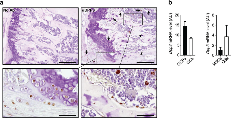
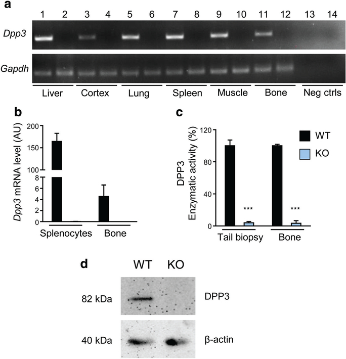
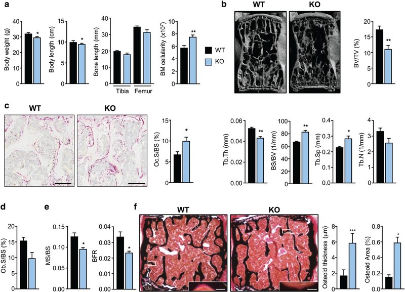
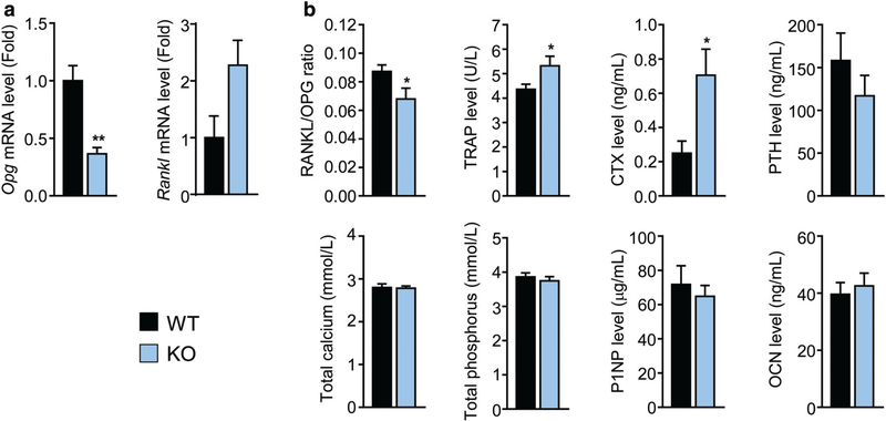
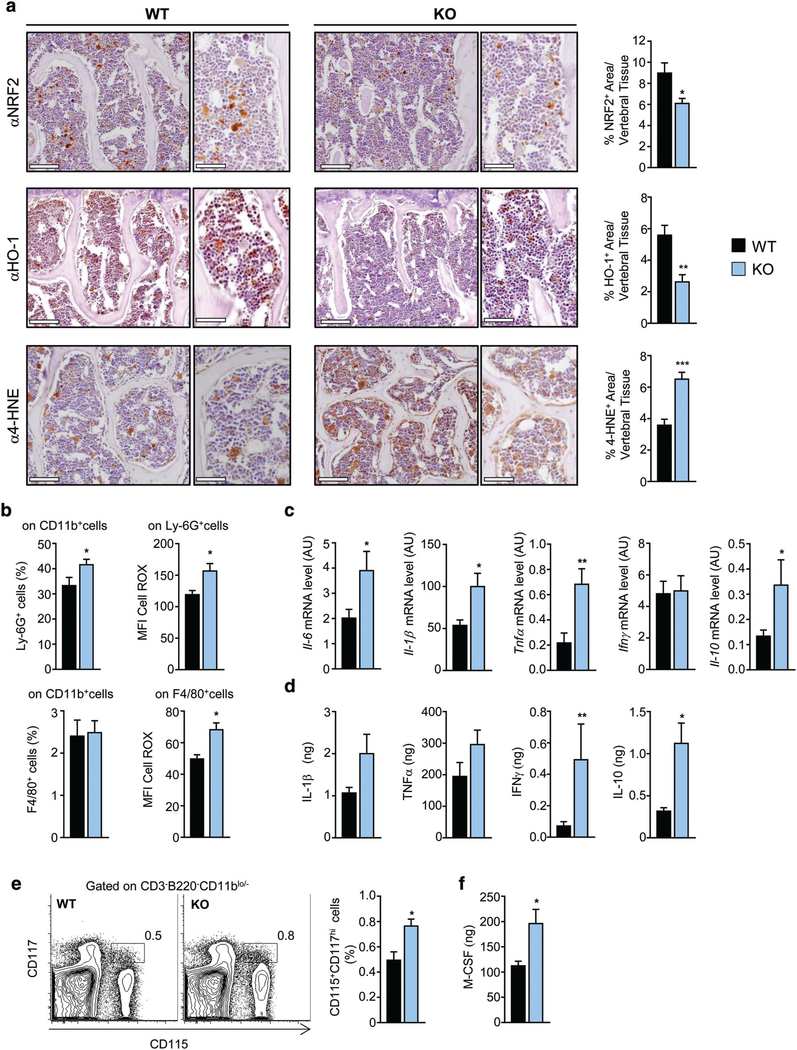
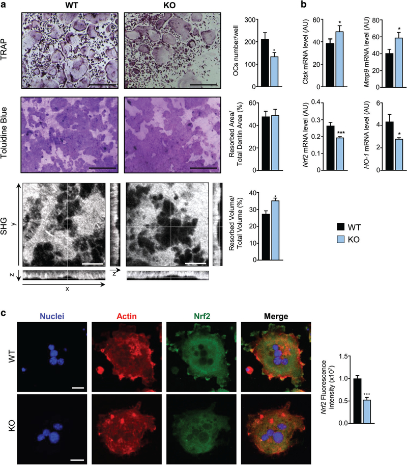
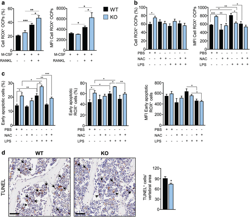
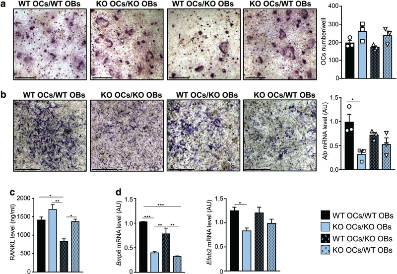
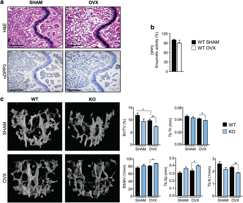
Controlling oxidative stress through the activation of antioxidant pathways is crucial in bone homeostasis, and impairments of the cellular defense systems involved contribute to the pathogenesis of common skeletal diseases. In this work we focused on the dipeptidyl peptidase 3 (DPP3), a poorly investigated ubiquitous zinc-dependent exopeptidase activating the Keap1-Nrf2 antioxidant pathway. We showed Dpp3 expression in bone and, to understand its role in this compartment, we generated a Dpp3 knockout (KO) mouse model and specifically investigated the skeletal phenotype. Adult Dpp3 KO mice showed a mild growth defect, a significant increase in bone marrow cellularity, and bone loss mainly caused by increased osteoclast activity. Overall, in the mouse model, lack of DPP3 resulted in sustained oxidative stress and in alterations of bone microenvironment favoring the osteoclast compared to the osteoblast lineage. Accordingly, in vitro studies revealed that Dpp3 KO osteoclasts had an inherent increased resorptive activity and ROS production, which on the other hand made them prone to apoptosis. Moreover, absence of DPP3 augmented bone loss after estrogen withdrawal in female mice, further supporting its relevance in the framework of bone pathophysiology. Overall, we show a nonredundant role for DPP3 in the maintenance of bone homeostasis and propose that DPP3 might represent a possible new osteoimmunological player and a marker of human bone loss pathology. © 2019 American Society for Bone and Mineral Research.

Keywords: BONE LOSS; DPP3; OSTEOPOROSIS; OXIDATIVE STRESS.


Figures

Fig. 1.
Fig. 1.
DPP3 is expressed in the bone tissue. (A) Representative images of immunohistochemical analysis of DPP3 expression in the femur of WT C57BL/6J mouse. Upper left, negative control; upper right and lower left and right, staining with mouse antibody αDPP3 at different magnification. Scale bars = upper panels 200 μm; lower panels 50 μm. Arrows indicate representative positive cells. (B) qPCR for murine Dpp3 expression in in vitro osteoclastogenesis (n = 3) and osteoblastogenesis (n = 2). In the latter, bars indicate the range. For each evaluation, n ≥ 2. DPP3 = dipeptidyl peptidase 3; OCP = osteoclast precursor; OC = osteoclast; MSC = mesenchymal stromal cell; OB = osteoblast.
Fig. 2.
Fig. 2.
Dpp3 gene expression and enzymatic activity are effectively shut down in the Dpp3 KO mouse model. (A) Representative RT-PCR for Dpp3 gene expression in several tissues obtained from WT (odd numbers) and Dpp3 KO (even numbers) mice. (B) qPCR of Dpp3 gene on splenocytes and total bone from WT and Dpp3 KO mice (n = 3 per genotype). (C) Evaluation of DPP3 enzymatic activity in WT. (E) Dpp3 KO mice in protein extracts from tail biopsies and total bone (n ≥ 3 per genotype). The Arg-Arg-2-naphthylamide peptide was used as reaction substrate. The enzymatic activity was quantified by spectrophotometric analysis. Data are presented as the mean ± SE. ***p < .001. (D) Representative Western blot analysis of Dpp3 protein in total bone tissue from WT and Dpp3 KO mice. β-actin = loading control; Gapdh = housekeeping control.
Fig. 3.
Fig. 3.
The Dpp3 KO mouse model displays a bone loss phenotype. (A) Body weight, body length, long-bone length, and BM cellularity from one femur and one tibia per mouse, in WT and Dpp3 KO mice. (B) μCT analysis of lumbar vertebrae: 3D reconstruction and static histomorphometric analysis of trabecular bone in WT and Dpp3 KO mice. (C) Representative images of decalcified paraffin-embedded TRAP-stained sections of WT and Dpp3 KO bone, and quantification of Oc.S/BS. Scale bar = 200 μm. (D) Ob.S/BS ratio, as assessed through ALP-staining of WT and Dpp3 KO lumbar vertebrae. (E) Dynamic histomorphometric analysis. (F) Representative images of mineralized resin-embedded von Kossa-stained vertebrae of WT and Dpp3 KO mice, and corresponding quantization of osteoid thickness and area. Scale bar = 150 μm and 5 μm, for the lower magnification and the inset, respectively. All the evaluations were performed in 6-month-old male mice. For each evaluation, n ≥ 6 per genotype. *p < .05, **p < .01, ***p < .001. BM = bone marrow; 3D = three-dimensional; Oc.S/BS = osteoclast surface/bone surface; Ob.S/BS = osteoblast surface/bone surface.
Fig. 4.
Fig. 4.
Lack of DPP3 alters bone metabolism. (A) qPCR of Opg and Rankl gene expression in total bone from WT and Dpp3 KO mice. (B) Serological analysis of markers of bone metabolism. For each evaluation, n ≥ 6 per genotype. *p < .05, **p < .01.
Fig. 5.
Fig. 5.
Absence of DPP3 leads to increased oxidative stress and inflammation in the bone tissue. (A) Representative images, and corresponding high magnification, of sections of decalcified paraffin-embedded vertebrae of WT and Dpp3 KO stained with antibodies against Nrf2, HO-1, and 4-HNE, and corresponding quantization. Scale bar = 100 μm for lower magnification, and 50 μm for higher magnification images. (B) Upper panels: frequency of PMNs (as percentage of Ly-6G+ after gating on CD11b+ cells) and corresponding amount of ROS production as MFI in the BM of WT and Dpp3 KO mice. Lower panels: frequency of macrophages (as percentage of F4/80+ after gating on CD11b+ cells) and corresponding amount of ROS production as MFI, in the BM of WT and Dpp3 KO mice. (C) qPCR of genes coding for inflammatory cytokines in the BM cells of WT and Dpp3 KO mice. (D)Quantification of amount of inflammatory cytokines in the BM supernatant of WT and Dpp3 KO mice. (E) Representative plot of FACS analysis of CD115+CD117hi cells in WT and Dpp3 KO BM, and corresponding quantification. (F) Quantification of M-CSF amount in the BM supernatant of WT and Dpp3 KO mice. For each evaluation, n ≥ 6 per genotype. *p < .05, **p < .01. PMN = polymorphonuclear cell; MFI = meanfluorescence intensity.
Fig. 6.
Fig. 6.
Loss of DPP3 increases in vitro osteoclast resorption activity and impairs Nrf2 signaling. (A) Upper panels: representative images of TRAP-stained in vitro differentiated osteoclasts from WT and Dpp3 KO osteoclast precursors. Middle panels: Toluidine blue–stained resorption pits on dentin. Scale bar = 400 μm. Lower panels: SHG of dentin collagen in the different projections. Scale bar = 40 μm. Graphs on the right represent quantification analysis for each evaluation. (B) qPCR analysis of osteoclast functional genes and Nrf2 pathway in in vitro–differentiated WT and Dpp3 KO osteoclasts. For each evaluation, n ≥ 6 per genotype. (C) Representative images of immunofluorescence analysis of in vitro–differentiated WT and Dpp3 KO osteoclasts stained as indicated, and Nrf2 fluorescence intensity. *p < .05, ***p < .001. SHG = second harmonic generation.
Fig. 7.
Fig. 7.
Dpp3 KO osteoclast precursor cells display higher ROS production and proneness to apoptosis. (A, B) Percentage of ROS+ WT and Dpp3 KO OCPs and amount of ROS production expressed as MFI of ROS+ OCPs, as assessed by FACS analysis in the presence of the indicated stimuli. (C) Percentage of WT and Dpp3 KO early apoptotic cells and measurements of ROS production in this cell population, assessed as in A and B. (D) Representative images of PFA-fixed paraffin-embedded bone of WT and Dpp3 KO mice stained with TUNEL, and corresponding quantification. Scale bar = 50 μm. Arrows indicate representative positive cells. For each evaluation, n ≥ 4 per genotype per group. *p < .05, **p < .01, ***p < .001. OPC = osteoclast precursor cell; MFI = mean fluorescence intensity.
Fig. 8.
Fig. 8.
Lack of DPP3 causes altered osteoblast/osteoclast crosstalk. (A) Representative images of TRAP staining after osteoclast differentiation on dentin in the indicated co-culture conditions (scale bar = 400 μm), and corresponding osteoclast count. (B) Representative images of ALP staining in the indicated co-culture conditions (scale bar = 1 mm), and corresponding levels of Alp mRNA expression. (C) Quantization of RANKL cytokine in the co-culture supernatants. (D) Expression analysis of Bmp6 and Efnb2 as relevant genes for osteoclast/osteoblast crosstalk. For each experimental condition, n = 3. *p < .05, **p < .01, ***p < .001.
Fig. 9.
Fig. 9.
DPP3 activity correlates with estrogen status and lack of DPP3 augments bone loss after estrogen withdrawal. (A) Representative images of sections of decalcified paraffin-embedded femurs of SHAM and OVX WT mice stained with H&E (upper panels) and with anti-DPP3 antibody. Scale bar = 500 μm. (B) Evaluation of DPP3 enzymatic activity in SHAM and OVX WT mice in BM protein extracts; n ≥ 5 per group. (C) μCT analysis of the spine: 3D reconstruction and static histomorphometric analysis in SHAM and OVX WT and Dpp3 KO mice. For each evaluation, n ≥ 5 per genotype per group. Only statistically significant comparisons are depicted; mean values ± SD for all the parameters measured, as well as detailed statistical analysis are reported in Supporting Table 2. *p < .05, **p < .01.

References

    1. Florencio-Silva R, Sasso GR, Sasso-Cerri E, Simoes MJ, Cerri PS. Biology of bone tissue: structure, function, and factors that influence bone cells. Biomed Res Int. 2015;2015:421746. - PMC - PubMed
    1. Han Y, You X, Xing W, Zhang Z, Zou W. Paracrine and endocrine actions of bone—the functions of secretory proteins from osteoblasts, osteocytes, and osteoclasts. Bone Res. 2018;6:16. - PMC - PubMed
    1. Kenkre JS, Bassett J. The bone remodelling cycle. Ann Clin Biochem. 2018;55(3):308–27. - PubMed
    1. Medina-Gomez C, Kemp JP, Trajanoska K, et al. Life-course genome-wide association study meta-analysis of total body BMD and assessment of age-specific effects. Am J Hum Genet. 2018;102(1):88–102. - PMC - PubMed
    1. Domazetovic V, Marcucci G, Iantomasi T, Brandi ML, Vincenzini MT. Oxidative stress in bone remodeling: role of antioxidants. Clin Cases Miner Bone Metab. 2017;14(2):209–16. - PMC - PubMed

Publication types

MeSH terms

Substances