Review
. 2019 Sep 14:709:134373.
doi: 10.1016/j.neulet.2019.134373. Epub 2019 Jul 8.

New insights into regulation and function of planar polarity in the inner ear

Affiliations
Review

New insights into regulation and function of planar polarity in the inner ear

Basile Tarchini et al. Neurosci Lett. .

Abstract

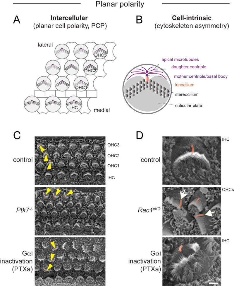
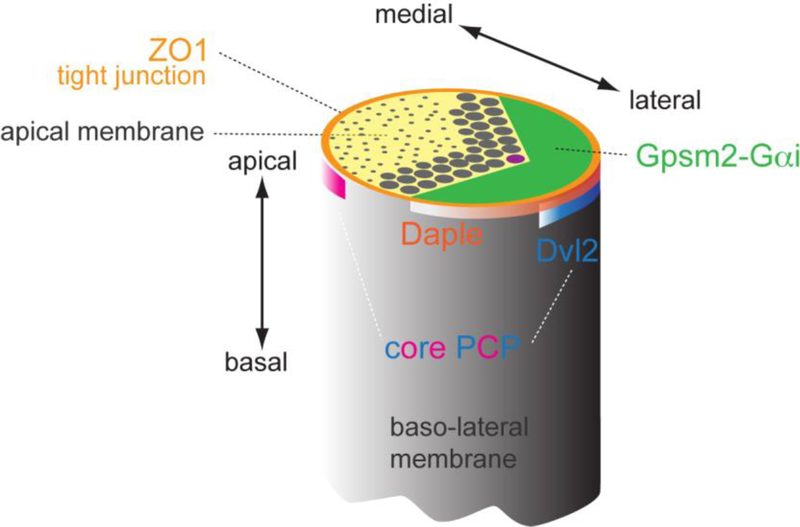
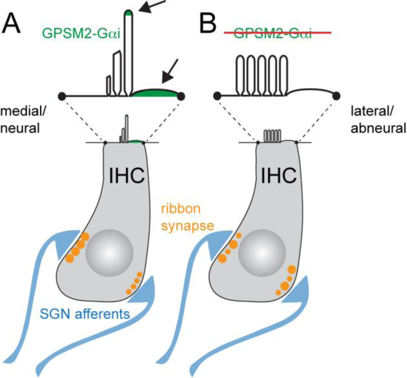
Acquisition of cell polarity generates signaling and cytoskeletal asymmetry and thus underpins polarized cell behaviors during tissue morphogenesis. In epithelial tissues, both apical-basal polarity and planar polarity, which refers to cell polarization along an axis orthogonal to the apical-basal axis, are essential for epithelial morphogenesis and function. A prime example of epithelial planar polarity can be found in the auditory sensory epithelium (or organ of Corti, OC). Sensory hair cells, the sound receptors, acquire a planar polarized apical cytoskeleton which is uniformely oriented along an axis orthogonal to the longitudinal axis of the cochlear duct. Both cell-intrinsic and tissue-level planar polarity are necessary for proper perception of sound. Here we review recent insights into the novel roles and mechanisms of planar polarity signaling gained from genetic analysis in mice, focusing mainly on the OC but also with some discussions on the vestibular sensory epithelia.

Keywords: Cochlea; Deafness; Hair bundle; Hair cell; Kinocilium; Planar cell polarity; Stereocilia.


Figures

Figure 1.
Figure 1.. Two levels of planar polarity in the organ of Corti.
A) Diagram depicting tissue-level, intercellular planar cell polarity (PCP), manifested by the uniform orientation of HCs along the medial-lateral axis of the cochlear duct. PCP is regulated by intercellular communication through asymmetric core PCP protein complexes at HC-SC junctions. B) By contrast, cell-intrinsic planar polarity refers to cytoskeleton asymmetry along the planar axis in individual HCs. Different structures displaying intracellular planar polarity are indicated. This behavior involves protein modules acting cell-intrinsically. C) Scanning electron microscopy (SEM) images showing PCP defects in representative mouse mutants. Yellow arrowheads indicate OHC orientation. D) SEM images showing cell-intrinsic planar polarity defects in representative mouse mutants. The kinocilium is highlighted in orange. Note OHCs in Rac1 mutants with off-center kinocilium relative to an abnormally flat hair bundle (arrows). PTXa mutants show IHCs where the kinocilium is disconnected from a split hair bundle. Ptk7 mutants strictly exhibit orientation (PCP) defects, whereas Rac1 and PTXa mutants exhibit both orientation and cell-intrinsic planar polarity defects. PTXa mutants have Cre-induced expression of the catalytic subunit of Pertussis toxin (PTXa) in HCs. Ptk7 and Rac1 SEM images are modified from [36, 66].
Figure 2.
Figure 2.. A molecular blueprint for planar polarization of the apical cytoskeleton.
A) SEM images of individual OHCs representative of different stages of apical differentiation. The kinocilium is highlighted in pink and the approximate OHC junction indicated in red. B) Diagram depicting changes at the HC apex from the onset of differentiation (E15.5, left) to around birth (P0, right). Initially, the aPKC kinase is uniformly enriched at the apical membrane, which is covered with microvilli, and the kinocilium occupies a central position. The first morphological evidence of planar asymmetry is the approximately lateral position of the kinocilium, which occurs at about the time the Insc-Gpsm2-Gαi complex becomes planar polarized at the lateral aspect of the cell. The Insc-Gpsm2-Gαi complex expands in surface area and labels the bare zone, the lateral region of apical membrane devoid of stereocilia or microvilli (asterisks). Insc-Gpsm2-Gαi prevents aPKC enrichment at the bare zone, establishing a molecular blueprint at the apical membrane that helps position and coordinate the hair bundle and the kinocilium. The expansion of the bare zone coincides with a relocalization of the kinocilium, from its post-migration position juxtaposed to the lateral junction to a more central position at the vertex of the chevron-shaped hair bundle around birth.
Figure 3.
Figure 3.. Deiters’ cell phalangeal processes are planar polarized along the longitudinal axis of the cochlea.
A) Diagram depicting one Deiters’ cell (green) that extends a slanted phalangeal process towards the luminal surface further towards the cochlear apex compared to its cell body position. B) A maximum-intensity projection of confocal Z stacks of a P18 cochlea whole-mount stained for F-actin (magenta) and alpha-tubulin (green). Note the planar polarized phalangeal processes of the three rows of Deiters’ cells (arrowheads). Arrows indicate the direction of the cochlear apex.
Figure 4.
Figure 4.. Shared protein modules regulate HC planar polarity and other polarized processes.
A) During oriented cell division, the Gpsm2-Gαi complex, and in some cases the Par3-Par6-aPKC complex, are polarized at the cell cortex and recruit effectors to pull on astral microtubules and position the mitotic spindle. Gαi is myristoylated and anchored at the membrane. Gpsm2-Gαi can interact with the Par3 complex through binding to the Insc adapter, or recruit the minus-end microtubule motor dynein through the Numa adapter. Lis1 is an activator for dynein required for spindle orientation. This evolutionary conserved process controls daughter cell positioning and tissue architecture across multiple tissues. B) During HC planar polarization, dynein-mediated pulling forces on apical microtubules may play a role in the lateral placement of the basal body. Junctional proteins including Dvl2, Daple and Par3, may serve as cortical dynein tethers to anchor pulling forces, and/or stabilize microtubule cortical attachment by activating Rac-Pak signaling. The Insc-Gpsm2-Gαi complex provides a blueprint on the HC apical membrane, and may also plays a role in basal body placement. C) During chemotaxis, for example in neutrophils, motility requires G protein coupled receptors (GPCRs) to sense extracellular cues in the environment and activate heterotrimeric G proteins. Gβγ dissociated from activated, GTP-bound Gαi in turn activates downstream effectors to regulate actin assembly and pseudopod extension at the leading edge. In addition to canonical GPCR signaling, it has been proposed that Gαi-GDP bound to Gpsm2 might more specifically regulate sustained directionality by signaling through Insc and Par3. In other cell types, sustained directionality was shown to require microtubules and Lis1/dynein to regulate transport and polarized activation of signaling molecules such as Rac1 GTPase. It is interesting to speculate that control over Gαi guanine nucleotide exchange involving GEF proteins like GPCRs or Daple might ultimately interconnect different polarity modules that act in concert in HCs.
Figure 5.
Figure 5.. Distinct subcellular localizations for different polarity modules at the HC apex.
Proteins identified as planar polarized at the apical HC surface have been shown to occupy distinct subcellular domains. Core PCP proteins are enriched at adherens junctions and form antagonistic lateral (e.g. Dvl2, blue band) or medial (pink band) protein complexes. In contrast, Par3 and Daple proteins largely coincide with lateral tight junctions (orange), slightly more apical than core PCP proteins. Finally, Insc-Gpsm2-Gαi are enriched above tight junctions at the bare zone, the region of apical membrane devoid of stereocilia or microvilli (green). Close vicinity between Gpsm2-Gαi, Daple-Par3 and Dvl2 at the lateral aspect of HCs suggests that cross-talk between these protein modules and compartments may integrate intercellular PCP and cell-intrinsic cytoskeleton polarization.
Figure 6.
Figure 6.. Novel roles for Gpsm2-Gαi and planar polarity in hair cells.
A) At the IHC apical membrane, Gpsm2-Gαi is required for stereocilia elongation and graded heights across stereocilia rows. Gpsm2 and Gαi proteins (green, arrows) are localized at the tip and near the base of stereocilia in the first row, being planar polarized at the lateral bare zone. This dual distribution suggests that planar polarity might play a role in the restriction of Gpsm2-Gαi trafficking to first row stereocilia, establishing their tallest identity and regulating hair bundle staircase-like architecture. At the basolateral IHC membrane, both Gpsm2 and Gαi are required for the medial-lateral gradient of ribbon synapse (orange) size and synaptic activity. The IHC ribbon synapse releases neurotransmitters onto spiral ganglion neuron (SGN) afferent terminals (blue). B) In absence of Gpsm2-Gαi function, stereocilia fail to elongate at postnatal stages and the hair bundle retains an immature morphology with an excess number of rows, and the gradient of ribbon synapse size is abolished. It remains unclear how Gpsm2-Gαi proteins influence synapse size and activity.

References

    1. Andreeva A, Lee J, Lohia M, Wu X, Macara IG, Lu X, PTK7-Src signaling at epithelial cell contacts mediates spatial organization of actomyosin and planar cell polarity, Dev Cell 29 (2014) 20–33. - PMC - PubMed
    1. Appler JM, Goodrich LV, Connecting the ear to the brain: Molecular mechanisms of auditory circuit assembly, Prog Neurobiol 93 (2011) 488–508. - PMC - PubMed
    1. Aznar N, Midde KK, Dunkel Y, Lopez-Sanchez I, Pavlova Y, Marivin A, Barbazán J, Murray F, Nitsche U, Janssen K-P, Willert K, Goel A, Abal M, Garcia-Marcos M, Ghosh P, Daple is a novel non-receptor GEF required for trimeric G protein activation in Wnt signaling, eLife 4 (2015) e07091. - PMC - PubMed
    1. Beer-Hammer S, Lee SC, Mauriac SA, Leiss V, Groh IAM, Novakovic A, Piekorz RP, Bucher K, Chen C, Ni K, Singer W, Harasztosi C, Schimmang T, Zimmermann U, Pfeffer K, Birnbaumer L, Forge A, Montcouquiol M, Knipper M, Nurnberg B, Ruttiger L, Galphai Proteins are Indispensable for Hearing, Cell Physiol Biochem 47 (2018) 1509–1532. - PMC - PubMed
    1. Belyantseva IA, Boger ET, Naz S, Frolenkov GI, Sellers JR, Ahmed ZM, Griffith AJ, Friedman TB, Myosin-XVa is required for tip localization of whirlin and differential elongation of hair-cell stereocilia, Nat Cell Biol 7 (2005) 148–156. - PubMed

Publication types

LinkOut - more resources