. 2019 Oct:130:104528.
doi: 10.1016/j.nbd.2019.104528. Epub 2019 Jul 8.

(-)-Phenserine and the prevention of pre-programmed cell death and neuroinflammation in mild traumatic brain injury and Alzheimer's disease challenged mice

Affiliations

(-)-Phenserine and the prevention of pre-programmed cell death and neuroinflammation in mild traumatic brain injury and Alzheimer's disease challenged mice

Daniela Lecca et al. Neurobiol Dis. 2019 Oct.

Abstract

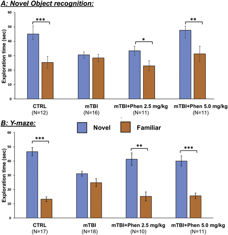
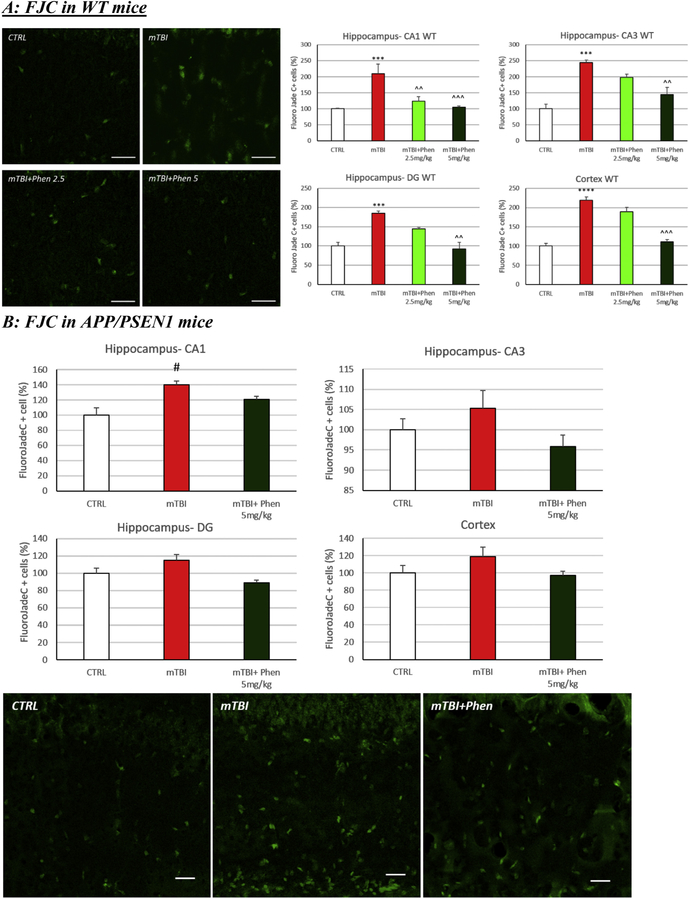
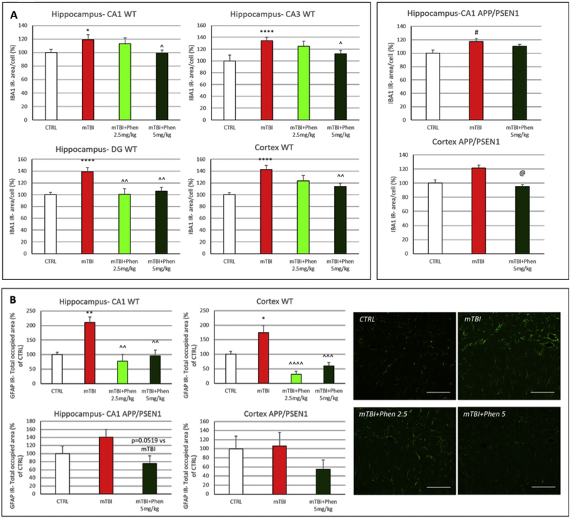
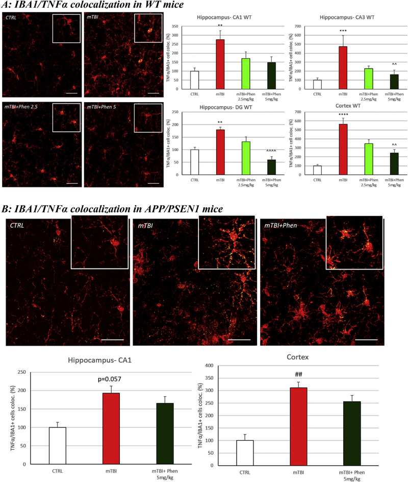
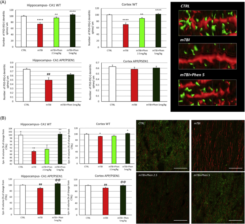
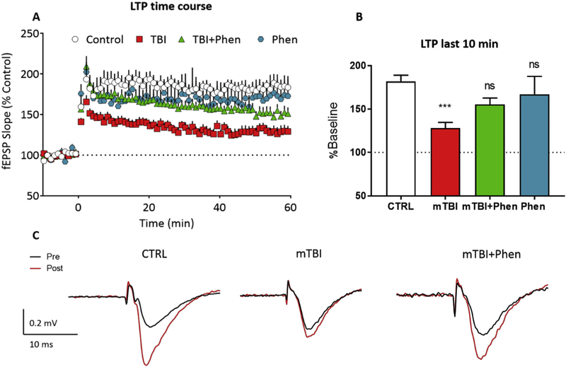
Mild traumatic brain injury (mTBI) is a risk factor for neurodegenerative disorders, such as Alzheimer's disease (AD) and Parkinson's disease (PD). TBI-derived neuropathologies are promoted by inflammatory processes: chronic microgliosis and release of pro-inflammatory cytokines that further promote neuronal dysfunction and loss. Herein, we evaluated the effect on pre-programmed cell death/neuroinflammation/synaptic integrity and function of (-)-Phenserine tartrate (Phen), an agent originally developed for AD. This was studied at two clinically translatable doses (2.5 and 5.0 mg/kg, BID), in a weight drop (concussive) mTBI model in wild type (WT) and AD APP/PSEN1 transgenic mice. Phen mitigated mTBI-induced cognitive impairment, assessed by Novel Object Recognition and Y-maze behavioral paradigms, in WT mice. Phen fully abated mTBI-induced neurodegeneration, evaluated by counting Fluoro-Jade C-positive (FJC+) cells, in hippocampus and cortex of WT mice. In APP/PSEN1 mice, degenerating cell counts were consistently greater across all experimental groups vs. WT mice. mTBI elevated FJC+ cell counts vs. the APP/PSEN1 control (sham) group, and Phen similarly mitigated this. Anti-inflammatory effects on microglial activation (IBA1-immunoreactivity (IR)) and the pro-inflammatory cytokine TNF-α were evaluated. mTBI increased IBA1-IR and TNF-α/IBA1 colocalization vs. sham, both in WT and APP/PSEN1 mice. Phen decreased IBA1-IR throughout hippocampi and cortices of WT mice, and in cortices of AD mice. Phen, likewise, reduced levels of IBA1/TNF-α-IR colocalization volume across all areas in WT animals, with a similar trend in APP/PSEN1 mice. Actions on astrocyte activation by mTBI were followed by evaluating GFAP, and were similarly mitigated by Phen. Synaptic density was evaluated by quantifying PSD-95+ dendritic spines and Synaptophysin (Syn)-IR. Both were significantly reduced in mTBI vs. sham in both WT and APP/PSEN1 mice. Phen fully reversed the PSD-95+ spine loss in WT and Syn-IR decrease in both WT and APP/PSEN1 mice. To associate immunohistochemical changes in synaptic markers with function, hippocampal long term potentiation (LTP) was induced in WT mice. LTP was impaired by mTBI, and this impairment was mitigated by Phen. In synopsis, clinically translatable doses of Phen ameliorated mTBI-mediated pre-programmed cell death/neuroinflammation/synaptic dysfunction in WT mice, consistent with fully mitigating mTBI-induced cognitive impairments. Phen additionally demonstrated positive actions in the more pathologic brain microenvironment of AD mice, further supporting consideration of its repurposing as a treatment for mTBI.

Keywords: (-)-Phenserine; Alzheimer's disease; Cognitive impairment; Long term potentiation; Neurodegeneration; Neuroinflammation; Synaptic proteins; TNF-α; Therapeutic intervention; Traumatic brain injury.


Conflict of interest statement

Declaration of competing interest

Becker RE has an issued patent on the use of (−)-Phenserine in neurodegenerative disorders. All other authors report no conflicts of interest.

Figures

Fig. 1.
Fig. 1.
Post-injury treatment with Phen mitigates mTBI-induced cognitive impairments, as evaluated by NOR and Y-maze paradigms 7 days following injury. mTBI challenge induced significant behavioral deficits in NOR (A) and Y-maze (B) with mTBI-vehicle mice failing to differentiate between the novel and familiar objects or arms, respectively. In contrast, animals treated with Phen (2.5 and 5 mg/kg, BID x 5 days) explored the novel object/arm for significantly longer than the familiar one, similar to sham animals. Data are presented as mean ± S.E.M. of N observations, *p < .05, **p < .01, ***p < .001 comparing the novel to familiar object/arm.
Fig. 2.
Fig. 2.
Phen reverses mTBI-induced neuronal loss in WT and APP/PSEN1 mice. Degenerating neuronal cells were quantified using the marker Fluorojade C (FJC: green). (A) An increased number of FJC+ cells were observed in vehicle administered mTBI (mTBI-VEH) vs. sham control (CTRL) WT mice across the hippocampus (CA1, CA3 and DG) and cerebral cortex (***p < .001, ****p < .0001 vs. CTRL by Tukey’s post hoc test). (B) A similar elevation was induced by mTBI in APP/PSEN1 vehicle administered mice that reached significance in the hippocampus (#p < .05 vs CTRL by Mann-Whitney rank test in CA1). Post treatment with Phen abated the neuronal loss, in which FJC+ cell counts were no different from values of sham control (CTRL) mice. Importantly in WT animals, values in the Phen 5 mg/kg group were significantly lower than the mTBI vehicle group (^^p < .01, ^^^p < .001 vs. mTBI by Tukey’s post hoc test). Representative images and graphs from hippocampus and cortex. Data shown as mean ± S.E.M. Scale bar = 30 μm.
Fig. 3.
Fig. 3.
Phen ameliorates mTBI-induced neuroinflammation in WT and APP/PSEN1 mice. (A) Following mTBI injury, ionized calcium binding adaptor molecule 1 (IBA1) immunoreactivity (IR) (a microglia/macrophage-specific marker) was increased in mTBI vehicle vs. sham control (CTRL) mice, in all analyzed brain regions of WT animals and in the hippocampus of APP/PSEN1 mice. In contrast, IBA1 IR values throughout hippocampal and cortical areas for mTBI challenged mice administered Phen were not statistically different from their respective sham (CTRL) groups across WT and APP/PSEN1 mice. In mTBI-challenged WT mice, Phen (2.5 and 5 mg/kg BID) significantly lowered microglial activation throughout hippocampus and lateral cortex compared to the vehicle group. In APP/PSEN1 mTBI mice, this was achieved by Phen in cortex. IBA1 IR was measured as the area occupied by IBA1+ cells in the analyzed brain regions. Graphs from hippocampus (CA1 for APP/PSEN1 mice) and cortex. (B) Glial fibrillary acidic protein (GFAP) IR (an astrocyte marker) was significantly increased in hippocampus and cortex of mTBI-challenged WT mice, and a similar trend, although not statistically significant, was evident in APP/PSEN1. Treatment with Phen fully reversed this GFAP IR increase in hippocampus and cortex of WT mice. A similar trend that did not reach statistical significance was evident in APP/PSEN1 transgenic animals. GFAP IR was quantified as total area occupied by GFAP + fluorescent signal in hippocampus and cortex. Representative confocal images from hippocampus - CA1 of WT mice. Graphs from hippocampus - CA1 and cortex. Scale bar = 100 μm. *p < .05, **p < .01, ****p < .0001 vs. CTRL by Tukey’s post hoc test; ^p < .05, ^^p < .01, ^^^p < .001, ^^^^p < .0001 vs. mTBI by Tukey’s post hoc test. #p < .05 vs. CTRL by Mann-Whitney rank test; @p < .05 vs. mTBI by Mann-Whitney rank test. Data shown as mean ± S.E.M.
Fig. 4.
Fig. 4.
Phen inhibits mTBI-induced TNF-α generation within activated microglial in WT and APP/PSEN1 mice. mTBI induced an elevation in immunoreactivity for the pro-inflammatory cytokine TNF-α within IBA1+ cells across all analyzed brain areas in WT animals (Fig. 4A) and in the cortex of APP/PSEN1 mice (Fig. 4B), as evaluated by TNF-α/Iba1 co-localization. By contrast, mTBI Phen-treated mTBI mice had values no different from the sham (CTRL) group. Administration of Phen (2.5 and 5 mg/kg, BID) reduced the levels of IBA1/TNF-α IR co-localization volume across all hippocampal and cortical areas in WT mTBI-challenged mice, as compared to the mTBI vehicle group. Representative confocal images showing co-localized elements (yellow) in IBA1 (red) positive cells in cortex. Percentage of co-localization of TNF-α IR in IBA1 positive cells. **p < .01, ***p < .001, ****p < .0001 vs. CTRL by Tukey’s post hoc test; ^^p < .01, ^^^^p < .0001 vs. mTBI by Tukey’s post hoc test. ##p < .01 vs. CTRL by Mann-Whitney rank test. Data shown as mean ± S.E.M. Scale bar = 30 μm.
Fig. 5.
Fig. 5.
Phen mitigates mTBI-induced loss of pre- and post synaptic elements in WT and APP/PSEN1 mice. (A) Postsynaptic density protein 95 (PSD-95): mTBI induced a loss of PSD-95+ dendritic spines across all analyzed areas in WT mice, and in hippocampus of AD mice, as compared to the sham (CTRL) group. By contrast, Phen treated mTBI mice were not statistically different from the sham (CTRL) group. WT mTBI mice treated with Phen (2.5 and 5 mg/kg) possessed a greater number of PSD-95+ dendritic spines across both hippocampus and cortex, as compared to the mTBI vehicle group. Representative images showing PSD-95+ spines (green) in MAP2+ dendrites. Data are expressed as number of PSD-95+ dendritic spines/μm. (B) Synaptophysin: the total volume occupied by the presynaptic marker synaptophysin IR was evaluated across WT and APP/PSEN1 mice and found to be significantly reduced in the mTBI vehicle group, as compared to their respective sham (CTRL) group. In contrast, mTBI Phen treated mice had synaptophysin IR levels no different from sham (CTRL) mice. Importantly, Phen treatment of mTBI-challenged mice resulted in significantly higher amounts of synaptophysin IR, compared to the mTBI vehicle group, across all analyzed brain areas in both WT and AD mice. *p < .05, **p < .01, ****p < .0001 vs. CTRL by Tukey’s post hoc test; ^p < .05, ^^p < .01, ^^^^p < .0001 vs. mTBI by Tukey’s post hoc test. ##p < .01 vs. CTRL by Mann-Whitney rank test. Data shown as mean ± S.E.M. @@p < .01 vs. mTBI by Mann-Whitney rank test. Data shown as mean ± S.E.M. Scale bar = 20 μm.
Fig. 6.
Fig. 6.
Phen mitigates mTBI-induced deficits in LTP in WT mice. (A) Time course of the mTBI-induced impairment in LTP (initiated by HFS at time = 0 on the x-axis) across the 60 min time course. The fEPSP slope is expressed as a % of the control baseline, collected prior to HFS. (B) LTP magnitude, evaluated over the last 10 min of recording in each of the groups shown in A. (C) Representative averaged traces of a control slice, a slice from a mTBI mouse, a slice from a mTBI+Phen 5 mg/kg mouse, and a slice from control mouse treated with 5 mg/kg Phen showing the mitigating effect of Phen treatment on the mTBI-induced LTP impairment. Traces are averaged from the period immediately prior to (pre) and 60min following (post) high-frequency stimulation. LTP in mTBI-Veh mice was significantly decreased as compared with the sham (CTRL) group. Note that treatment of mTBI-challenged mice with Phen (5 mg/kg, BID) mitigated the LTP impairment, and that the mTBI Phen group did not statistically differ from sham (CTRL) animals. Data shown as mean ± S.E.M. ***p < .001, one-way ANOVA (F (3,49) = 6.053), and Tukey’s post-hoc test.

References

    1. Acosta SA, Tajiri N, Shinozuka K, Ishikawa H, Grimmig B, Diamond DM, Sanberg PR, Bickford PC, Kaneko Y, Borlongan CV, 2013. Long-term upregulation of inflammation and suppression of cell proliferation in the brain of adult rats exposed to traumatic brain injury using the controlled cortical impact model. PLoS One 8, e53376 10.1371/journal.pone.0053376. - DOI - PMC - PubMed
    1. Anderson WW, Collingridge GL, 2007. Capabilities of the WinLTP data acquisition program extending beyond basic LTP experimental functions. J. Neurosci. Methods 162, 346–356. 10.1016/J.JNEUMETH.2006.12.018. - DOI - PubMed
    1. Ansari MA, Roberts KN, Scheff SW, 2008. A time course of contusion-induced oxidative stress and synaptic proteins in cortex in a rat model of TBI. J. Neurotrauma 25, 513–526. 10.1089/neu.2007.0451. - DOI - PubMed
    1. Bader M, Li Y, Lecca D, Rubovitch V, Tweedie D, Glotfelty E, Rachmany L, Kim HK, Choi H-I, Hoffer BJ, Pick CG, Greig NH, Kim DS, 2019. Pharmacokinetics and efficacy of PT302, a sustained-release Exenatide formulation, in a murine model of mild traumatic brain injury. Neurobiol. Dis 124, 439–453. 10.1016/J.NBD.2018.11.023. - DOI - PMC - PubMed
    1. Bains M, Hall ED, 2012. Antioxidant therapies in traumatic brain and spinal cord injury. Biochim. Biophys. Acta 1822, 675–684. 10.1016/j.bbadis.2011.10.017. - DOI - PMC - PubMed

Publication types

MeSH terms