Review
. 2019 Jul 10;20(14):3377.
doi: 10.3390/ijms20143377.

Protective Effects of Euthyroidism Restoration on Mitochondria Function and Quality Control in Cardiac Pathophysiology

Affiliations
Review

Protective Effects of Euthyroidism Restoration on Mitochondria Function and Quality Control in Cardiac Pathophysiology

Francesca Forini et al. Int J Mol Sci. .

Abstract

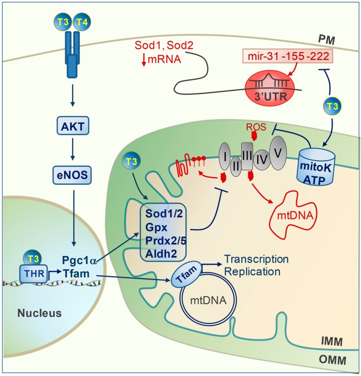
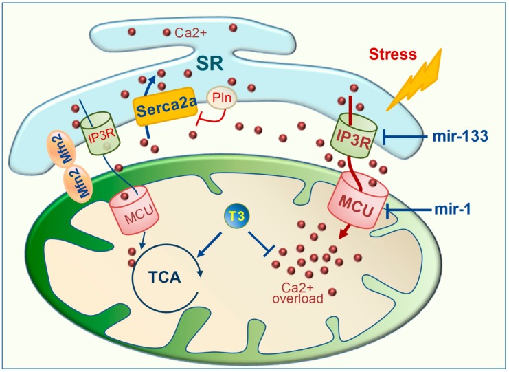
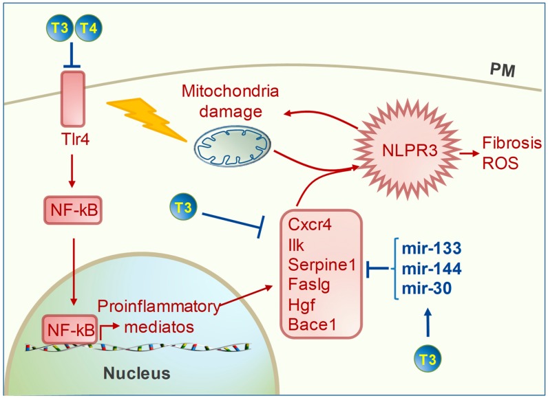
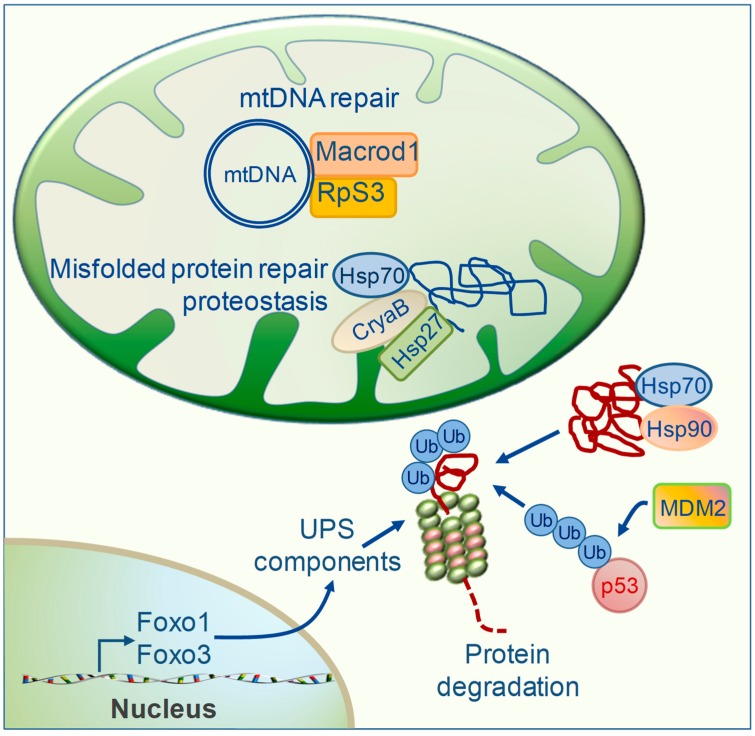
Mitochondrial dysfunctions are major contributors to heart disease onset and progression. Under ischemic injuries or cardiac overload, mitochondrial-derived oxidative stress, Ca2+ dis-homeostasis, and inflammation initiate cross-talking vicious cycles leading to defects of mitochondrial DNA, lipids, and proteins, concurrently resulting in fatal energy crisis and cell loss. Blunting such noxious stimuli and preserving mitochondrial homeostasis are essential to cell survival. In this context, mitochondrial quality control (MQC) represents an expanding research topic and therapeutic target in the field of cardiac physiology. MQC is a multi-tier surveillance system operating at the protein, organelle, and cell level to repair or eliminate damaged mitochondrial components and replace them by biogenesis. Novel evidence highlights the critical role of thyroid hormones (TH) in regulating multiple aspects of MQC, resulting in increased organelle turnover, improved mitochondrial bioenergetics, and the retention of cell function. In the present review, these emerging protective effects are discussed in the context of cardiac ischemia-reperfusion (IR) and heart failure, focusing on MQC as a strategy to blunt the propagation of connected dangerous signaling cascades and limit adverse remodeling. A better understanding of such TH-dependent signaling could provide insights into the development of mitochondria-targeted treatments in patients with cardiac disease.

Keywords: calcium handling; cardiac disease; inflammation; mitochondrial quality control; oxidative stress; thyroid hormone homeostasis.


Conflict of interest statement

The authors declare no conflicts of interest.

Figures

Figure 1
Figure 1
Antioxidant effect of thyroid hormones (TH). Reactive oxygen species (ROS) produced at electron transport chain (ETC) complex I and III can damage mitochondrial DNA, lipids, and proteins. T3 and T4 decrease reactive oxygen species (ROS) production by multiple mechanisms: (1) membrane-initiated pathways as for the AKT/eNOS axis; (2) transcriptional regulation of Pgc1α and Tfam; (3) post-transcriptional regulation via inhibition of mir-31, -155, and -222; (4) activation of the mitoK-ATP protective channel. Effectors modulated by TH are marked in blue. (AKT: protein kinase B; Aldh2:aldehyde dehydrogenase 2; eNOS: nitric oxide synthase; IMM: inner mitochondrial membrane; Gpx; Glutathione peroxidase mtDNA: mitochondrial DNA; OMM: outer mitochondrial membrane; Pgc1α: PPARG coactivator 1 alpha; PM: plasma membrane; Prdx2/5: peroxiredoxin 2/5; Sod1/2: superoxide dismutase1/2; Tfam: mitochondrial transcription factor A; THR: thyroid hormone receptor).
Figure 2
Figure 2
Effect of T3 on Ca2+ handling. Under stress conditions, Ca2+ dis-homeostasis is determined by a reduced function of the Serca2a channel and increased activity of the Serca2a inhibitor Pln. Excessive activation of IP3R and MCU channels contributes to mitochondrial Ca2+ overload, mitochondrial dysfunction, and cell death. T3 limits mitochondrial Ca2+ overload and favors energy production through several mechanisms: (1) promoting Ca2+ uptake within the SR by up-regulating the Serca2a channel and down-regulating Pln; (2) enhancing the expression level of mir-1 and mir-133, established suppressors of IP3R and MCU, respectively; (3) stimulating the activation of the Ca2+ dependent TCA enzymes by Mfn2-dependent tethering of the mitochondrial membrane and SR, thus prompting the formation of Ca2+ microdomains. (IP3R: inositol 1,4,5-trisphosphate receptors; MCU: mitochondrial calcium uniporter; Mfn2: mitofusin2; Pln; phospholambam; Serca2a: SR Ca2+ ATPase 2a; SR: sarcoplasmic reticulum; TCA: tricarboxylic acid cycle).
Figure 3
Figure 3
Anti-inflammatory effect of thyroid hormones (TH). Excessive reactive oxygen species (ROS) formation and Ca2+ overload lead to mitochondrial dysfunction and activation of the inflammasome NLPR3, which exacerbates mitochondrial impairment and ROS production and activates the fibrotic response. TH limits the inflammasome formation by: (1) blunting the pro-inflammatory Tlr4/NF-kB axis; (2) down-regulating the expression levels of an array of pro-inflammatory mediators; (3) up-regulating the expression levels of mir-133, -144, and -30, predicted suppressors of such pro-inflammatory mediators. (Bace 1: beta secretase 1; Cxcr4: C-X-C motif chemokin receptor 4; Faslg: fas ligand; Hgf: hepatocyte growth factor; Ilk: integrin-linked kinase; NF-kB: nuclear factor kappa B; NLPR3: inflammasome; Tlr4: toll like receptor 4).
Figure 4
Figure 4
Effects of thyroid hormones (TH) on mitochondrial protein repair and proteostasis. Accumulation of damaged mitochondrial proteins and mutated mtDNA favors mitochondrial impairments and cell dysfunction. TH acts at multiple levels to avoid such dangerous effects. (1) T3 favors the repair of misfolded proteins or their chaperone-mediated degradation by enhancing the protein levels of small (HSP27, CryaB) and large (HSP 70 and 90) heat shock proteins; (2) T3 and T4 up-regulate Foxo1 and Foxo3, transcription factors that orchestrate the expression of components of the ubiquitin protesome system (UPS) for protein degradation; (3) T3 may favor the ubiquitination of p53 under cardiac stress conditions via the MDM2 pathway; (4) T3 favors the mitochondrial accumulation of DNA repair systems such as Macrod1 and Rps3. (CryaB: α-crystallin B; HSP: heat shock protein; Foxo1/3: forkhead box O 1/3; Macrod1: Mono-ADP Ribosylhydrolase; Mdm2: mouse double minute 2 homolog; mtDNA: mitochondrial DNA; RpS3: 40s ribosomal protein S3; ub: ubiquitin).
Figure 5
Figure 5
Effect of T3 on mitochondrial protein import and mitochondrial clearance. On the left, T3 enhances mitochondrial protein import by up-regulating the expression and protein levels of translocases of the outer (Tom) and inner mitochondrial membrane (Tim). Also, T3 increases the activity of Hsp70 that is involved in recognition of the mitochondrial pre-proteins, sliding of pre-proteins within translocase complexes and folding the imported pre-proteins. On the right, T3 favors the selective degradation of irreparably damaged mitochondria via mitophagy. (1) T3 up-regulates a set of molecules involved in phagophore formation and elongation including Lc3, beclin and Ulk1; (2) T3 enhances Park-dependent and independent recognition of damaged mitochondria by the Lc3 component of the phagophore; (3) T3 regulates mir-144 and mir-222 thus blunting the inhibitory effect of the mTOR pathway on the mitophagy program. (Bnip3: BCL2 interacting protein 3; Fundc1: FUN14 domain-containing protein 1; Hsp70: heat shock protein 70; Lc3: light chain3; Mfn2: mitofusin2; mTOR: mammalian target of rapamycin; p62: ubiquitin protein binding p62; Park: parkin; Pink1: PTEN induced kinase 1; Ulk1: unc-51 like autophagy activating kinase1).

References

    1. Moyzis A.G., Sadoshima J., Gustafsson A.B. Mending a broken heart: The role of mitophagy in cardioprotection. Am. J. Physiol. Heart. Circ. Physiol. 2015;308:H183–H192. doi: 10.1152/ajpheart.00708.2014. - DOI - PMC - PubMed
    1. Suliman H.B., Piantadosi C.A. Mitochondrial Quality Control as a Therapeutic Target. Pharmacol. Rev. 2016;68:20–48. doi: 10.1124/pr.115.011502. - DOI - PMC - PubMed
    1. Gustafsson A.B., Gottlieb R.A. Heart mitochondria: Gates of life and death. Cardiovasc. Res. 2008;77:334–343. doi: 10.1093/cvr/cvm005. - DOI - PubMed
    1. Yoboue E.D., Devin A. Reactive Oxygen Species-Mediated Control of Mitochondrial Biogenesis. Int. J. Cell. Biol. 2012;2012:1–8. doi: 10.1155/2012/403870. - DOI - PMC - PubMed
    1. Di Meo S., Reed T.T., Venditti P., Victor V.M. Role of ROS and RNS Sources in Physiological and Pathological Conditions. Oxid. Med. Cell. Longev. 2016;2016:1–44. doi: 10.1155/2016/1245049. - DOI - PMC - PubMed