Review
. 2019 Jul 10;20(14):3379.
doi: 10.3390/ijms20143379.

Cellular Responses to Proteasome Inhibition: Molecular Mechanisms and Beyond

Affiliations
Review

Cellular Responses to Proteasome Inhibition: Molecular Mechanisms and Beyond

Nicolas Albornoz et al. Int J Mol Sci. .

Abstract

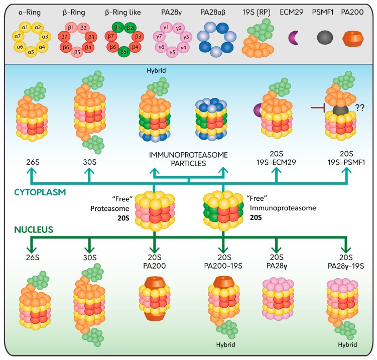
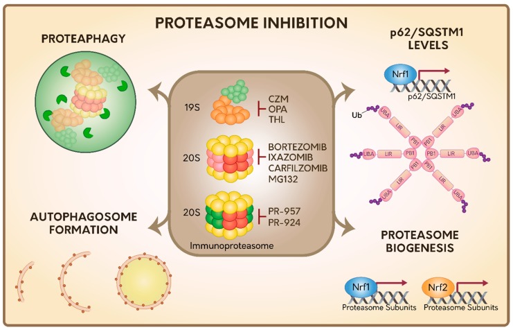
Proteasome inhibitors have been actively tested as potential anticancer drugs and in the treatment of inflammatory and autoimmune diseases. Unfortunately, cells adapt to survive in the presence of proteasome inhibitors activating a variety of cell responses that explain why these therapies have not fulfilled their expected results. In addition, all proteasome inhibitors tested and approved by the FDA have caused a variety of side effects in humans. Here, we describe the different types of proteasome complexes found within cells and the variety of regulators proteins that can modulate their activities, including those that are upregulated in the context of inflammatory processes. We also summarize the adaptive cellular responses activated during proteasome inhibition with special emphasis on the activation of the Autophagic-Lysosomal Pathway (ALP), proteaphagy, p62/SQSTM1 enriched-inclusion bodies, and proteasome biogenesis dependent on Nrf1 and Nrf2 transcription factors. Moreover, we discuss the role of IRE1 and PERK sensors in ALP activation during ER stress and the involvement of two deubiquitinases, Rpn11 and USP14, in these processes. Finally, we discuss the aspects that should be currently considered in the development of novel strategies that use proteasome activity as a therapeutic target for the treatment of human diseases.

Keywords: autophagy; immunoproteasome; proteasome.


Conflict of interest statement

The authors declare no conflict of interest.

Figures

Figure 1
Figure 1
Proteasome assembly and its regulators. The constitutive 20S proteasome is assembled in α (Yellow) and β (Red) heptameric rings, constituting subunits β1, β2, β5 of the catalytic core (Light Red); meanwhile the 20S immunoproteasome catalytic core has three inducible beta subunits named β1i, β2i and β5i (Dark Green). Both types of proteasomes are found without regulators, indicated as “free” or can be associated with 19S RP (Light Orange and Light Green) either at one or both ends forming the 26S or 30S proteasome, respectively; or with the PA28 complexes either at one or both ends; PA28αβ (Blue and Light Blue; cytoplasm) or PA28γ (Pink; nucleus). Hybrid proteasome complexes (Hybrid) are also found when the catalytic core is simultaneously associated with 19S RP and another type of regulator (PA28αβ, PA28γ and PA200). Specific regulators in the cytoplasm are ECM29 (Purple) and PSMF1 (Grey), which can modify the assembly or activity of the proteasome; while in the nucleus the main proteasome regulator is PA200 (Dark Orange).
Figure 2
Figure 2
Summary of the cellular responses during proteasome inhibition. Several proteasome inhibitors have been described targeting 19S RP, 20S proteasome and immunoproteasome. In response to proteasome inhibition, several responses are activated specifically related with the 20S proteasome, including the Autophagic-Lysosomal Pathway (ALP), proteaphagy, the transcriptional upregulation of the autophagy Ub receptor p62/SQSTM1, and proteasome genes, by Nrf1 and Nrf1/Nrf2 transcription factors, respectively. The activation of the autophagy Ub receptor p62/SQSTM1 is dependent on specific post-translational modifications based on ubiquitylation. However, it is still unknown which responses could be activated under the inhibition of 19S RP or immunoproteasome.

References

    1. Gidalevitz T., Prahlad V., Morimoto R.I. The stress of protein misfolding: From single cells to multicellular organisms. Cold Spring Harb. Perspect. Biol. 2011;3 doi: 10.1101/cshperspect.a009704. - DOI - PMC - PubMed
    1. Chiti F., Dobson C.M. Protein misfolding, amyloid formation, and human disease: A summary of progress over the last decade. Annu. Rev. Biochem. 2017;86:27–68. doi: 10.1146/annurev-biochem-061516-045115. - DOI - PubMed
    1. Kwon Y.T., Ciechanover A. The Ubiquitin Code in the Ubiquitin-Proteasome System and Autophagy. Trends Biochem. Sci. 2017;42:873–886. doi: 10.1016/j.tibs.2017.09.002. - DOI - PubMed
    1. Collins G.A., Goldberg A.L. The logic of the 26S proteasome. Cell. 2017;169:792–806. doi: 10.1016/j.cell.2017.04.023. - DOI - PMC - PubMed
    1. Ciechanover A., Kwon Y.T. Degradation of misfolded proteins in neurodegenerative diseases: Therapeutic targets and strategies. Exp. Mol. Med. 2015;47:e147. doi: 10.1038/emm.2014.117. - DOI - PMC - PubMed

MeSH terms

Substances

LinkOut - more resources