. 2019 Jul 10;12(14):2218.
doi: 10.3390/ma12142218.

Understanding the Influence of a Bifunctional Polyethylene Glycol Derivative in Protein Corona Formation around Iron Oxide Nanoparticles

Affiliations

Understanding the Influence of a Bifunctional Polyethylene Glycol Derivative in Protein Corona Formation around Iron Oxide Nanoparticles

Amalia Ruiz et al. Materials (Basel). .

Abstract

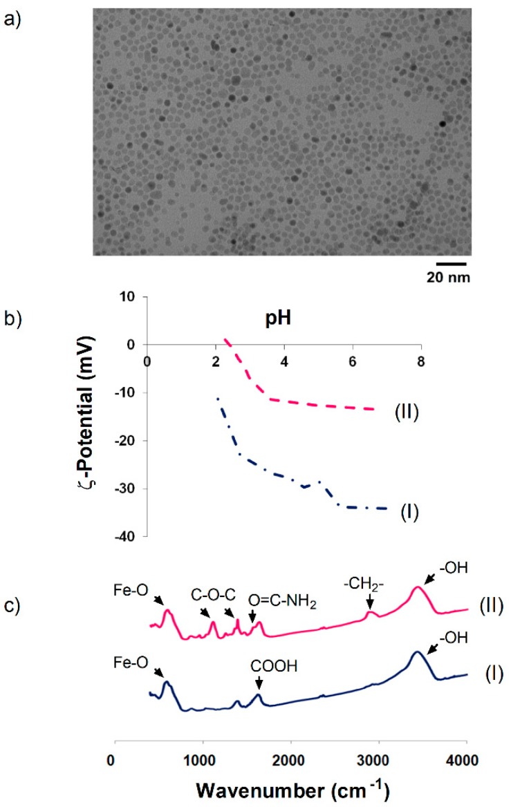
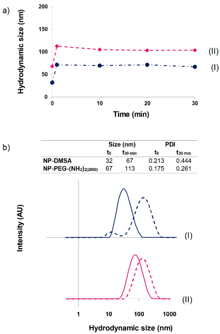
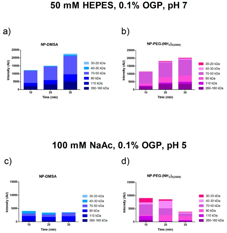
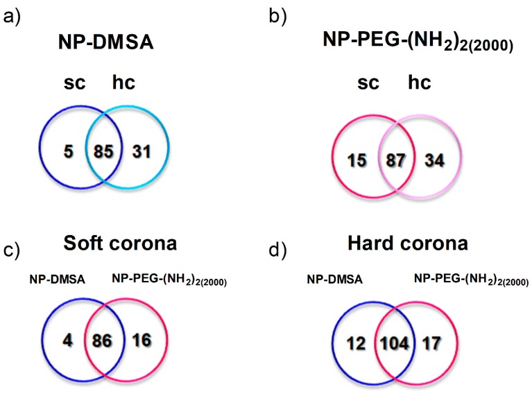
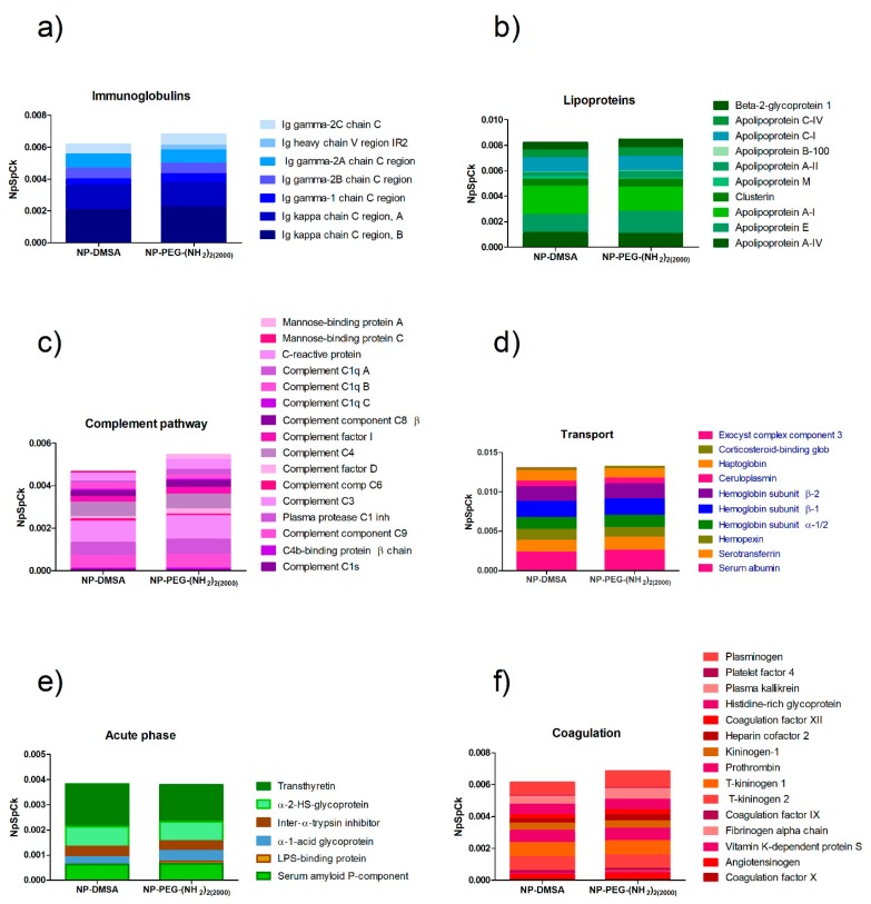
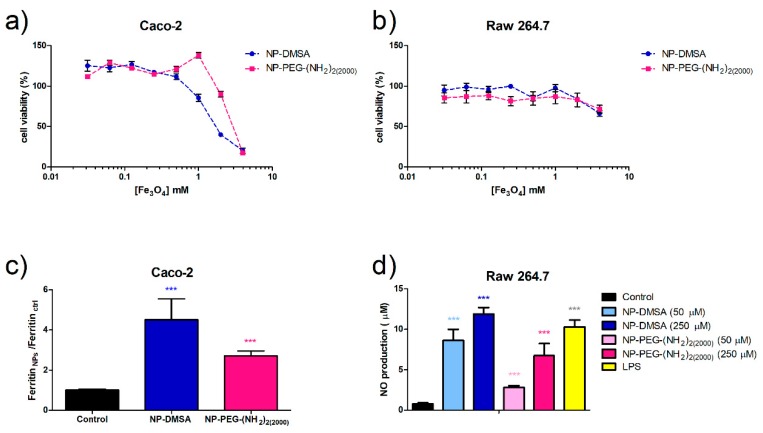
Superparamagnetic iron oxide nanoparticles are one of the most prominent agents used in theranostic applications, with MRI imaging the main application assessed. The biomolecular interface formed on the surface of a nanoparticle in a biological medium determines its behaviour in vitro and in vivo. In this study, we have compared the formation of the protein corona on highly monodisperse iron oxide nanoparticles with two different coatings, dimercaptosuccinic acid (DMSA), and after conjugation, with a bifunctional polyethylene glycol (PEG)-derived molecule (2000 Da) in the presence of Wistar rat plasma. The protein fingerprints around the nanoparticles were analysed in an extensive proteomic study. The results presented in this work indicate that the composition of the protein corona is very difficult to predict. Proteins from different functional categories-cell components, lipoproteins, complement, coagulation, immunoglobulins, enzymes and transport proteins-were identified in all samples with very small variability. Although both types of nanoparticles have similar amounts of bonded proteins, very slight differences in the composition of the corona might explain the variation observed in the uptake and biotransformation of these nanoparticles in Caco-2 and RAW 264.7 cells. Cytotoxicity was also studied using a standard 3-(4,5-dimethylthiazol-2-yl)-2,5-diphenyl tetrazolium bromide assay. Controlling nanoparticles' reactivity to the biological environment by deciding on its surface functionalization may suggest new routes in the control of the biodistribution, biodegradation and clearance of multifunctional nanomedicines.

Keywords: PEG-coated nanoparticles; iron oxide nanoparticles; protein corona.


Conflict of interest statement

The authors declare no conflict of interest.

Figures

Figure 1
Figure 1
(a) Transmission electron microscopy (TEM) images of 10 nm nanoparticles. (b) Evolution of ζ-potential as a function of pH. (c) FTIR spectra. (I) NP-DMSA, (II) NP-PEG-(NH2)2(2000).
Figure 2
Figure 2
(a) Nanoparticle hydrodynamic size evolution up to 30 min in presence of plasma. (b) Hydrodynamic sizes of nanoparticles in aqueous suspension (solid line) and after 30 min of incubation with plasma (dotted line). (I) NP-DMSA, (II) NP-PEG-(NH2)2(2000).
Figure 3
Figure 3
Densitometric analysis of the Coomassie blue-stained SDS-PAGE bands. Proteins from particles incubated at 10, 20 and 30 min at 37 °C were eluted sequentially with buffers at different pH. Top panel: Proteins eluted after incubation in buffer 50 mM HEPES 0.1% OGP, pH 7 (soft corona). Lower panel: Proteins eluted after incubation in buffer 100 mM NaAc 0.1% OGP, pH 5 (hard corona). (a and c) NP-DMSA (b and d) NP-PEG-(NH2)2 (2000).
Figure 4
Figure 4
Proteins identified by mass spectrometry on the surface of the nanoparticles. Individual analysis of the protein corona around: (a) NP-DMSA. (b) NP-PEG-(NH2)2(2000). Sc and hc represent specific proteins detected in the soft and hard corona of the particles, respectively. Overlapped areas represent proteins detected in common in their soft and hard corona. (c) Analysis of the proteins identified in the soft corona of both nanoparticles. (d) Analysis of the proteins identified in the hard corona of the nanoparticles. Blue represents specific proteins detected in NP-DMSA and pink represents specific proteins detected in NP-PEG-(NH2)2(2000). Overlapped areas represent proteins detected in common in those fractions.
Figure 5
Figure 5
Semiquantitative analysis of the protein corona composition of NP-DMSA and NP-PEG-(NH2)2(2000). (a) Inmunoglobulins; (b) Lipoproteins; (c) Complement pathway; (d) Transport; (e) Acute phase; (f) Coagulation.
Figure 6
Figure 6
Relative abundance of opsonins (immunoglobulins and apolipoproteins) versus dysopsonins (albumin) on the protein corona composition of NP-DMSA and NP-PEG-(NH2)2(2000).
Figure 7
Figure 7
Evaluation of cell viability by MTT assay. (a) Caco-2 cells. (b) RAW 264.7 cells. (c) Ferritin formation measured by ELISA in Caco-2 cells exposed to 250 µM of NP-DMSA or NP-PEG-(NH2)2(2000). (d) Evaluation of macrophages activation by measurement of nitric oxide production. Data represent means ± SD (n = 3). * Shows statistical significance compared with the control (one-way ANOVA, Bonferroni´s post-hoc test * p < 0.05, ** p < 0.01, *** p < 0.001, **** p < 0.0001).

References

    1. Clinicaltrials.gov [(accessed on 6 February 2019)]; Available online: https://www.clinicaltrials.gov/ct2/results?term=iron+oxide+nanoparticles.
    1. Magro M., Baratella D., Bonaiuto E., de A. Roger J., Vianello F. New Perspectives on Biomedical Applications of Iron Oxide Nanoparticles. Curr. Med. Chem. 2018;25:540–555. doi: 10.2174/0929867324666170616102922. - DOI - PubMed
    1. Arias L., Pelim Pessan J., Miranda Vieira A., Toito de Lima T., Botazzo Delbem A., Monteiro D. Iron Oxide Nanoparticles for Biomedical Applications: A Perspective on Synthesis, Drugs, Antimicrobial Activity, and Toxicity. Antibiotics. 2018;7:46. doi: 10.3390/antibiotics7020046. - DOI - PMC - PubMed
    1. Veronese F.M., Pasut G. PEGylation, Successful Approach to Drug Delivery. Drug Discov. Today. 2005;10:1451–1458. doi: 10.1016/S1359-6446(05)03575-0. - DOI - PubMed
    1. Tkachenko A.G., Xie H., Liu Y., Coleman D., Ryan J., Glomm W.R., Shipton M.K., Franzen S., Feldheim D.L. Cellular Trajectories of Peptide-Modified Gold Particle Complexes: Comparison of Nuclear Localization Signals and Peptide Transduction Domains. Bioconjug. Chem. 2004;15:482–490. doi: 10.1021/bc034189q. - DOI - PubMed