Review
. 2019 Jul 10;8(7):701.
doi: 10.3390/cells8070701.

The Centrosome and the Primary Cilium: The Yin and Yang of a Hybrid Organelle

Affiliations
Review

The Centrosome and the Primary Cilium: The Yin and Yang of a Hybrid Organelle

Vladimir Joukov et al. Cells. .

Abstract

Centrosomes and primary cilia are usually considered as distinct organelles, although both are assembled with the same evolutionary conserved, microtubule-based templates, the centrioles. Centrosomes serve as major microtubule- and actin cytoskeleton-organizing centers and are involved in a variety of intracellular processes, whereas primary cilia receive and transduce environmental signals to elicit cellular and organismal responses. Understanding the functional relationship between centrosomes and primary cilia is important because defects in both structures have been implicated in various diseases, including cancer. Here, we discuss evidence that the animal centrosome evolved, with the transition to complex multicellularity, as a hybrid organelle comprised of the two distinct, but intertwined, structural-functional modules: the centriole/primary cilium module and the pericentriolar material/centrosome module. The evolution of the former module may have been caused by the expanding cellular diversification and intercommunication, whereas that of the latter module may have been driven by the increasing complexity of mitosis and the requirement for maintaining cell polarity, individuation, and adhesion. Through its unique ability to serve both as a plasma membrane-associated primary cilium organizer and a juxtanuclear microtubule-organizing center, the animal centrosome has become an ideal integrator of extracellular and intracellular signals with the cytoskeleton and a switch between the non-cell autonomous and the cell-autonomous signaling modes. In light of this hypothesis, we discuss centrosome dynamics during cell proliferation, migration, and differentiation and propose a model of centrosome-driven microtubule assembly in mitotic and interphase cells. In addition, we outline the evolutionary benefits of the animal centrosome and highlight the hierarchy and modularity of the centrosome biogenesis networks.

Keywords: cell cycle; cell differentiation; cell signaling; centriole; centrosome; microtubule cytoskeleton; microtubule nucleation; mitosis; organelle biogenesis; primary cilia.


Conflict of interest statement

The authors declare no conflict of interest.

Figures
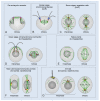
Figure 1
Figure 1
Centrosomes and the basal body apparatus in different eukaryotic lineages. (A) Putative pre-eukaryotic ancestor, which had circular chromosomes (dark grey loops) associated with a precursor centrosome with a dual centrosome and kinetochore function (purple and orange half-circles, respectively). The precursor centrosome was still attached to the surface membrane [42]. Microtubules (MTs) are in green. (BG) Eukaryotes of different lineages. Centrioles/basal bodies are in blue or light purple; flagella are in grey; microtubules (MTs) are in green; pericentriolar material (PCM) is in yellow; the centrin-containing structures are in red. Higher plants (E) lack Polo-like kinase 1 (PLK1) and the apparent orthologs of the PCM proteins involved in γ-tubulin ring complex (γ-TuRC) anchoring and activation in animals. Conceivably, plant-specific γ-TuRC-anchoring and activating factors form centrosome-like MT-organizing centers (MTOCs), which organize spindle poles in higher plants [(yellow circles in (E)] [66]. Taxonomic supergroups are indicated in square brackets. SAR: stramenopiles, alveolates, and Rhizaria.
Figure 2
Figure 2
Centrosomes and the basal body apparatus in certain lineages of the Amorphea supergroup. A schematic illustration of cells in early interphase. Centrioles and basal bodies are in blue, flagella are in grey, microtubules (MTs) are in green, and the pericentriolar material (PCM) [which presumably originates from the ancestral nucleus-associated MT-organizing center (MTOC)] is in yellow. The ancestral centrin-containing nucleus-basal body connector and other centrin-containing structures are in red. Dashed red lines indicate that evidence of a nucleus-basal body connection is incomplete. In apusomonads, the basal bodies are connected to the nucleus with a striated fibrous root, rhizostyle, but it is unclear if it contains centrin or not [70]. In choanoflagellates, prior to cell division, the basal bodies duplicate and migrate to poles of the nucleus [71]. For Physarum polycephalum and Rhizophydium sphaerotheca, interphase cells of two different life cycle stages are shown. It is unclear if the basal bodies are surrounded by the PCM in these organisms.
Figure 3
Figure 3
Schematic of the centrosome cycle. The inner (proximal to the centriole) pericentriolar material (PCM) layer, which contains centrosomal protein of 192 kDa (CEP192) and polo-like kinase 1 (PLK1), is highlighted by a purple line. See text for details.
Figure 4
Figure 4
Schematic structure of a primary cilium. Panels on the right are schematic cross-sections of a typical primary cilium and a motile cilium. GPCR: G protein-coupled receptor; RTK: receptor tyrosine kinase; IFT: intraflagellar transport. MT: microtubule; PCM: pericentriolar material.
Figure 5
Figure 5
Molecular pathways underlying centriole and pericentriolar material (PCM) assembly in the vertebrates. Protein kinases are in red. The centrosomal protein of 192 kDa (CEP192) and the inner (proximal to the centriole) PCM layer, which contains CEP192 and polo-like kinase 1 (PLK1), are in purple. Distinct modules, which are repurposed and used in centrosome-independent processes, are highlighted in grey. Module 1: Basal body assembly module used in multiciliated cells. Module 2: pericentrin (PCNT)-CEP215 module used for the assembly of non-centrosomal microtubule (MT)-organizing centers (MTOCs) in postmitotic cells. Module 3: A putative PCNT-CEP192 module used for the assembly of acentriolar mitotic MTOCs in mouse oocytes and early embryos. This module relies on the CEP192-mediated, autocatalytic mechanism of Aurora A (AurA)-PLK1 activation, and PCM protein recruitment. Dashed arrows/lines indicate inferred interactions/effects that need to be experimentally validated. Module 4: PCM scaffold assembly module used in D. melanogaster cells. STIL: SCL-interrupting locus protein [anastral spindle 2 (Ana-2) in D. melanogaster; spindle assembly abnormal protein 5 (SAS-5) in C. elegans]; γ-TuRC: γ-tubulin ring complex; CPAP: centrosomal P4.1-associated protein [also known as centromere protein J (CENPJ); SAS-4 in D. melanogaster and C. elegans]; NEDD1: developmentally down-regulated protein 1; CKAP5: cytoskeleton-associated protein 5 [also known as colonic and hepatic tumor overexpressed protein (chTOG) and Xenopus MT-associated protein of 215 kDa (XMAP215)]. See text for details.
Figure 6
Figure 6
The centrosomal protein of 192 kDa (CEP192) organizes Aurora A (AurA) and Polo-like kinase 1 (PLK1) in a kinase cascade that drives microtubule (MT)-organizing center (MTOC) formation. (A) Schematic of the cascade. (B) Artificial centrosomes formed by magnetic beads coated with a recombinant N-terminal fragment of CEP192 (amino acids 1–1000) wild type (wt) (CEP1921-1000-wt), which binds AurA, PLK1, NEDD1-γ-TuRC (developmentally down-regulated protein 1-γ-tubulin ring complex), and Xenopus MT-associated protein of 215 kDa (XMAP215) [also known as cytoskeleton-associated protein 5 (CKAP5)] in a metaphase-arrested Xenopus egg extract. Beads coated with glutathione S-transferase (GST) are shown as a control. The extract was supplemented with rhodamine-labeled α/β-tubulin to visualize MTs. (C) Western blots of proteins retrieved from a metaphase-arrested Xenopus egg extract with beads coated with CEP1921-1000-wt or with its mutant counterparts lacking the binding sites for PLK1 (T46A) or AurA (δAurA). (D) Western blots of proteins retrieved from a metaphase-arrested Xenopus egg extract with beads coated with CEP1921-1000-wt or with its mutant counterparts lacking one (S→A1), two (S→A2), or five (S→A5) NEDD1-γ-TuRC-binding serines. AurA(pT295) and PLK1(pT201): AurA and PLK1 isoforms phosphorylated at the conserved threonine residue in the T loop. The graphs in (C) and (D) show a relative efficiency of MT nucleation (proportion of bead-induced MT asters). Extracts analyzed by Western blotting in (C,D) were supplemented with nocodazole to prevent MT assembly. All images are adapted from [134].
Figure 7
Figure 7
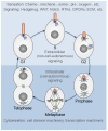
The centrosome cycle in proliferating cells. The primary cilium is formed by the mother centriole-centrosome complex in G1 phase and progressively shortens thereafter. In the interphase (upper part), the primary cilium serves as an “antenna” that senses extracellular cues and relays the signals to the cell’s interior. The ciliary membrane (red) differs in its composition from the plasma membrane and is enriched in specific ion channels and receptors for various extracellular regulatory factors (see Figure 4). After mitotic commitment (G2), the two centrosomes separate, recruit additional PCM components and form microtubule (MT) asters—the nascent spindle poles (centrosome maturation). The mother centrosome internalizes with the primary cilium while retaining the ciliary membrane, which may act as a cell fate determinant. The ciliary disassembly completes at the end of mitosis, although the timing may differ between cell types [208,211,214,216]. WNT: wingless-type MMTV integration site family; RTKs: receptor tyrosine kinases; GPCRs: G protein-coupled receptors; ECM: extracellular matrix.
Figure 8
Figure 8
Hypothetical model of centrosome-driven microtubule (MT) assembly in interphase and postmitotic cells. See also Figure 5. CDK1: cyclin-dependent kinase 1; PCNT: pericentrin; CEP: centrosomal protein.

References

    1. Fletcher D.A., Mullins R.D. Cell mechanics and the cytoskeleton. Nature. 2010;463:485–492. doi: 10.1038/nature08908. - DOI - PMC - PubMed
    1. Dogterom M., Koenderink G.H. Actin-microtubule crosstalk in cell biology. Nat. Rev. Mol. Cell Biol. 2018 doi: 10.1038/s41580-018-0067-1. - DOI - PubMed
    1. Kollman J.M., Merdes A., Mourey L., Agard D.A. Microtubule nucleation by gamma-tubulin complexes. Nat. Rev. Mol. Cell Biol. 2011;12:709–721. doi: 10.1038/nrm3209. - DOI - PMC - PubMed
    1. Teixido-Travesa N., Roig J., Luders J. The where, when and how of microtubule nucleation—One ring to rule them all. J. Cell Sci. 2012;125:4445–4456. doi: 10.1242/jcs.106971. - DOI - PubMed
    1. Martin M., Akhmanova A. Coming into Focus: Mechanisms of Microtubule Minus-End Organization. Trends Cell Biol. 2018;28:574–588. doi: 10.1016/j.tcb.2018.02.011. - DOI - PubMed

LinkOut - more resources