

microRNA-367-3p regulation of GPRC5A is suppressed in ischemic stroke
- PMID: 31296130
- PMCID: PMC7238381
- DOI: 10.1177/0271678X19858637
microRNA-367-3p regulation of GPRC5A is suppressed in ischemic stroke
Abstract
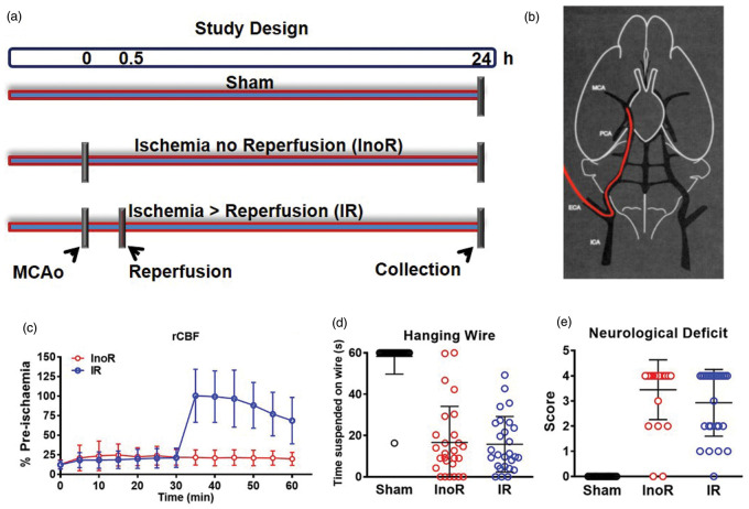
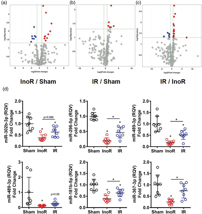
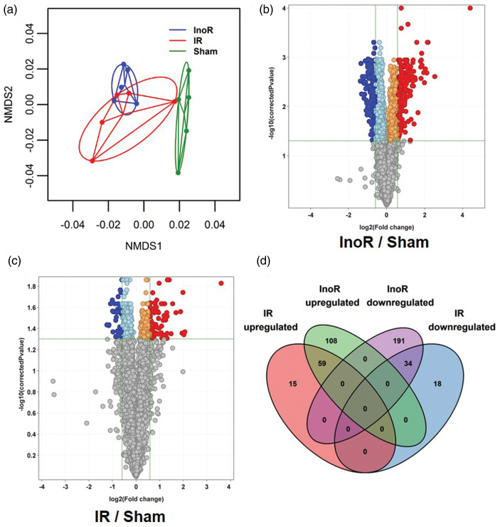
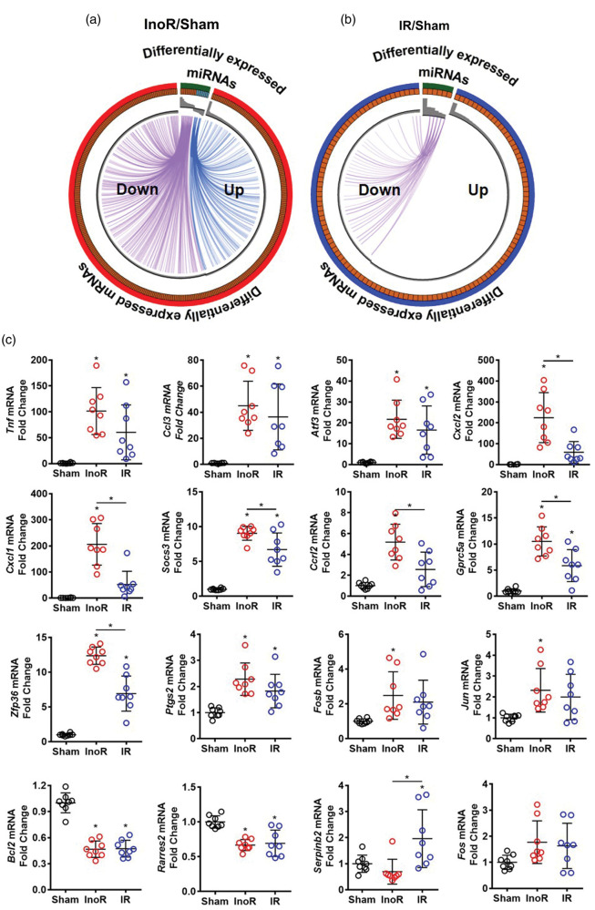
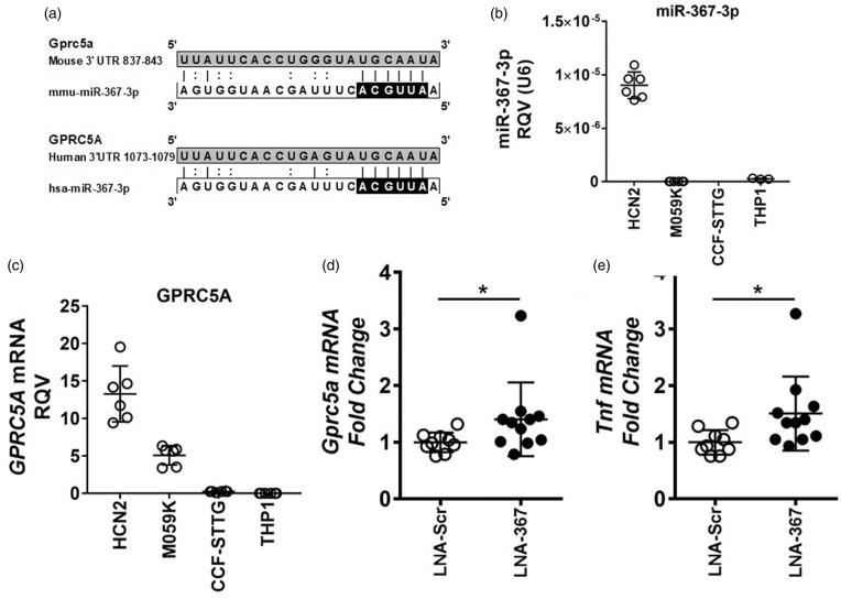
Ischemic stroke is a major cause of mortality and long-term disability with limited treatment options, and a greater understanding of the gene regulatory mechanisms underlying ischemic stroke-associated neuroinflammation is required for new therapies. To study ischemic stroke in vivo, mice were subjected to sustained ischemia by intraluminal filament-induced middle cerebral artery occlusion (MCAo) for 24 h without reperfusion or transient ischemia for 30 min followed by 23.5 h reperfusion, and brain miRNA and mRNA expression changes were quantified by TaqMan OpenArrays and gene (mRNA) expression arrays, respectively. Sustained ischemia resulted in 18 significantly altered miRNAs and 392 altered mRNAs in mouse brains compared to Sham controls; however, the transient ischemic condition was found to impact only 6 miRNAs and 126 mRNAs. miR-367-3p was found to be significantly decreased in brain homogenates with sustained ischemia. G protein-coupled receptor, family C, group 5, member A (Gprc5a), a miR-367-3p target gene, was found to be significantly increased with sustained ischemia. In primary neurons, inhibition of endogenous miR-367-3p resulted in a significant increase in Gprc5a expression. Moreover, miR-367-3p was found to be co-expressed with GPRC5A in human neurons. Results suggest that loss of miR-367-3p suppression of GPRC5A may contribute to neuroinflammation associated with ischemic stroke.
Keywords: Microrna; brain; ischemia; neuroinflammation; neurons; stroke.
Figures
Similar articles
-
Inhibition of microRNA-9-5p and microRNA-128-3p can inhibit ischemic stroke-related cell death in vitro and in vivo.IUBMB Life. 2020 Nov;72(11):2382-2390. doi: 10.1002/iub.2357. Epub 2020 Aug 14. IUBMB Life. 2020. PMID: 32797712
-
Intracerebral overexpression of miR-669c is protective in mouse ischemic stroke model by targeting MyD88 and inducing alternative microglial/macrophage activation.J Neuroinflammation. 2020 Jun 19;17(1):194. doi: 10.1186/s12974-020-01870-w. J Neuroinflammation. 2020. PMID: 32560730 Free PMC article.
-
MicroRNA-488-3p Regulates Neuronal Cell Death in Cerebral Ischemic Stroke Through Vacuolar Protein Sorting 4B (VPS4B).Neuropsychiatr Dis Treat. 2021 Jan 7;17:41-55. doi: 10.2147/NDT.S255666. eCollection 2021. Neuropsychiatr Dis Treat. 2021. PMID: 33442254 Free PMC article.
-
microRNA as a therapeutic for ischemic stroke.Neurochem Int. 2023 Feb;163:105487. doi: 10.1016/j.neuint.2023.105487. Epub 2023 Jan 16. Neurochem Int. 2023. PMID: 36657721 Review.
-
The role of microRNA in neuronal inflammation and survival in the post ischemic brain: a review.Neurol Res. 2023 Sep;45(9):1-9. doi: 10.1080/01616412.2017.1327505. Epub 2017 May 27. Neurol Res. 2023. PMID: 28552032 Review.
Cited by
-
CX3CL1/CX3CR1 axis attenuates early brain injury via promoting the delivery of exosomal microRNA-124 from neuron to microglia after subarachnoid hemorrhage.J Neuroinflammation. 2020 Jul 14;17(1):209. doi: 10.1186/s12974-020-01882-6. J Neuroinflammation. 2020. PMID: 32664984 Free PMC article.
-
Identification of Dysregulated Mechanisms and Potential Biomarkers in Ischemic Stroke Onset.Int J Gen Med. 2021 Aug 22;14:4731-4744. doi: 10.2147/IJGM.S327594. eCollection 2021. Int J Gen Med. 2021. PMID: 34456585 Free PMC article.
-
Dysregulated expression of miR‑367 in disease development and its prospects as a therapeutic target and diagnostic biomarker (Review).Biomed Rep. 2023 Oct 10;19(6):91. doi: 10.3892/br.2023.1673. eCollection 2023 Dec. Biomed Rep. 2023. PMID: 37901877 Free PMC article. Review.
-
MicroRNA-367-3p suppresses sevoflurane-induced adult rat astrocyte apoptosis by targeting BCL2L11.Exp Ther Med. 2022 Jan;23(1):9. doi: 10.3892/etm.2021.10931. Epub 2021 Oct 28. Exp Ther Med. 2022. PMID: 34815761 Free PMC article.
-
MicroRNA Profiles in Critically Ill Patients.Curr Med Chem. 2024;31(41):6801-6825. doi: 10.2174/0929867331666230726095222. Curr Med Chem. 2024. PMID: 37496239 Review.
References
Publication types
MeSH terms
Substances
Grants and funding
LinkOut - more resources
Full Text Sources
Medical
