

Long-term impairment of neurovascular coupling following experimental subarachnoid hemorrhage
- PMID: 31296132
- PMCID: PMC7238370
- DOI: 10.1177/0271678X19863021
Long-term impairment of neurovascular coupling following experimental subarachnoid hemorrhage
Abstract
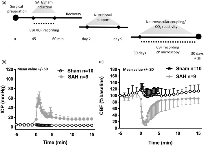
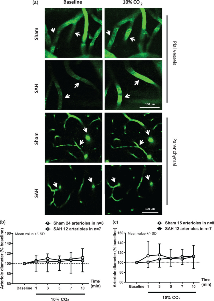
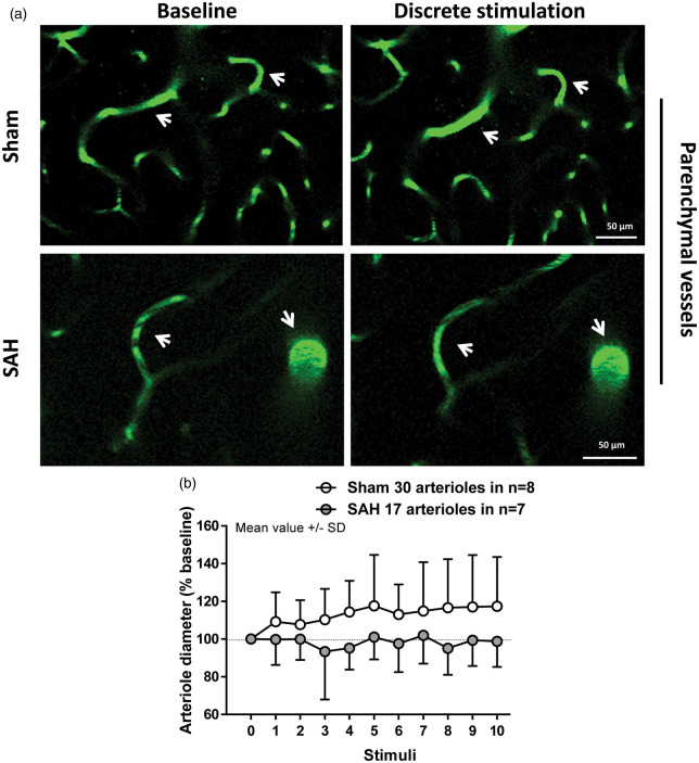
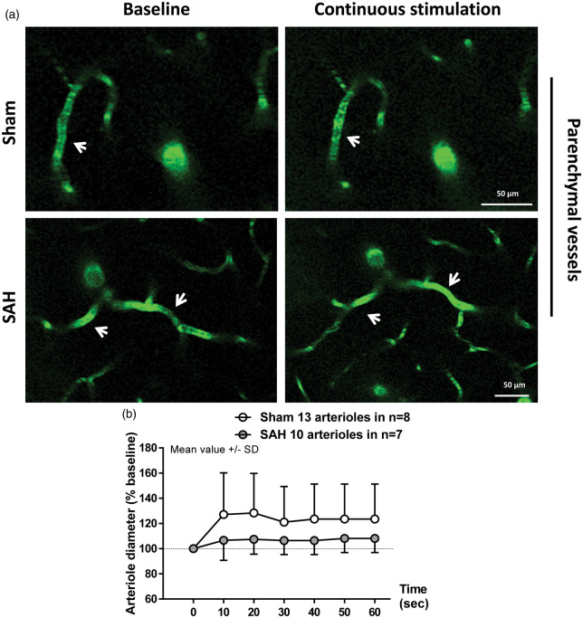
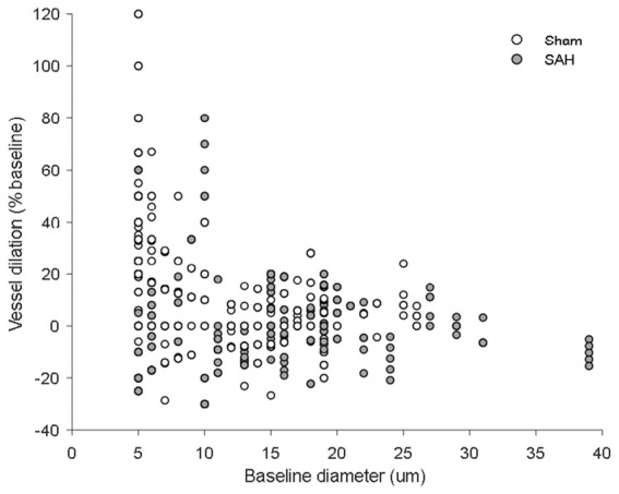
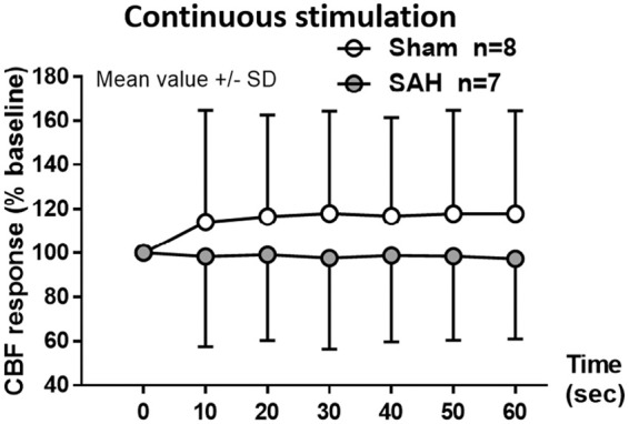
CO2-reactivity and neurovascular coupling are sequentially lost within the first 24 h after subarachnoid hemorrhage (SAH). Whether and when these impairments recover is not known. Therefore, we investigated the reactivity of pial and intraparenchymal vessels by in vivo two-photon microscopy one month after experimental SAH. C57BL/6 mice were subjected to either sham surgery or SAH by filament perforation. One month later, cerebral blood flow following CO2-challenge and forepaw stimulation was assessed by laser Doppler fluxmetry. Diameters of pial and intraparenchymal arterioles were quantified by in vivo two-photon microscopy. One month after SAH, pial and parenchymal vessels dilated in response to CO2. Neurovascular coupling was almost completely absent after SAH: vessel diameter did not change upon forepaw stimulation compared to a 20% increase in sham-operated mice. The current results demonstrate that neurovascular function differentially recovers after SAH: while CO2-reactivity normalizes within one month after SAH, neurovascular coupling is still absent. These findings show an acute and persistent loss of neurovascular coupling after SAH that may serve as a link between early brain injury and delayed cerebral ischemia, two distinct pathophysiological phenomena after SAH that were so far believed not to be directly related.
Keywords: Subarachnoid hemorrhage; in vivo; mice; neurovascular coupling; two-photon microscopy.
Figures
Similar articles
-
Acute changes in neurovascular reactivity after subarachnoid hemorrhage in vivo.J Cereb Blood Flow Metab. 2017 Jan;37(1):178-187. doi: 10.1177/0271678X15621253. Epub 2015 Dec 16. J Cereb Blood Flow Metab. 2017. PMID: 26676226 Free PMC article.
-
Inversion of neurovascular coupling after subarachnoid hemorrhage in vivo.J Cereb Blood Flow Metab. 2017 Nov;37(11):3625-3634. doi: 10.1177/0271678X16686595. Epub 2017 Jan 23. J Cereb Blood Flow Metab. 2017. PMID: 28112024 Free PMC article.
-
Astrocyte Ca2+ Signaling Drives Inversion of Neurovascular Coupling after Subarachnoid Hemorrhage.J Neurosci. 2015 Sep 30;35(39):13375-84. doi: 10.1523/JNEUROSCI.1551-15.2015. J Neurosci. 2015. PMID: 26424885 Free PMC article.
-
Subarachnoid hemorrhage: a review of experimental studies on the microcirculation and the neurovascular unit.Transl Stroke Res. 2014 Apr;5(2):174-89. doi: 10.1007/s12975-014-0323-4. Epub 2014 Feb 11. Transl Stroke Res. 2014. PMID: 24510780 Review.
-
Neuroinflammation and Microvascular Dysfunction After Experimental Subarachnoid Hemorrhage: Emerging Components of Early Brain Injury Related to Outcome.Neurocrit Care. 2019 Oct;31(2):373-389. doi: 10.1007/s12028-019-00710-x. Neurocrit Care. 2019. PMID: 31012056 Free PMC article. Review.
Cited by
-
Non-invasive Assessment of Neurovascular Coupling After Aneurysmal Subarachnoid Hemorrhage: A Prospective Observational Trial Using Retinal Vessel Analysis.Front Neurol. 2021 Jun 14;12:690183. doi: 10.3389/fneur.2021.690183. eCollection 2021. Front Neurol. 2021. PMID: 34194387 Free PMC article.
-
From Mechanisms to Medicine: Neurovascular Coupling in the Diagnosis and Treatment of Cerebrovascular Disorders: A Narrative Review.Cells. 2024 Dec 27;14(1):16. doi: 10.3390/cells14010016. Cells. 2024. PMID: 39791717 Free PMC article. Review.
-
The urotensin II receptor triggers an early meningeal response and a delayed macrophage-dependent vasospasm after subarachnoid hemorrhage in male mice.Nat Commun. 2024 Sep 29;15(1):8430. doi: 10.1038/s41467-024-52654-2. Nat Commun. 2024. PMID: 39341842 Free PMC article.
-
CX3CL1/CX3CR1 axis attenuates early brain injury via promoting the delivery of exosomal microRNA-124 from neuron to microglia after subarachnoid hemorrhage.J Neuroinflammation. 2020 Jul 14;17(1):209. doi: 10.1186/s12974-020-01882-6. J Neuroinflammation. 2020. PMID: 32664984 Free PMC article.
-
MiR-340-5p alleviates neuroinflammation and neuronal injury via suppressing STING in subarachnoid hemorrhage.Brain Behav. 2022 Sep;12(9):e2687. doi: 10.1002/brb3.2687. Epub 2022 Aug 11. Brain Behav. 2022. PMID: 35957622 Free PMC article.
References
-
- Weaver JP, Fisher M. Subarachnoid hemorrhage: an update of pathogenesis, diagnosis and management. J Neurol Sci 1994; 125: 119–131. - PubMed
-
- van Gijn J, Rinkel GJ. Subarachnoid haemorrhage: diagnosis, causes and management. Brain 2001; 124(Pt 2): 249–278. - PubMed
-
- Mayer SA, Kreiter KT, Copeland D, et al.Global and domain-specific cognitive impairment and outcome after subarachnoid hemorrhage. Neurology 2002; 59: 1750–1758. - PubMed
-
- Passier PE, Visser-Meily JM, Rinkel GJ, et al.Life satisfaction and return to work after aneurysmal subarachnoid hemorrhage. J Stroke Cerebrovasc Dis 2011; 20: 324–329. - PubMed
Publication types
MeSH terms
LinkOut - more resources
Full Text Sources
