. 2019 Jul 11;16(1):144.
doi: 10.1186/s12974-019-1534-0.

Long-term voluntary wheel running does not alter vascular amyloid burden but reduces neuroinflammation in the Tg-SwDI mouse model of cerebral amyloid angiopathy

Affiliations

Long-term voluntary wheel running does not alter vascular amyloid burden but reduces neuroinflammation in the Tg-SwDI mouse model of cerebral amyloid angiopathy

Lisa S Robison et al. J Neuroinflammation. .

Abstract

Background: Cardiovascular exercise (CVE) has been shown to be protective against cognitive decline in aging and the risk for dementias, including Alzheimer's Disease (AD). CVE has also been shown to have several beneficial effects on brain pathology and behavioral impairments in mouse models of AD; however, no studies have specifically examined the effects of CVE on cerebral amyloid angiopathy (CAA), which is the accumulation of amyloid-beta (Aβ) in the cerebral vasculature. CAA may be uniquely susceptible to beneficial effects of CVE interventions due to the location and nature of the pathology. Alternatively, CVE may exacerbate CAA pathology, due to added stress on already compromised cerebral vasculature.

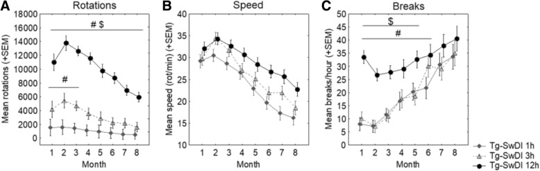
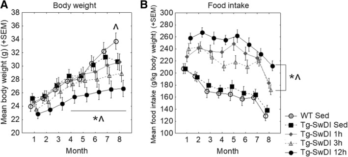
Methods: In the current study, we examined the effects of CVE over many months in mice, thereby modeling a lifelong commitment to CVE in humans. We assessed this voluntary CVE in Tg-SwDI mice, a transgenic mouse model of CAA that exhibits behavioral deficits, fibrillar vascular Aβ pathology, and significant perivascular neuroinflammation. Various "doses" of exercise intervention (0 h ("Sedentary"), 1 h, 3 h, 12 h access to running wheel) were assessed from ~ 4 to 12 months of age for effects on physiology, behavior/cognitive performance, and pathology.

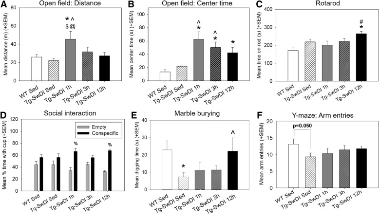
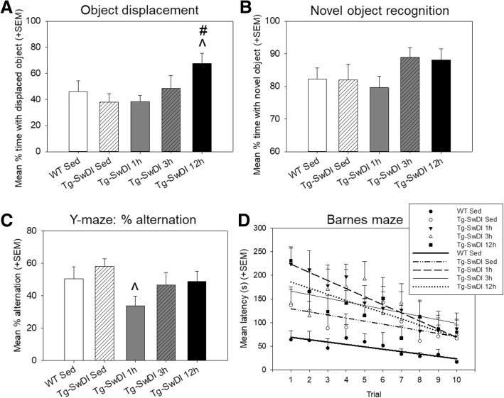
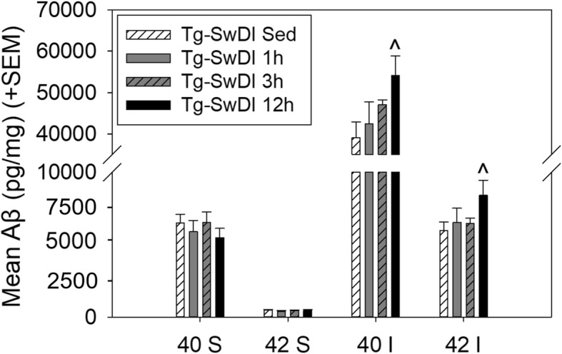
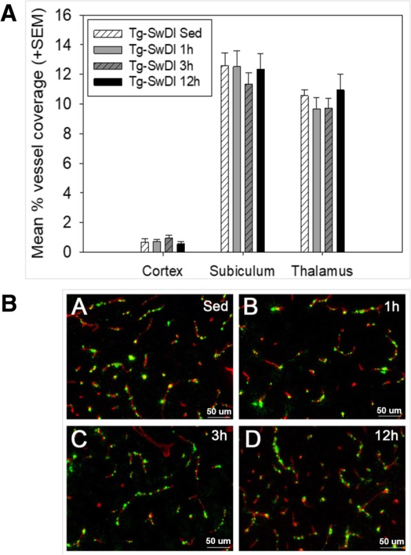
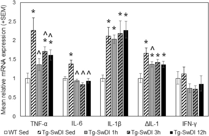
Results: The 12 h group performed the greatest volume of exercise, whereas the 1 h and 3 h groups showed high levels of exercise intensity, as defined by more frequent and longer duration running bouts. Tg-SwDI mice exhibited significant cerebral vascular Aβ pathology and increased expression of pro-inflammatory cytokines as compared to WT controls. Tg-SwDI mice did not show motor dysfunction or altered levels of anxiety or sociability compared to WT controls, though Tg-SwDI animals did appear to exhibit a reduced tendency to explore novel environments. At all running levels, CAA pathology in Tg-SwDI mice was not significantly altered, but 12-h high-volume exercise showed increased insoluble Aβ burden. However, CVE attenuated the expression of pro-inflammatory cytokines TNF-α and IL-6 and was generally effective at enhancing motor function and reducing anxiety-like behavior in Tg-SwDI mice, though alterations in learning and memory tasks were varied.

Conclusions: Taken together, these results suggest that CAA can still develop regardless of a lifespan of substantial CVE, although downstream effects on neuroinflammation may be reduced and functional outcomes improved.

Keywords: Aerobic; Alzheimer’s disease; Anti-inflammatory; Beta amyloid; Cardiovascular; Cerebral amyloid angiopathy; Exercise; Fitness; Inflammation.


Conflict of interest statement

The authors declare that they have no competing interests.

Figures

Fig. 1
Fig. 1
Monthly wheel running parameters over the course of the 8-month exercise intervention period. a The number of rotations performed by each mouse was recorded with computer software in 1-minute bins during each exercise session. The total number of rotations performed was summed during each session as a measure of running volume. b Running speed (rotations/minute) was calculated by averaging the number of rotations performed in 1-min bins that were not counted as a “break,” essentially averaging the number of rotations performed per minute when the number of rotations performed in that bin was greater than five. c Breaks per hour was calculated to determine the number of 1-min bins per hour during which the mouse was not running. A “break” was a 1-min bin in which the animal performed less than five wheel rotations. #p < 0.05 versus Tg-SwDI 1 h; $p < 0.05 versus Tg-SwDI 3 h
Fig. 2
Fig. 2
Body weight and food intake over the course of the exercise intervention period. a Monthly body weights. b Average daily food intake by month of exercise intervention. *p < 0.05 versus WT sedentary; ^p < 0.05 versus Tg-SwDI sedentary
Fig. 3
Fig. 3
Performance on behavioral tasks assessing motor function, exploratory behavior, and temperament. a Distance traveled in the open field arena during the open field test, in meters. b Time spent in the center of the arena during the open field test, in seconds. c Rotarod performance, as measured by time spent on the rod, in seconds (max time = 300 s). d Percentage of time spent with empty versus conspecific cup in the social interaction test. e Time spent digging in the defensive burying task, in seconds. f Exploration in the Y-maze task, as measured by number of arm entries. *p < 0.05 versus WT sedentary; ^p < 0.05 versus Tg-SwDI sedentary, #p < 0.05 versus Tg-SwDI 1 h; $p < 0.05 versus Tg-SwDI 3 h; @p < 0.05 versus Tg-SwDI 12 h; %p < 0.05 more time spent with conspecific cup vs. empty cup
Fig. 4
Fig. 4
Performance on cognitive tasks of learning and memory. a Performance on the object displacement task, as measured by percentage of time spent with the displaced object. b Performance on the novel object recognition task, as measured by percentage of time spent with the novel object. c Performance on the Y-maze task, as measured by percent alternation between arms. d Performance on the Barnes maze task by trial, as measured by latency to find the escape hole (max time = 300 s). Data points for each group were fit with a linear line to quantify the rate of learning. ^p < 0.05 versus Tg-SwDI sedentary, #p < 0.05 versus Tg-SwDI 1 h
Fig. 5
Fig. 5
Levels of beta-amyloid species in forebrain homogenate of Tg-SwDI mice by ELISA. Brains were collected following the 8-month exercise intervention period and completion of behavior testing. Forebrain homogenate was assayed for levels of beta-amyloid (Aβ) 40 and 42 in soluble (S) and insoluble (I) fractions. ^p < 0.05 versus Tg-SwDI sedentary
Fig. 6
Fig. 6
Quantitative and histological illustration of vascular amyloid burden. a Vascular amyloid burden in Tg-SwDI mice, represented as percent of the vasculature covered by fibrillar amyloid in the cortex, subiculum, and thalamus. This measure was calculated as [(ThioflavinS+ stain/Collagen IV+ stain) × 100]. WT mice were not included in this analysis, as only Tg-SwDI mice exhibit measurable amyloid pathology. Brains were collected following the 8-month exercise intervention period and completion of behavior testing. ^p < 0.05 versus Tg-SwDI sedentary. b Representative images of histological staining from thalamus (scale bars = 50 μm) for fibrillar Aβ (Thioflavin-S; green) and blood vessels (collagen IV; red) in sagittal sections from Tg-SwDI mouse brains
Fig. 7
Fig. 7
Quantitative polymerase chain reaction (qPCR) results showing inflammatory cytokine expression. Expression levels of TNF-α, IL-6, IL-1β, IL-1, and IFN-γ were assessed in forebrain homogenate, collected following the 8-month exercise regimen and behavior testing. Statistical analyses were performed on ΔCt values (one-way ANOVA (between-subjects factor: group)), and data are plotted as fold change from WT sedentary mice. *p < 0.05 vs. WT sedentary controls. ^p < 0.05 vs. Tg-SwDI sedentary mice

References

    1. Radak Z, Hart N, Sarga L, Koltai E, Atalay M, Ohno H, Boldogh I. Exercise plays a preventive role against Alzheimer's disease. J Alzheimers Dis. 2010;20:777–783. doi: 10.3233/JAD-2010-091531. - DOI - PubMed
    1. Lange-Asschenfeldt C, Kojda G. Alzheimer’s disease, cerebrovascular dysfunction and the benefits of exercise: from vessels to neurons. Exp Gerontol. 2008;43:499–504. doi: 10.1016/j.exger.2008.04.002. - DOI - PubMed
    1. Weuve J, Kang JH, Manson JE, Breteler MM, Ware JH, Grodstein F. Physical activity, including walking, and cognitive function in older women. Jama. 2004;292:1454–1461. doi: 10.1001/jama.292.12.1454. - DOI - PubMed
    1. Kulmala J, Solomon A, Kåreholt I, Ngandu T, Rantanen T, Laatikainen T, Soininen H, Tuomilehto J, Kivipelto M. Association between mid-to late life physical fitness and dementia: evidence from the CAIDE study. J Intern Med. 2014;276:296–307. doi: 10.1111/joim.12202. - DOI - PubMed
    1. Sofi F, Valecchi D, Bacci D, Abbate R, Gensini GF, Casini A, Macchi C. Physical activity and risk of cognitive decline: a meta-analysis of prospective studies. J Intern Med. 2011;269:107–117. doi: 10.1111/j.1365-2796.2010.02281.x. - DOI - PubMed

Substances

LinkOut - more resources