. 2019 Jul 11;7(1):112.
doi: 10.1186/s40478-019-0766-7.

Complement-independent bystander injury in AQP4-IgG seropositive neuromyelitis optica produced by antibody-dependent cellular cytotoxicity

Affiliations

Complement-independent bystander injury in AQP4-IgG seropositive neuromyelitis optica produced by antibody-dependent cellular cytotoxicity

Tianjiao Duan et al. Acta Neuropathol Commun. .

Abstract

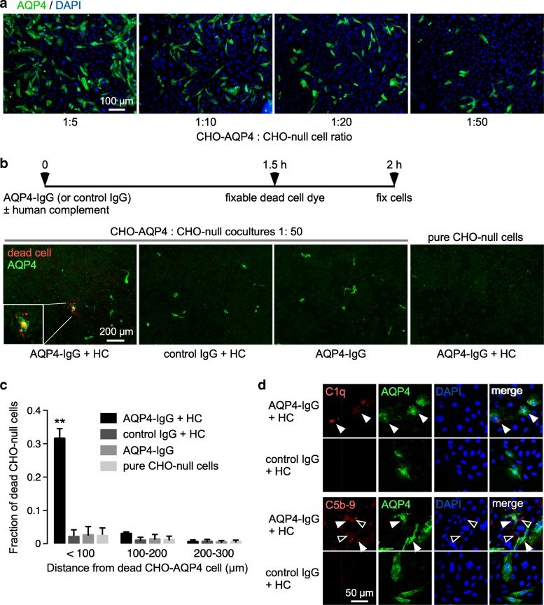
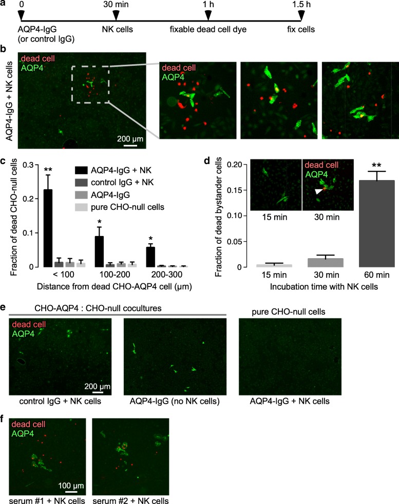
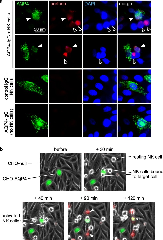
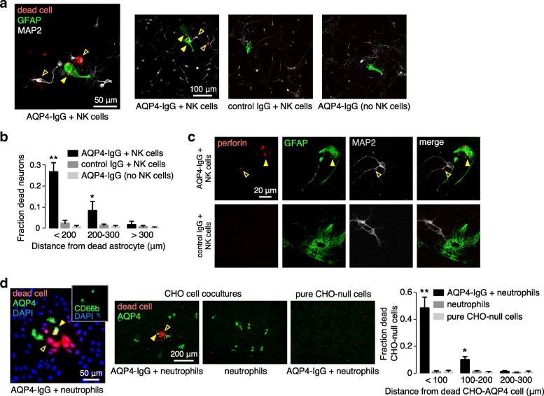
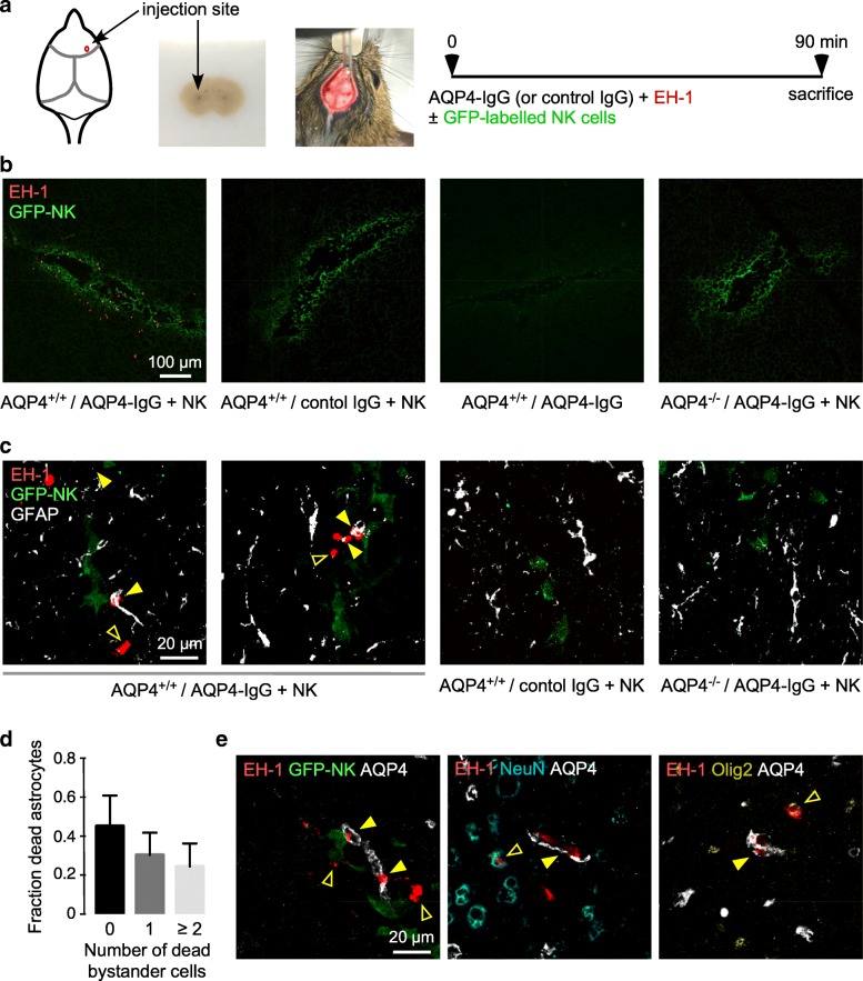
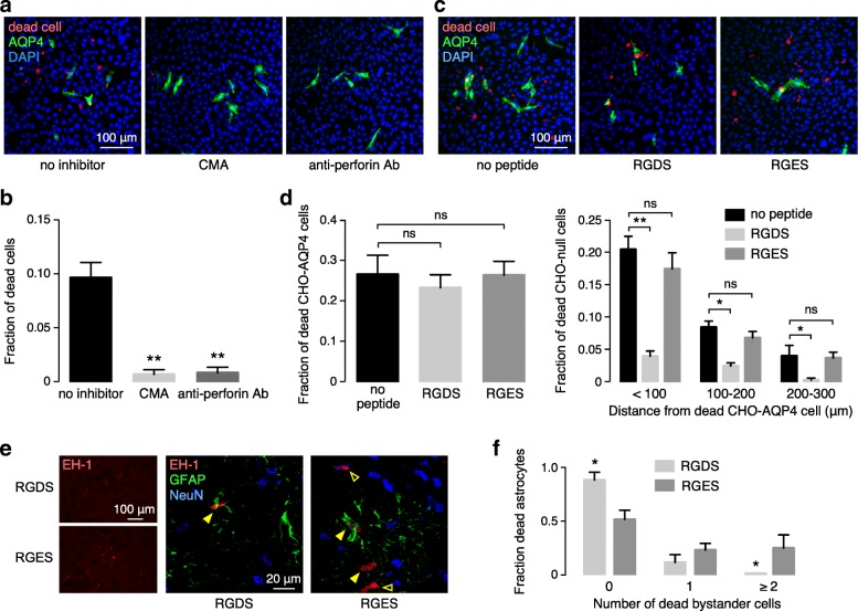
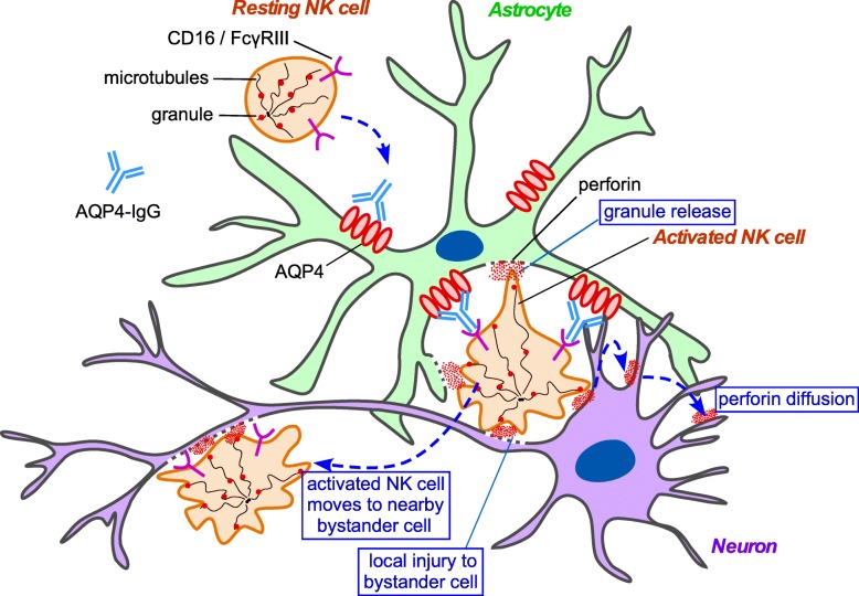
Cellular injury in AQP4-IgG seropositive neuromyelitis spectrum disorder (herein called NMO) involves AQP4-IgG binding to astrocytes, resulting in astrocyte injury by complement-dependent cytotoxicity (CDC) and antibody-dependent cellular cytotoxicity (ADCC) mechanisms. The rapid disease progression, severe tissue damage, and abundant leukocyte infiltration seen in some NMO patients suggest a more direct mechanism for demyelination and neurologic deficit than secondary injury from astrocyte loss. Here, we report evidence for an 'ADCC bystander mechanism' in NMO involving injury to nearby cells by leukocytes following their activation by AQP4-bound AQP4-IgG on astrocytes. In model cocultures containing AQP4-expressing and null CHO cells, AQP4-IgG and complement killed bystander null cells to ~ 100 μm away from AQP4-expressing cells; AQP4-IgG and NK cells produced bystander killing to ~ 300 μm, with perforin deposition seen on injured null cells. Bystander cytotoxicity was also seen with neutrophil-mediated ADCC and in astrocyte-neuron cocultures. Mechanistic studies, including real-time imaging, suggested that leukocytes activated by an AQP4-dependent ADCC mechanism injure bystander cells by direct targeted exocytosis on neighboring cells and not by diffusion of soluble granule contents. In support of this conclusion, ADCC bystander injury was preferentially reduced by an RGDS peptide that inhibits integrin adhesion. Evidence for ADCC bystander injury to oligodendrocytes and neurons was also found in mice following intracerebral injection of AQP4-IgG and NK cells, which was inhibited by RGDS peptide. These results establish a novel cellular pathogenesis mechanism in AQP4-IgG seropositive NMO and provide evidence that inflammatory mechanisms can cause widespread tissue damage in NMO independently of the secondary effects from astrocyte loss.

Keywords: ADCC; Aquaporin-4; Astrocyte; Leukocyte; NMO.


Conflict of interest statement

The authors declare that they have no competing interests.

Figures

Fig. 1
Fig. 1
Complement bystander cytotoxicity in cocultures containing AQP4-expressing and null CHO cells. a AQP4 immunofluorescence (green) with DAPI counterstain (blue) in cocultures of CHO-AQP4 and CHO-null cells plated at different cell ratios. b Complement-dependent cytotoxicity. Cocultures at 1:50 cell ratio was incubated with 10 μg/ml AQP4-IgG (or control IgG) and 5% human complement (HC), with fixable red-fluorescent dead cell marker (left). AQP4 immunofluorescence (green) with dead cell stain (red) for cells incubated with AQP4-IgG and HC, with controls including cells incubated with control IgG and HC, with AQP4-IgG alone, and pure CHO-null cells incubated with AQP4-IgG and HC (right). c Fraction of red-stained dead CHO-null cells as a function of distance from dead CHO-AQP4 cells (mean ± S.E.M., 3 slides with > 50 dead cells analyzed, ** P<0.01 comparing AQP4-IgG + HC vs. control IgG + HC or AQP4-IgG or pure CHO-null cells by two-way ANOVA). d C1q (top) and C5b-9 (bottom) immunofluorescence (red) in cocultures incubated as in (b), costained with AQP4 (green) and DAPI (blue). White filled arrows indicate C5b-9 or C1q on CHO-AQP4 cells, white open arrows show C5b-9 on CHO-null cells
Fig. 2
Fig. 2
ADCC bystander killing in cocultures containing AQP4-expressing and null CHO cells. a Cocultures at 1:50 CHO-AQP4:CHO-null cell ratio were pre-incubated for 30 min with AQP4-IgG (or control IgG) then washed followed by addition of NK cells with a fixable dead cell stain at 30 min prior to fixation. b AQP4 immunofluorescence (green) with dead cell stain (red) at low magnification (left) in cocultures incubated with 5 μg/ml AQP4-IgG and NK cells at an effector:target cell ratio of 5:1. Three fields at high magnification are shown (right). c Fraction of red-stained, dead CHO-null cells as a function of distance from dead CHO-AQP4 cells (mean ± S.E.M., 5 slides with > 80 dead cells analyzed, ** P<0.01, *P<0.05 comparing AQP4-IgG + NK vs. control IgG + NK or AQP4-IgG or pure CHO-null cells by two-way ANOVA). d Fraction of dead bystander cells, as in (c), as a function of incubation time with NK cells (mean ± S.E.M., 5 slides, ** P<0.01 by unpaired t test). Inset shows representative fields at 15 min and 30 min. White filled arrow indicates dead CHO-AQP4 cell at 30 min. e Control studies including CHO-AQP4 and CHO-null cocultures incubated with 5 μg/ml control IgG and NK cells at an effector:target cell ratio of 5:1, with AQP4-IgG alone, and pure CHO-null cells incubated with AQP4-IgG and NK cells. f Cocultures were incubated with NK cells and 1% serum from two seropositive NMO patients, and immunostained as in panel (b)
Fig. 3
Fig. 3
Mechanism of ADCC bystander killing. a Perforin immunofluorescence of CHO-AQP4 and CHO-null cocultures after pre-coating with AQP4-IgG and incubation NK cells, as in Fig. 2b, with controls including control IgG and NK cells, and AQP4-IgG alone. White filled arrows indicate perforin on AQP4-expressing CHO cells, white open arrows show perforin on CHO-null cells. b Time-lapse imaging of coculture of CHO-AQP4 cells (labeled green with cell tracker) and CHO-null cells, pre-incubated for 30 min AQP4-IgG, then at indicated times after NK cell addition (for explanation see text and Additional file 2: Video S2)
Fig. 4
Fig. 4
ADCC bystander killing in astrocyte-neuron cocultures. a Astrocyte-neuron cocultures following 30 min pre-coating with AQP4-IgG and 2 h incubation with NK cells at an effector:target cell ratio of 5:1, with fixable dead cell marker added for the final 30 min. GFAP (astrocyte) and MAP2 (neuron) immunofluorescence, with dead cells stained red, shown at high (left) and low (right) magnifications. Yellow filled arrows indicate dead astrocytes, yellow open arrows show dead neurons. b Fraction of dead neurons at different distances from the center of dead astrocytes (mean ± S.E.M., 3 slides with > 30 dead cells analyzed, ** P<0.01, *P<0.05 comparing AQP4-IgG + NK vs. control IgG + NK or AQP4-IgG without NK cells by two-way ANOVA). c Perforin immunofluorescence (red) of cocultures treated as in a. with GFAP (green) and MAP2 (gray) immunofluorescence. Yellow filled arrows indicate perforin on astrocytes, yellow open arrows show perforin on neurons. d AQP4 immunofluorescence (green) with dead cell stain (red) and DAPI (blue) at high (left) and low (center) magnifications in CHO-AQP4 and CHO-null cocultures following 5 μg/ml AQP4-IgG and neutrophils at an effector:target cell ratio of 5:1. Yellow filled arrows indicate dead CHO-AQP4 cells, yellow open arrows show dead CHO-null cells. (Right) Fraction of dead CHO-null cells at different distances from the center of dead CHO-AQP4 cells (mean ± S.E.M., 3 slides with > 50 dead cells analyzed, ** P<0.01, *P<0.05 comparing AQP4-IgG + neutrophils vs. neutrophils or pure CHO-null cells by two-way ANOVA)
Fig. 5
Fig. 5
ADCC bystander killing in mouse brain. a Mice were administered AQP4-IgG or control IgG (4 μg) and dead cell stain EH-1 (3 μM) with or without GFP-NK cells (104 cells) by intracerebral injection and sacrificed at 90 min. b Low-magnification confocal microscopy showing dead cells (red EH-1 fluorescence) and green immunostained NK cells. c High-magnification confocal images of AQP4+/+ or AQP4-/- mice treated as in a, showing dead cells (red), GFP-NK cells (green), and astrocytes (white). Yellow filled arrows show dead astrocytes, yellow open arrows show dead bystander cells. d Fraction of dead astrocytes associated with 0, 1 or ≥ 2 dead bystander cells (mean ± S.E.M., 5 slides with > 30 dead cells analyzed). e High-magnification confocal images of mice treated as in a, with indicated stain combinations. Yellow filled arrows show dead astrocytes, yellow open arrows show dead bystander cells some of which were NeuN-positive (neurons) or Olig2-positive (oligodendrocytes)
Fig. 6
Fig. 6
Inhibition of ADCC bystander killing by small molecule, antibody and peptide inhibitors. a CMA and anti-perforin antibody. Cocultures of CHO-AQP4 and CHO-null cell were incubated for 30 min with AQP4-IgG then washed and exposed for 1 h to NK cells with or without CMA (10 nM), and with or without added anti-perforin antibody (10 μg/ml), with fixable dead cell stain added 30 min prior to fixation. b Fraction of dead cells in cocultures from studies as in a (mean ± S.E.M., 4 slides with > 60 dead cells analyzed, ** P<0.01 by unpaired t test). c RGDS peptide. Cocultures were pre-incubated with AQP4-IgG then washed and incubated with RGDS peptide (or control RGES peptide) (200 μM) for 1 h, then exposed to NK cells with dead cell stain added 30 min prior to fixation. d (Left) Cultures treated as in c, showing fraction of dead CHO-AQP4 cells in cocultures with no peptide or RGDS or RGES (mean ± S.E.M., 4 slides with > 30 dead cells analyzed). (Right) Fraction of red-stained, dead CHO-null cells as a function of distance from dead CHO-AQP4 cells (mean ± S.E.M., 4 slides with > 60 dead cells analyzed, ** P<0.01, *P<0.05 comparing no-peptide vs. RGDS or RGES by two-way ANOVA). e Mice were injected with RGDS and RGES peptides (on contralateral side), together with AQP4-IgG (4 μg), NK cells (104 cells) and dead cell stain EH-1 (3 μM). (Left) Low magnification showing EH-1 positive cells. (Right) High magnification confocal images showing dead cells (red), astrocytes (green) and neurons (blue). Yellow filled arrows show dead astrocytes, yellow open arrows show dead bystander cells. f Fraction of dead astrocytes associated with 0, 1 or ≥ 2 dead bystander cells in sections of brains from RGDS and RGES treated mice (mean ± S.E.M., 5 slides with > 30 dead cells analyzed)
Fig. 7
Fig. 7
Proposed mechanisms of ADCC bystander killing in AQP4-IgG seropositive NMOSD. Diagram shows AQP4-IgG binding to AQP4 on astrocytes, with binding and activation of NK cells through Fcγ receptors. Activated NK cells can induce bystander cell damage via three mechanisms, including: (i) targeted lytic protein secretion onto immediately adjacent bystander cells following activation; (ii) sequential attack of target cells followed by release and binding/attack of a nearby bystander cell; and (iii) release of soluble granule contents following activation that then diffuse through the extracellular medium and are deposited on nearby cells

Similar articles

Cited by

References

    1. Akiyama H, Kawamata T, Yamada T, Tooyama I, Ishii T, McGeer PL. Expression of intercellular adhesion molecule (ICAM)-1 by a subset of astrocytes in Alzheimer disease and some other degenerative neurological disorders. Acta Neuropathol. 1993;85:628–634. doi: 10.1007/BF00334673. - DOI - PubMed
    1. Ando K, Hiroishi K, Kaneko T, Moriyama T, Muto Y, Kayagaki N, Yagita H, Okumura K, Imawari M. Perforin, Fas/Fas ligand, and TNF-alpha pathways as specific and bystander killing mechanisms of hepatitis C virus-specific human CTL. J Immunol. 1997;158:5283–5291. - PubMed
    1. Anikeeva N, Steblyanko M, Fayngerts S, Kopylova N, Marshall DJ, Powers GD, Sato T, Campbell KS, Sykulev Y. Integrin receptors on tumor cells facilitate NK cell-mediated antibody-dependent cytotoxicity. Eur J Immunol. 2014;44:2331–2339. doi: 10.1002/eji.201344179. - DOI - PMC - PubMed
    1. Asavapanumas N, Ratelade J, Verkman AS. Unique neuromyelitis optica pathology produced in naive rats by intracerebral administration of NMO-IgG. Acta Neuropathol. 2014;127:539–551. doi: 10.1007/s00401-013-1204-8. - DOI - PMC - PubMed
    1. Avadhanula V, Rodriguez CA, Ulett GC, Bakaletz LO, Adderson EE. Nontypeable haemophilus influenzae adheres to intercellular adhesion molecule 1 (ICAM-1) on respiratory epithelial cells and upregulates ICAM-1 expression. Infect Immun. 2006;74:830–838. doi: 10.1128/IAI.74.2.830-838.2006. - DOI - PMC - PubMed

Publication types