Review
. 2019 Jul 16;74(2):238-256.
doi: 10.1016/j.jacc.2019.05.024.

Cardiac MRI Endpoints in Myocardial Infarction Experimental and Clinical Trials: JACC Scientific Expert Panel

Affiliations
Review

Cardiac MRI Endpoints in Myocardial Infarction Experimental and Clinical Trials: JACC Scientific Expert Panel

Borja Ibanez et al. J Am Coll Cardiol. .

Abstract

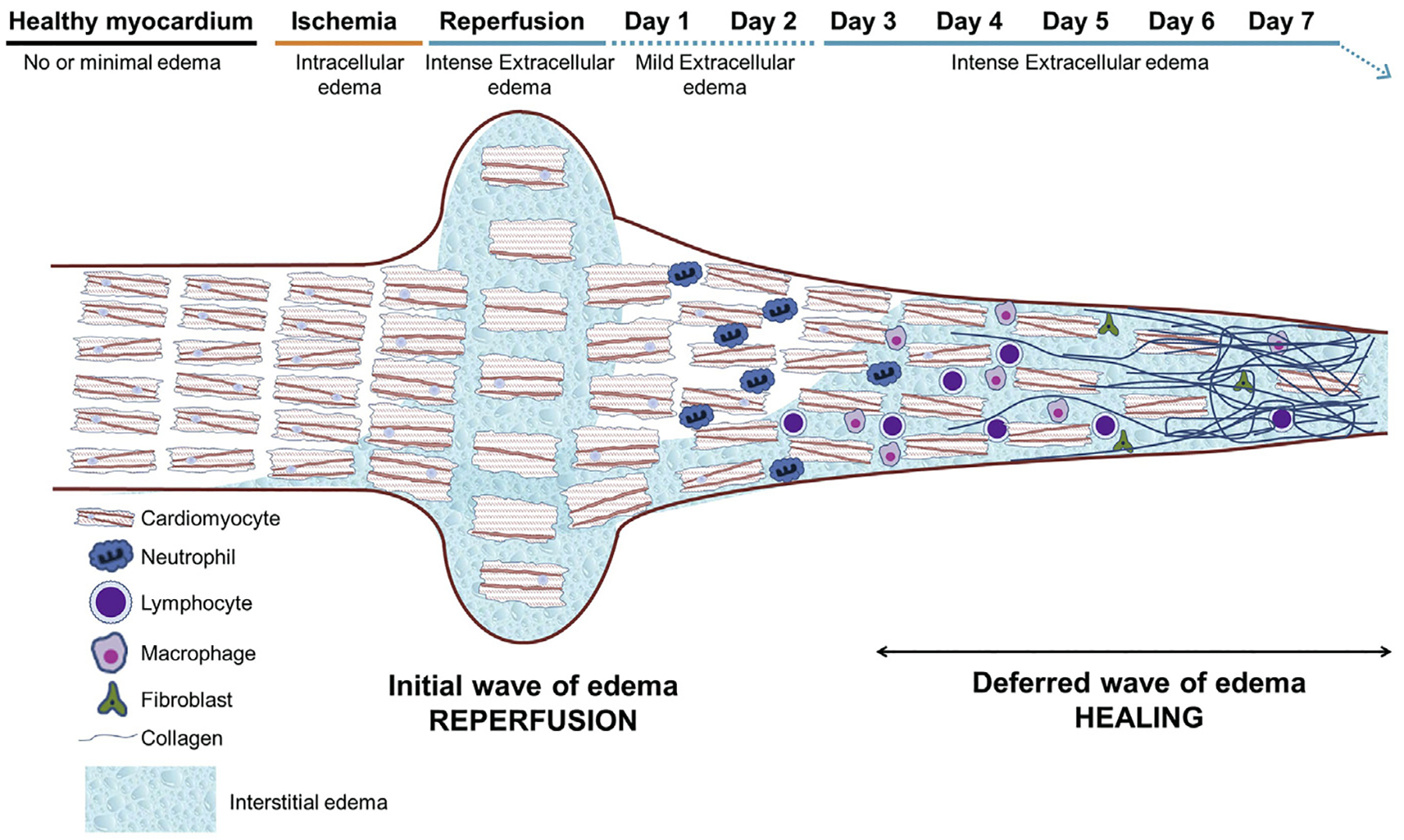
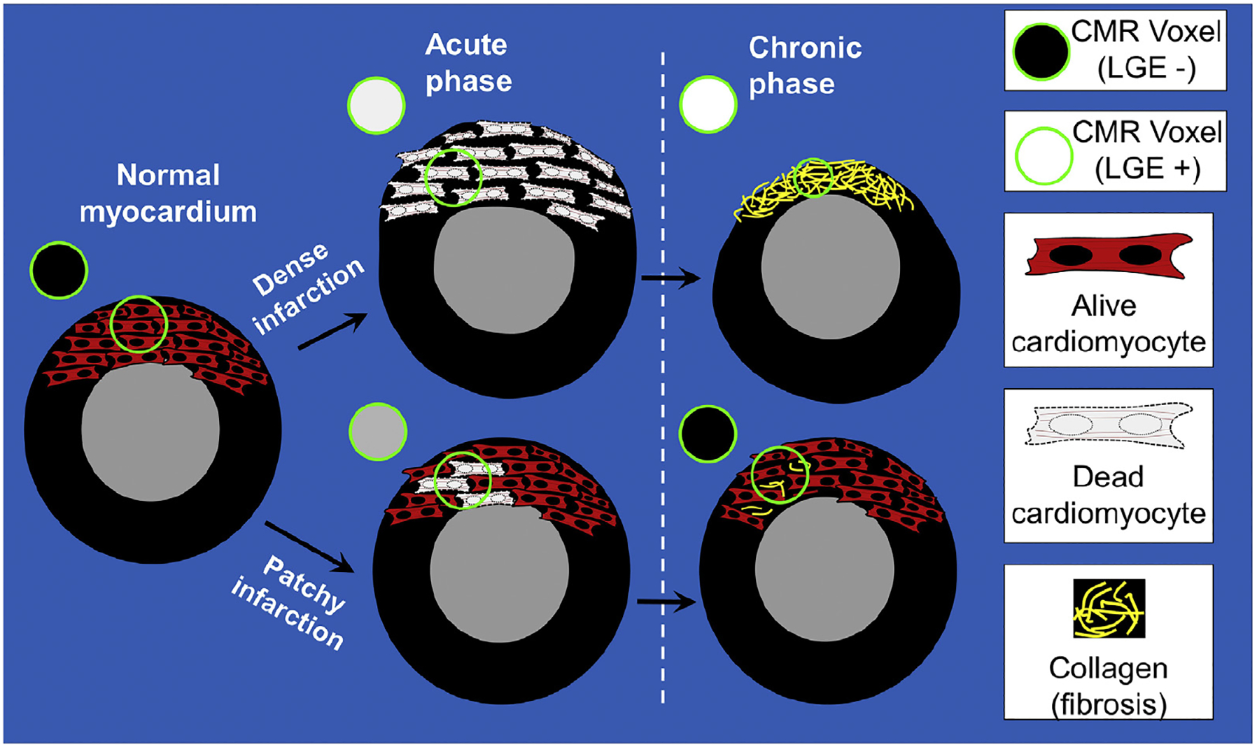
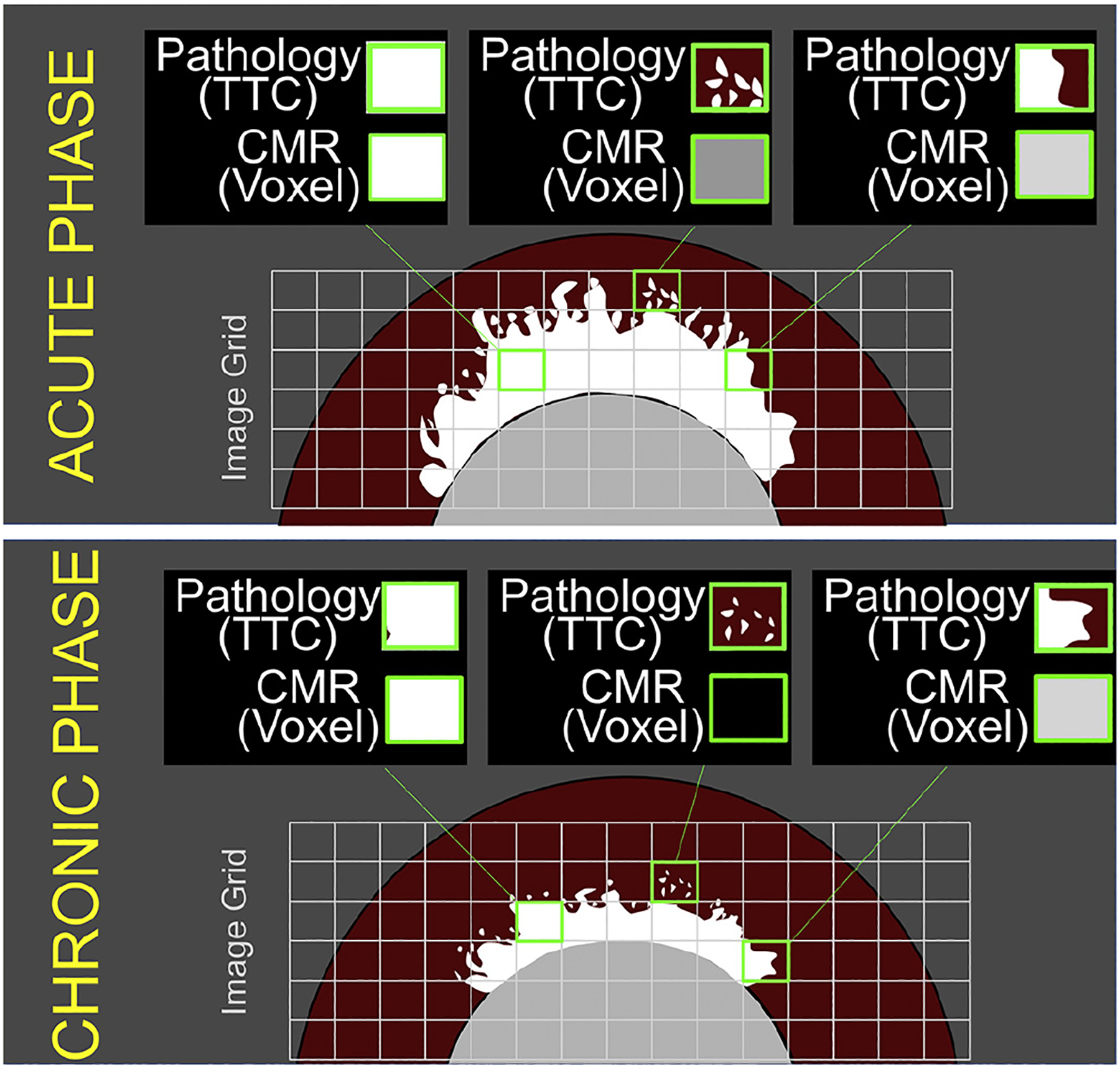
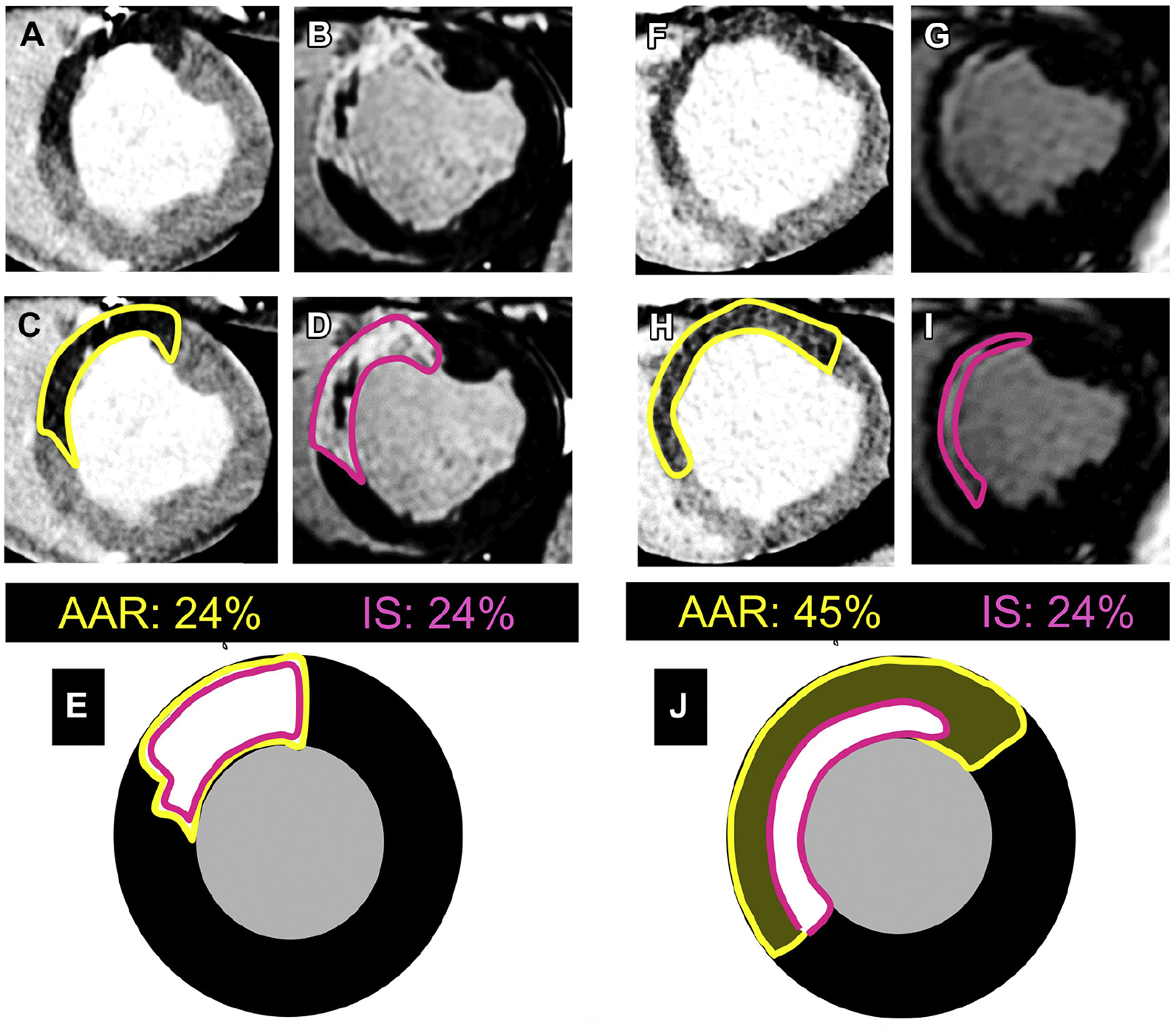
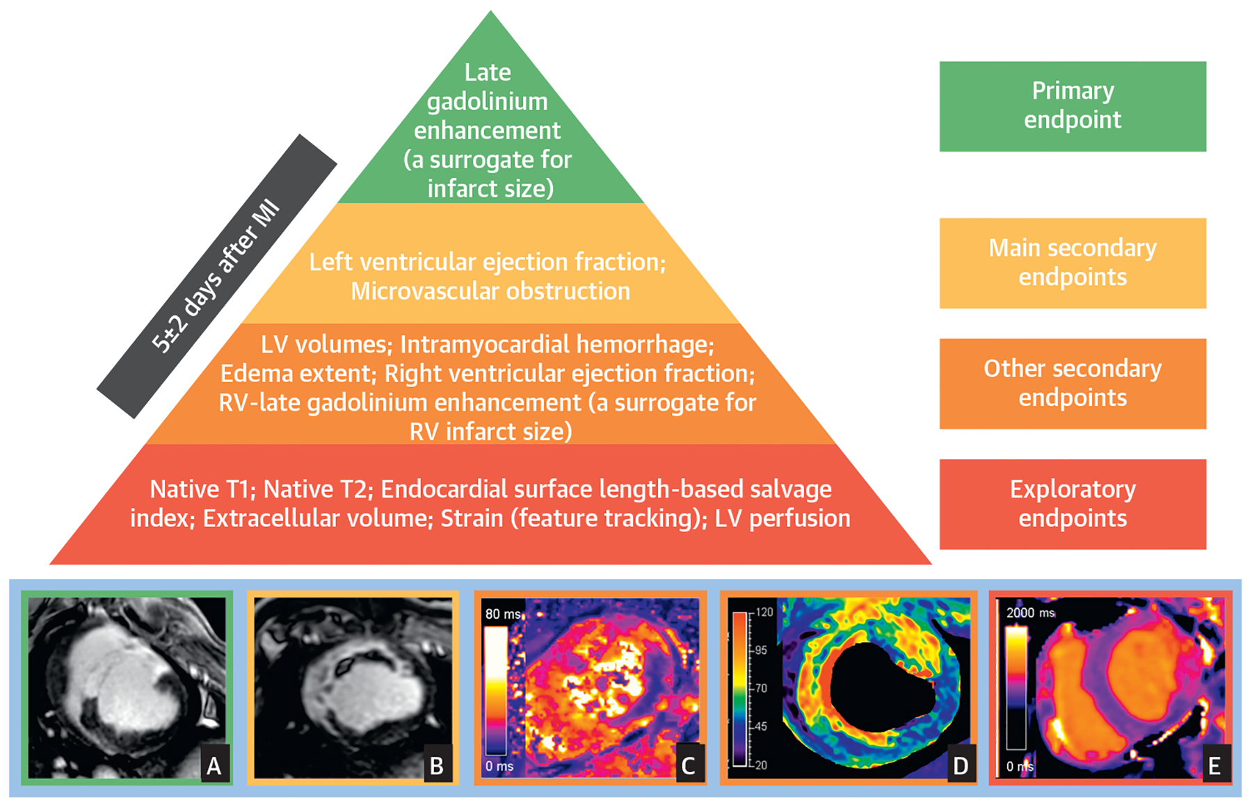
After a reperfused myocardial infarction (MI), dynamic tissue changes occur (edema, inflammation, microvascular obstruction, hemorrhage, cardiomyocyte necrosis, and ultimately replacement by fibrosis). The extension and magnitude of these changes contribute to long-term prognosis after MI. Cardiac magnetic resonance (CMR) is the gold-standard technique for noninvasive myocardial tissue characterization. CMR is also the preferred methodology for the identification of potential benefits associated with new cardioprotective strategies both in experimental and clinical trials. However, there is a wide heterogeneity in CMR methodologies used in experimental and clinical trials, including time of post-MI scan, acquisition protocols, and, more importantly, selection of endpoints. There is a need for standardization of these methodologies to improve the translation into a real clinical benefit. The main objective of this scientific expert panel consensus document is to provide recommendations for CMR endpoint selection in experimental and clinical trials based on pathophysiology and its association with hard outcomes.

Keywords: STEMI; area at risk; clinical trial; edema; endpoint; infarct size; magnetic resonance imaging; myocardial infarction.


Figures

FIGURE 1
FIGURE 1. Dynamic Tissue Composition Changes Occurring After Ischemia/Reperfusion
During ischemia, some degree of intracellular edema initiates. Reperfusion after prolonged ischemia results in a very rapid and intense extracellular edema formation. Cardiac fibers are separated by the extracellular edema and as a result there is a myocardial swelling (increase in myocardial thickness). The reperfusion-related edema is significantly attenuated within 24 h. Neutrophils, macrophages, and other inflammatory cells infiltrate the post-ischemic region, and a progressive replacement of cardiomyocyte debris by collagen and extracellular matrix takes place during the days following reperfusion. The replacement of cardiomyocytes by extracellular matrix results in a significant shrinkage of the myocardial thickness. The healing process results in the so called “deferred wave of edema,” which can last for days or weeks depending on remodeling processes.
FIGURE 2
FIGURE 2. Dynamics of LGE in Dense Versus Patchy Infarctions
In the normal (noninfarcted) myocardium, gadolinium washes out rapidly and there is no hyperenhancement on late gadolinium enhancement (LGE) images (voxel [circle] is black). Severe I/R injury leads to the death of all myocardial cells in the region. Regardless of myocardial swelling in the acute phase or shrinkage in the chronic phase, small regions of tissue in the infarct zone lack any viable cardiomyocytes and representative voxels on LGE (circles) are “bright” both in the acute and chronic phases. Conversely, patchy necrosis (bottom) can lead to LGE in the acute phase. The hyperenhancement in the acute phase is partially due to the edema-related increase in the size of the infarcted portion, occupying a large part of the voxel. In the chronic phase, LGE can disappear. This is partially due to the shrinkage of the patchy dead myocardium, with neighbor alive cardiomyocytes occupying a large part of the voxel. CMR = cardiac magnetic resonance.
FIGURE 3
FIGURE 3. Partial Volume Effect on LGE Images in the Acute and Chronic Phase
The border of the infarcted region can be highly irregular, with apparent islands and peninsulas of necrosis. This can lead to small regions of tissue that are only partially infarcted, but on the scale of a voxel on LGE imaging, are of sufficient extent to render the voxel as “bright.” In the chronic phase, infarct shrinkage will occur both in the infarct core and periphery. Islands of infarction at the border may no longer be of sufficient size to render a voxel of tissue as bright on LGE imaging. TTC = tetrazolium chloride; other abbreviations as in Figure 2.
FIGURE 4
FIGURE 4. Absolute Infarct Size (% LV) Versus Relative IS (% AAR)
Two subjects with the same absolute infarct size may have different amounts of salvage. The illustrated cases correspond to experimental ischemia/reperfusion (I/R) (pig model). (A and F) Arterial enhanced multidetector computed tomography (MDCT) perfusion scans during index coronary occlusion to delineate true area at risk (AAR) (dark areas not perfused). (C and H) The same as A and F, but with the AAR traced. (B and G) LGE CMR 1 week after I/R (in D and I, the infarct size [IS] has been traced). (E and J) Representation of AAR and IS overlaid (yellow corresponds to AAR and pink to IS). In the left case, the entire AAR is transmurally infarcted with no salvage at all. In the case to the right, AAR is much larger, but infarction occupies only the subendocardial area, suggesting large amount of salvaged myocardium. The case to the right corresponds to a subject undergoing a given cardioprotective strategy (IPC in this case). Abbreviations as in Figure 2.
CENTRAL ILLUSTRATION
CENTRAL ILLUSTRATION. Hierarchy of Recommended Cardiac Magnetic Resonance Endpoints in Experimental Studies and Clinical Trials
Recommended CMR endpoints and illustrative images. LGE predicts 1-year death and incident heart failure. The best time point for quantification is 3 to 7 days post-MI. The main criteria for selection of endpoints were associations with MACE, and consistent evidence in multiple studies. Secondary endpoints proposed are those with consistent links to MACE. Other secondary endpoints are those with associations with MACE that are inconsistent among studies and/or do not persist after adjustment for other CMR variables. Exploratory endpoints are those with no or anecdotal evidence of an association with MACE. Representative images for some surrogates are presented at the bottom of the figure (A: infarct size on LGE, B: extensive MVO within LGE area, C: IMH in the lateral wall on T2* mapping, D: intense edema in anteroseptal wall on T2 mapping; E: T1 mapping abnormalities in lateral wall). A, B, and D correspond to pig I/R experiments (images courtesy of Borja Ibanez and Rodrigo Fernández-Jiménez, CNIC), whereas C and E correspond to human post-STEMI cases (clinical images courtesy of Colin Berry and Peter McCartney, University of Glasgow). LV = left ventricle; RV = right ventricle.

References

    1. Ibanez B, Heusch G, Ovize M, Van de Werf F. Evolving therapies for myocardial ischemia/reperfusion injury. J Am Coll Cardiol 2015;65: 1454–71. - PubMed
    1. Puntmann VO, Valbuena S, Hinojar R, et al. Society for Cardiovascular Magnetic Resonance (SCMR) expert consensus for CMR imaging endpoints in clinical research: part I—analytical validation and clinical qualification. J Cardiovasc Magn Reson 2018;20:67. - PMC - PubMed
    1. Messroghli DR, Moon JC, Ferreira VM, et al. Clinical recommendations for cardiovascular magnetic resonance mapping of T1, T2, T2* and extracellular volume: a consensus statement by the Society for Cardiovascular Magnetic Resonance (SCMR) endorsed by the European Association for Cardiovascular Imaging (EACVI). J Cardiovasc Magn Reson 2017;19:75. - PMC - PubMed
    1. Kramer CM, Barkhausen J, Flamm SD, Kim RJ, Nagel E. Standardized cardiovascular magnetic resonance (CMR) protocols 2013 update. J Cardiovasc Magn Reson 2013;15:91. - PMC - PubMed
    1. Schulz-Menger J, Bluemke DA, Bremerich J, et al. Standardized image interpretation and post processing in cardiovascular magnetic resonance: Society for Cardiovascular Magnetic Resonance (SCMR) Board of Trustees Task Force on Standardized Post Processing. J Cardiovasc Magn Reson 2013;15:35. - PMC - PubMed

Publication types