. 2019 Jul 23;116(30):15297-15306.
doi: 10.1073/pnas.1908556116. Epub 2019 Jul 11.

The plastidial pentose phosphate pathway is essential for postglobular embryo development in Arabidopsis

Affiliations

The plastidial pentose phosphate pathway is essential for postglobular embryo development in Arabidopsis

Vasilios M E Andriotis et al. Proc Natl Acad Sci U S A. .

Abstract

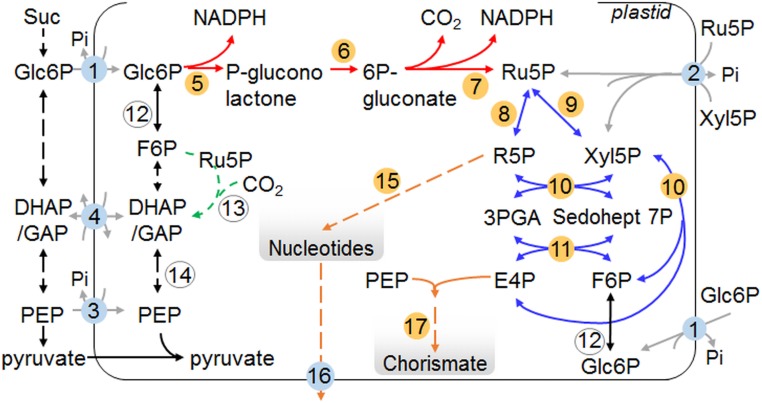
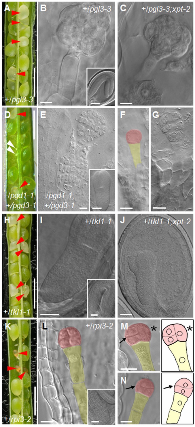
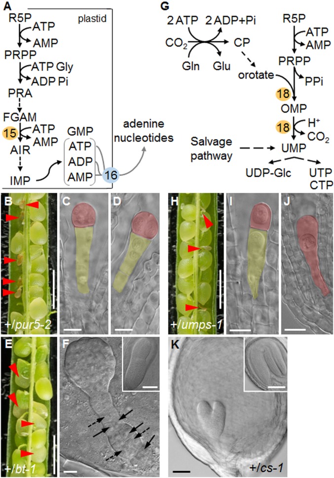
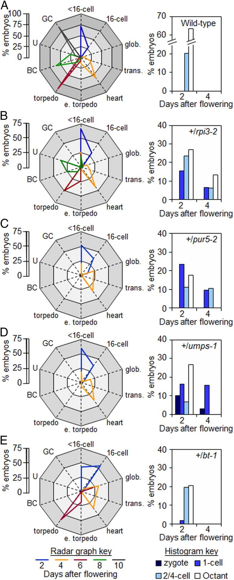
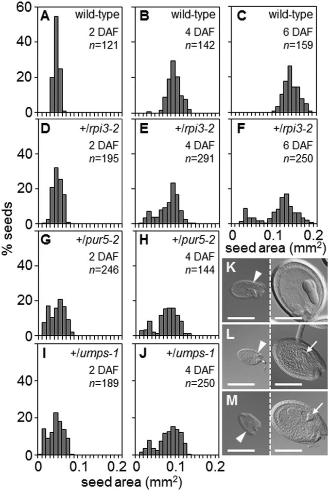
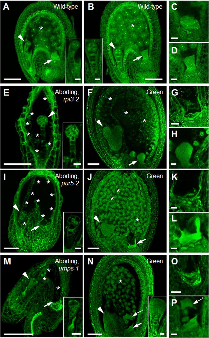
Large numbers of genes essential for embryogenesis in Arabidopsis encode enzymes of plastidial metabolism. Disruption of many of these genes results in embryo arrest at the globular stage of development. However, the cause of lethality is obscure. We examined the role of the plastidial oxidative pentose phosphate pathway (OPPP) in embryo development. In nonphotosynthetic plastids the OPPP produces reductant and metabolic intermediates for central biosynthetic processes. Embryos with defects in various steps in the oxidative part of the OPPP had cell division defects and arrested at the globular stage, revealing an absolute requirement for the production via these steps of ribulose-5-phosphate. In the nonoxidative part of the OPPP, ribulose-5-phosphate is converted to ribose-5-phosphate (R5P)-required for purine nucleotide and histidine synthesis-and subsequently to erythrose-4-phosphate, which is required for synthesis of aromatic amino acids. We show that embryo development through the globular stage specifically requires synthesis of R5P rather than erythrose-4-phosphate. Either a failure to convert ribulose-5-phosphate to R5P or a block in purine nucleotide biosynthesis beyond R5P perturbs normal patterning of the embryo, disrupts endosperm development, and causes early developmental arrest. We suggest that seed abortion in mutants unable to synthesize R5P via the oxidative part of the OPPP stems from a lack of substrate for synthesis of purine nucleotides, and hence nucleic acids. Our results show that the plastidial OPPP is essential for normal developmental progression as well as for growth in the embryo.

Keywords: Arabidopsis; embryo; nucleotide synthesis; pentose phosphate pathway; plastid.


Conflict of interest statement

The authors declare no conflict of interest.

Figures

Fig. 1.
Fig. 1.
Overview of the plastidial OPPP and related pathways. Dashed lines indicate multiple enzymatic steps that for simplicity are omitted. Arrows: red and blue, oxidative and nonoxidative sections of the OPPP, respectively; black, glycolysis and related reactions; green, partial Calvin/Benson cycle: in heterotrophic plastids Rubisco catalyses the conversion of CO2 lost in respiration to 3PGA, which can enter plastidial glycolysis (61); gray, plastid envelope transporters; orange, pathways for nucleotide and chorismate synthesis. Numbers: yellow, enzymes discussed in text; blue, transporters at the plastid envelope. 1: GPT1, Glc6P transporter; 2: XPT, pentose phosphate transporter; 3: PPT, phosphoenolpyruvate transporter; 4: TPT, triose phosphate transporter; 5: G6PDH, Glc6P dehydrogenase; 6: PGL3, 6-phosphogluconolactonase; 7: PGD, 6-phosphogluconate dehydrogenase; 8: RPI, ribose 5P isomerase; 9: RPE3, ribulose 5P epimerase; 10: TKL, transketolase; 11: transaldolase; 12: PGI, phosphoglucose isomerase; 13: Rubisco; 14: phosphoglyceromutase and enolase; 15: PUR5, 5-aminoimidazole ribonucleotide (AIR) synthase; 16: BT1, plastidial adenine nucleotide transporter; 17: CS, chorismate synthase.
Fig. 2.
Fig. 2.
Phenotype of seeds in siliques on heterozygous plants. (A) Silique from a +/pgl3-3 plant. (B) Raspberry-like embryo from a white seed from A (Inset: embryo from a green seed from A) and (C) from a white seed from a silique on a +/pgl3-3;xpt-2 plant. (D) Silique from a −/pgd1-1;+/pgd3-1 plant. (E) Raspberry-like embryo from a white seed in a silique from a −/pgd1-1;+/pgd3-1 plant (Inset: embryo at the torpedo stage from a green seed from D). (F) One-cell stage embryo and (G) octant stage embryo from aborted seeds in a silique from a −/pgd1-1;+/pgd3-1 plant. (H) Silique from a +/tkl1-1 plant. (I) Aborting torpedo-stage embryo from a white seed in H (Inset: embryo from a green seed from H) and (J) from a silique on a +/tkl1-1;xpt-2 plant. (K) Silique from a +/rpi3-2 plant. (L) Octant-stage embryo from an aborting seed in K (Inset: embryo from a green seed from K). (M) Embryo from an aborting seed as in K, in which the first division in the embryo proper was longitudinal (asterisk) and the second transverse (arrow). A drawing is included for clarity. (N) As in M, but for an embryo with transverse rather than longitudinal division planes (arrow). Red arrowheads in A, D, H, and K: abnormal or aborting seeds; open arrowheads in D: unfertilized or aborted ovules. Embryos in B, C, E, G, I, J, and LN were imaged with DIC optics. False color in F and LN highlights the embryo proper (red) and the suspensor (yellow). (Scale bars: A, D, H, and K are 1 mm; E is 7.5 μm; B, C, G, F, and LN are 10 μm; I and J are 25 μm; and Insets in B, I, and L are 50 μm.)
Fig. 3.
Fig. 3.
Nucleotide synthesis and embryo development. (A) Overview of purine biosynthesis. PPRP: 5-phosphoribosyl-1-pyrophosphate; PRA: 5-phosphoribosylamine; FGAM: formylglycinamidine ribotide; AIR: 5-aminoimidazole ribotide. 15: PUR5, AIR synthase. 16: BT1. (B) Silique from a +/pur5-2 plant. (C and D) Embryos from aborting seeds in B at the 1-cell (C) or 2-/4-cell (D) stage. Siblings from green seeds in the same silique were at the upturned-U stage. (E) Silique from a +/bt-1 plant. (F) Raspberry-like embryo from a white seed in E. Arrows and dashed arrows: abnormal longitudinal and oblique cell division planes, respectively, in the suspensor. (Inset) Torpedo-stage embryo from a green seed from E. (G) Overview of pyrimidine synthesis. For simplicity, plastidial, mitochondrial, and cytosolic compartmentation of individual reactions is not shown. CP: carbamyl phosphate, OMP: orotidine 5-monophosphate. 18: UMPSase. (H) Silique from a +/umps-1 plant. (I) Embryo at the 1-cell stage and (J) zygote from aborted seeds in H. Embryos from green seeds were at the torpedo stage. (K) Embryo from a pale green seed in a silique from a +/cs-1 plant (Inset: upturned-U stage embryo from a normal seed). Red arrowheads: abnormal or aborting seeds. Embryos in C, D, F, I, J, and K were imaged with DIC optics. False color highlights the embryo (red) and the suspensor (yellow). (Scale bars: B, E, and H are 1 mm; C, D, F, I, and J are 10 μm; and K and Insets in F and K are 50 μm.)
Fig. 4.
Fig. 4.
Developmental progression of embryos from wild-type and heterozygous plants. (Left) Radar graphs showing the developmental stages of embryos at particular time points (key at the base of the figure). Within each graph the white zone represents 0–25%, the light gray zone 25 to 50%, and the gray zone 50–75% of embryos examined. In A the dark gray zone represents 75 to 100% of embryos examined (for clarity the scale is 0 to 100% in A and 0 to 75% in BE). (Right) Histograms of the distribution of embryos at preglobular stages of development (referred to as <16 cells in the radar graphs) at 2 and 4 DAF. Seeds were from (A) wild-type, (B) +/rpi3-2, (C) +/pur5-2, (D) +/umps-1, (E) +/bt-1 plants. Abbreviations: BC, bent-cotyledon; e. torpedo, early torpedo; GC, green cotyledon; glob., globular; trans., transition; U, upturned-U. See SI Appendix, Table S4 for original data.
Fig. 5.
Fig. 5.
Seed size distribution. Seed areas were measured at 2, 4, and 6 DAF. Numbers (n) of seeds measured for each age and genotype are shown. (AC) Wild-type, (DF) +/rpi3-2, (G and H) +/pur5-2, and (I and J) +/umps-1. (K, L, and M) DIC images of aborting (Left) and normal (Right) seeds at 6 DAF from a silique on (K) +/rpi3-2, (L) +/pur5-2, (M) +/umps-1 plants. The normal seeds in L and M contain a heart-stage embryo (arrows). Arrowheads: micropylar end of the seeds. (Scale bars, 200 μm.)
Fig. 6.
Fig. 6.
Endosperm development in seeds destined for abortion. (A) Wild-type seed with embryo at the 2-/4-cell stage (Inset) and (B) at the 8-cell stage (Inset). (C) Close-up of the chalazal cyst from A and (D) from B. The chalazal cyst is normal. (E) Aborting seed from a +/rpi3-2 plant with embryo arrested at the 8-cell/dermatogen stage (Inset). (F) Green seed from the same silique as in E, with torpedo-stage embryo. The endosperm is cellularized. (G) Close-up of the chalazal-end of an aborting seed from a +/rpi3-2 plant. The chalazal cyst failed to develop. (H) Close-up of the chalazal cyst in F. The chalazal cyst is normal. (I) Aborting seed from a +/pur5-2 plant with embryo arrested at the 2-/4-cell stage (Inset). (J) Green seed from the same silique as in I, with a heart-stage embryo. (K) Close-up of the chalazal end of the seed in I. The chalazal cyst failed to develop. (L) Close-up of the chalazal cyst in J. The chalazal cyst is normal. (M) Aborting seed from a +/umps-1 plant with embryo arrested at the elongated zygote/1-cell stage (Inset). (N) Phenotypically normal seed from the same silique as in M with a late-globular stage embryo. Endosperm development is normal. (O) Close-up of the chalazal end of the seed in M. The chalazal cyst failed to develop. (P) Close-up of the chalazal cyst of the seed in N. The chalazal cyst is normal. Dashed arrows in N and P show part of the chalazal cyst that was detached from its original position during sample preparation. Throughout, arrowheads point to embryos, arrows to the chalazal cyst, asterisks indicate endosperm nuclei. Seeds were Feulgen-stained and imaged under a confocal microscope. Images of whole seeds are single (F) or maximum optical projections of (A) 15, (B) 8, (E) 19, (I) 38, (J) 10, (M) 14, (N) 30 z-slices covering the seed cavity. Images in C, D, G, H, K, L, O, and P are from a single z-slice. (Scale bars: A, B, E, F, I, J, M, and N are 50 μm; and C, D, G, H, K, L, O, and P and Insets in A, B, E, I, M, and N are 10 μm.)

References

    1. Meinke D. W., Embryo-lethal mutants of Arabidopsis thaliana: Analysis of mutants with a wide range of lethal phases. Theor. Appl. Genet. 69, 543–552 (1985). - PubMed
    1. Muralla R., Lloyd J., Meinke D., Molecular foundations of reproductive lethality in Arabidopsis thaliana. PLoS One 6, e28398 (2011). - PMC - PubMed
    1. Tzafrir I., et al. , The Arabidopsis SeedGenes Project. Nucleic Acids Res. 31, 90–93 (2003). - PMC - PubMed
    1. Tzafrir I., et al. , Identification of genes required for embryo development in Arabidopsis. Plant Physiol. 135, 1206–1220 (2004). - PMC - PubMed
    1. Mansfield S. G., Briarty L. G., Early embryogenesis in Arabidopsis thaliana. II. The developing embryo. Can. J. Bot. 69, 461–476 (1991).

Publication types

MeSH terms

LinkOut - more resources