. 2019 Aug 30;294(35):12957-12974.
doi: 10.1074/jbc.RA119.008100. Epub 2019 Jul 11.

Temporal and differential regulation of KAISO-controlled transcription by phosphorylated and acetylated p53 highlights a crucial regulatory role of apoptosis

Affiliations

Temporal and differential regulation of KAISO-controlled transcription by phosphorylated and acetylated p53 highlights a crucial regulatory role of apoptosis

Seo-Hyun Choi et al. J Biol Chem. .

Abstract

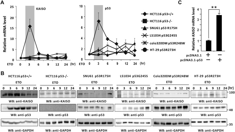
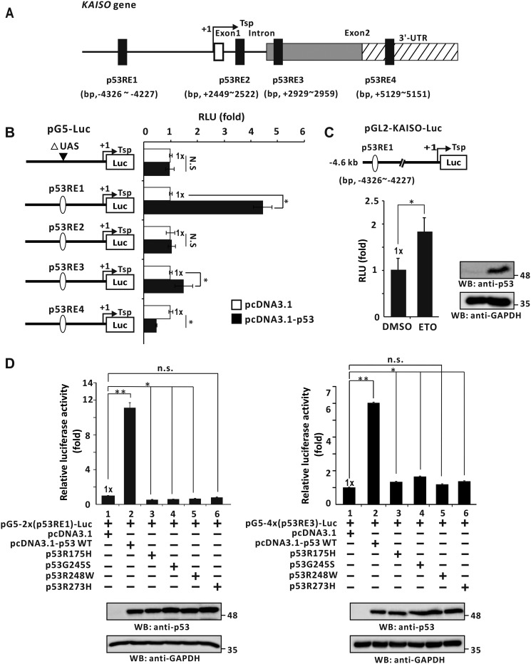
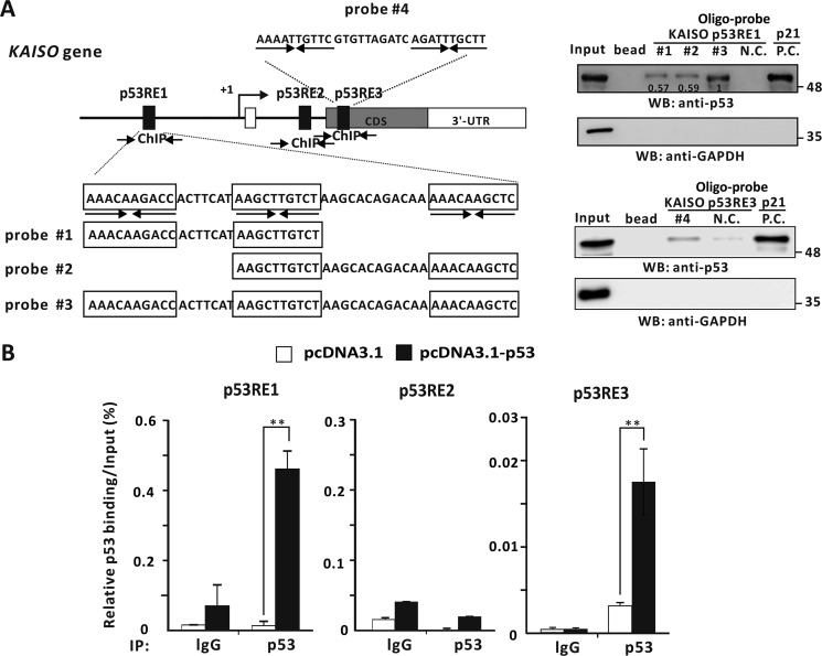
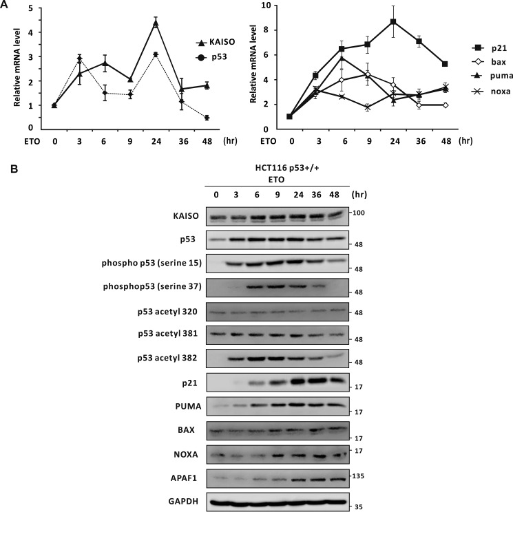
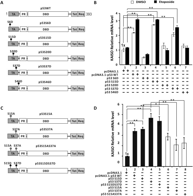
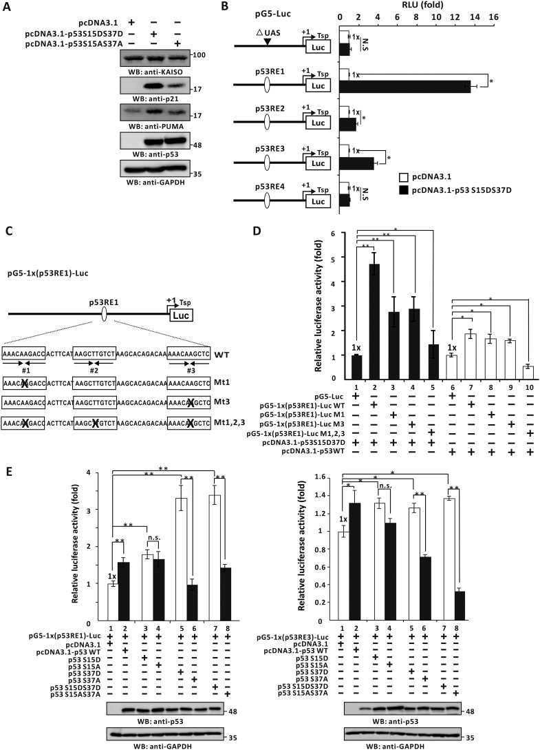
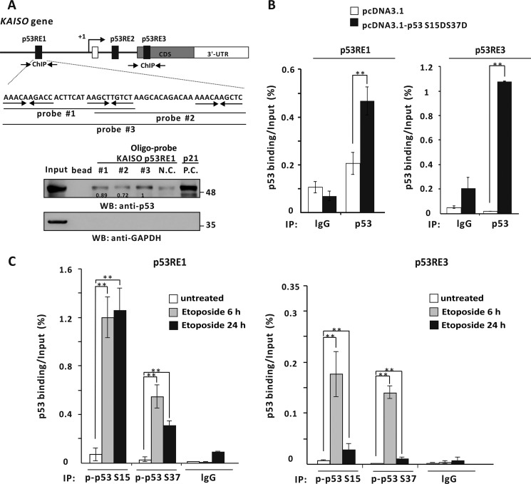
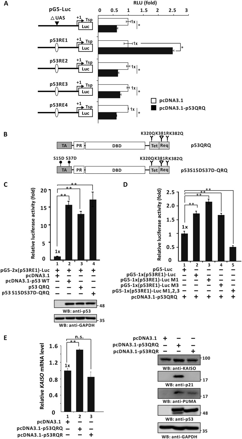
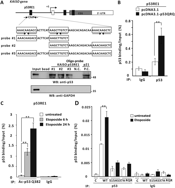
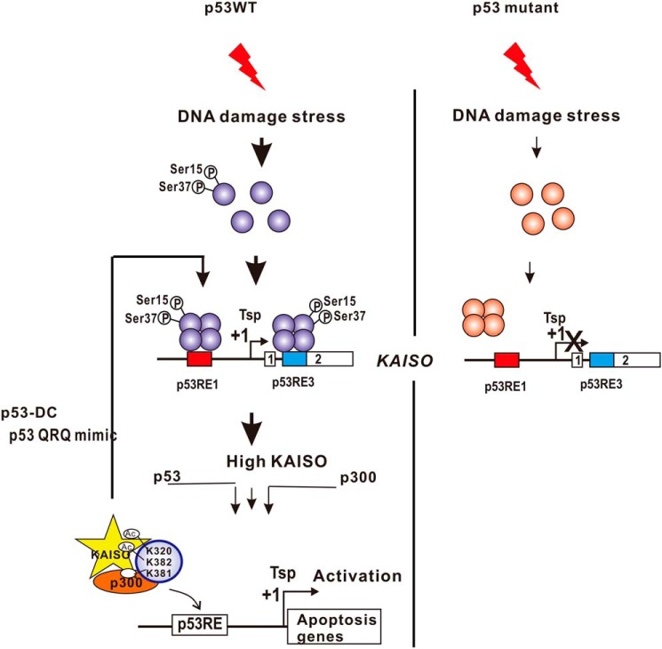
Transcriptional regulator KAISO plays a critical role in cell cycle arrest and apoptosis through modulation of p53 acetylation by histone acetyltransferase p300. KAISO potently stimulates apoptosis in cells expressing WT p53, but not in p53-mutant or p53-null cells. Here, we investigated how KAISO transcription is regulated by p53, finding four potential p53-binding sites (p53-responsive DNA elements; p53REs) located in a distal 5'-upstream regulatory element, intron 1, exon 2 coding sequence, and a 3'-UTR region. Transient transcription assays of pG5-p53RE-Luc constructs with various p53REs revealed that p53 activates KAISO (ZBTB33) transcription by acting on p53RE1 (-4326 to -4227) of the 5'-upstream region and on p53RE3 (+2929 to +2959) of the exon 2 coding region during early DNA damage responses (DDRs). ChIP and oligonucleotide pulldown assays further disclosed that p53 binds to the p53RE1 and p53RE3 sites. Moreover, ataxia telangiectasia mutated (ATM) or ATM-Rad3-related (ATR) kinase-mediated p53 phosphorylation at Ser-15 or Ser-37 residues activated KAISO transcription by binding its p53RE1 or p53RE3 sites during early DDR. p53RE1 uniquely contained three p53-binding half-sites, a structural feature important for transcriptional activation by phosphorylated p53 Ser-15·Ser-37. During the later DDR phase, a KAISO-mediated acetylated p53 form (represented by a p53QRQ acetyl-mimic) robustly activated transcription by acting on p53RE1 in which this structural feature is not significant, but it provided sufficient KAISO levels to confer a p53 "apoptotic code." These results suggest that the critical apoptosis regulator KAISO is a p53 target gene that is differently regulated by phosphorylated p53 or acetylated p53, depending on DDR stage.

Keywords: DNA damage; DNA damage response; DNA damage response (DDR); KAISO; apoptosis; gene transcription; mutant p53; p53; p53QRQ; post-translational modification (PTM); stress response; transcription factor; transcriptional regulation; zinc finger and BTB domain-containing 33 (ZBTB33).


Conflict of interest statement

The authors declare that they have no conflicts of interest with the contents of this article

Figures

Figure 1.
Figure 1.
Induction of KAISO expression by DNA damage is p53-dependent. A and B, RT-qPCR and Western blot analysis (WB) of KAISO and p53 expression. HCT116 p53+/+, HCT116 p53−/−, SNU61, Colo320DM, LS1034, and HT-29 cells were treated with etoposide (60 μm), harvested at the indicated times, and analyzed for mRNA by RT-qPCR and protein expression by Western blot analysis, respectively. GAPDH was used as control. C, RT-qPCR analysis. H1299 p53-null cells were transfected with p53 expression or control vector and analyzed for KAISO mRNA levels by RT-qPCR.
Figure 2.
Figure 2.
p53 activates KAISO gene transcription by acting on the p53RE1 located in the 5′-upstream regulatory region. A, four potential p53REs identified in the KAISO gene: 5′-upstream regulatory region, intron 1, exon 2 coding sequence, and 3′-UTR. B, structure of the four pG5-Luc reporter plasmid constructs with the KAISO p53REs and transient transcription assays. The upstream activation sequence (UAS) (bp +1 to +97) was removed from pG5-Luc reporter plasmid. H1299 p53-null cells were transiently co-transfected with pG5–1x(p53RE)-Luc reporter plasmid and WT p53 expression vector. All assays were performed in triplicate. Error bars, S.D. C, HCT116 p53+/+ cells were transfected with pGL2-KAISO-Luc-4.6 kb containing the KAISO promoter (bp −4441 to +165). After treatment with etoposide for 24 h, luciferase activity was measured and normalized to total cellular protein. Data presented are the average of three independent assays. Error bars, S.D. *, p < 0.05. D, transient transcription analysis of the p53REs of the KAISO gene fusion reporter plasmid constructs. pG5–2x(p53RE1)-Luc or pG5–4x(p53RE3)-Luc and WT p53 expression vector or four hot spot p53 mutant expression vectors were transiently co-transfected in H1299 p53-null cells, and luciferase activities were measured. Luciferase activities were normalized to co-expressed β-gal activity, and data are presented as the average of three independent assays. Error bars, S.D. *, p < 0.05; **, p < 0.01; n.s., not significant. WB, Western blotting.
Figure 3.
Figure 3.
p53 binds to the p53REs of the 5′-upstream regulatory region and coding region of KAISO. A, structure and oligonucleotide pulldown/Western blotting assay of p53REs of the KAISO gene. Endogenous p53REs of KAISO 5′-upstream regulatory region (ChIP#1, bp −4354 to −4254), intron 1 (ChIP#2, bp +2426 to +2556), and exon-coding sequence region (ChIP#3, bp +2885 to +2982) are shown above. Arrows, locations of the ChIP oligonucleotide primer sets spanning the p53-responsive elements (p53REs). Probe #1, #2, #3, and #4 represent the p53REs used for oligonucleotide pulldown assays. Shown is an oligonucleotide pulldown/Western blotting assay of p53 binding to the p53REs of KAISO 5′-upstream regulatory region and exon 2 coding sequence region. H1299 p53-null cell extracts with ectopic p53 were incubated with biotinylated double-stranded oligonucleotides. The mixtures were further incubated with streptavidin-agarose beads and precipitated by centrifugation. The precipitate was analyzed by a Western blotting assay (WB) using antibodies against p53 or GAPDH. B, ChIP assays of p53 binding to a potential p53RE of the endogenous KAISO 5′-upstream regulatory region and coding region. H1299 p53-null cells were transfected with a p53 expression vector and immunoprecipitated (IP) with anti-p53 antibody, followed by PCR amplification of the region flanking p53RE1, p53RE2, and p53RE3. **, p < 0.01. Error bars, S.D.
Figure 4.
Figure 4.
Temporal expression of mRNA and protein of KAISO, p53, p53 phosphorylated at Ser-15 or Ser-37, p53 acetylated at Lys-320, Lys-381, or Lys-382, and a p53 target gene. A and B, temporal expression of mRNA and protein of KAISO, p53, p53 phosphorylated at Ser-15 or Ser-37, or p53 acetylated at Lys-320, Lys-381, or Lys-382, and p53 target genes in HCT116 p53+/+ cells treated with etoposide (ETO). The HCT116 p53+/+ cells treated with etoposide (60 μm) were harvested at the indicated times and analyzed for mRNA by RT-qPCR and protein expression by Western blot analysis, respectively. GAPDH was used as control. Error bars, S.D.
Figure 5.
Figure 5.
Phosphorylated p53 at Ser-15 and/or Ser-37 residues activates KAISO transcription. A, WT p53 and mimics of phosphorylated p53 at serine residues. Filled circles, phosphomimics, p53 serine residue X; TA, transactivation domain (residues 1–42); PR, proline-rich domain (residues 40–92); DBD, DNA-binding domain (residues 101–306); Tet, tetramerization domain (residues 307–355); Reg, regulatory domain (residues 356–393). B, RT-qPCR analysis of KAISO mRNA expression. H1299 p53-null cells were transfected with WT p53 expression or p53 phosphomimic mutant expression vector and analyzed for KAISO mRNA, with or without etoposide (60 μm) treatment for 3 h. C, structures of p53, p53-S15D and/or -S37D, and p53-S15A and/or -S37A. p53-S15A/S37A is a dominant-negative form of p53-S15D/S37D. Filled circles, phosphorylation target p53 serine residue. Filled red circles, nonphosphorylated p53 serine residue. D, RT-qPCR analysis. H1299 p53-null cells were transfected with WT p53, p53 phosphomimic mutant (Ser → Asp), or p53 nonphosphorylatable Ser-15/37 → Ala-15/37 mutant expression vector and analyzed for KAISO mRNA for 3 h. Error bars, S.D.; *, p < 0.05; **, p < 0.01.
Figure 6.
Figure 6.
Identification of p53-response element and transcriptional regulation of KAISO by p53 and a p53-S15D·S37D, a mimic of phosphorylated p53 Ser-15 and Ser-37. A, Western blot analysis (WB) of KAISO, p53 target genes (p21, PUMA), and p53 expression of the cell extracts prepared from the H1299 p53-null cells transfected with control vector, p53-S15D·S37D, or p53-S15A·S37A. N.S., not significant. B, structure of the four pG5-Luc reporter plasmid constructs with the KAISO p53REs and transient transcription assays. H1299 p53-null cells were transiently co-transfected with pG5–1x(p53RE)-Luc reporter plasmid and p53-S15D·S37D expression vector. All assays were performed in triplicate. Error bars, S.D. C and D, structure of the pG5–1x(p53RE1)-Luc reporter plasmid constructs with WT p53RE1 and mutations at p53RE half-sites (Mt1, Mt2, Mt1,2,3) and transient transcription assays. H1299 p53-null cells were transfected with reporter plasmids, WT p53, and a mimic of phosphorylated p53-S15D·S37D expression vector and were analyzed for luciferase activity. Reporter activities were normalized to co-expressed β-gal activity. Data presented are the average of three independent assays. Error bars, S.D. *, p < 0.05; **, p < 0.01. E, pG5-Luc reporter plasmid constructs with p53RE1 or p53RE3 of the KAISO gene and transient transcription assays. H1299 p53-null cells were transfected with reporter plasmids and WT p53 or a phosphomimic mutant p53-S15D and/or S37D or p53 nonphosphorylatable p53-S15A or S37A expression vector and were analyzed for luciferase activity. Reporter activities were normalized to co-expressed β-gal activity. Data presented are the average of three independent assays. Error bars, S.D. *, p < 0.05; **, p < 0.01.
Figure 7.
Figure 7.
The endogenous phosphorylated p53 at Ser-15 or Ser-37 and p53-S15D·S37D, a mimic of phosphorylated p53 Ser-15 and Ser-37, bind to the p53RE1 and -RE3 of the KAISO gene. A, oligonucleotide pulldown/Western blotting assay of p53 binding to the p53RE1 of KAISO. Mimics of phosphorylated p53-S15D·S37D bind to the p53RE1 in vitro. Shown is the structure of p53RE1 and three probes used in oligonucleotide pulldown assays of p53 binding. H1299 p53-null cell extracts with ectopic p53-S15D·S37D expression were incubated with biotinylated double-stranded oligonucleotides, precipitated, and analyzed by a Western blotting assay (WB) using antibodies against p53 or GAPDH. B, ChIP assays of p53-S15D·S37D binding to the p53RE1 and -RE3 of endogenous KAISO. H1299 p53-null cells were transfected with a p53-S15D·S37D expression vector and immunoprecipitated (IP) with an anti-p53 antibody, followed by PCR amplification of the region flanking p53RE1 and -RE3. Shown is the average of three independent assays. Error bars, S.D. **, p < 0.05. C, ChIP assays of endogenously phosphorylated p53 Ser-15 or Ser-37 binding to p53RE1 and -RE3 of the endogenous KAISO gene. HCT116 p53+/+ cells treated with etoposide (60 μm) were harvested at 6 or 24 h, immunoprecipitated with a specific anti-phosphorylated p53 Ser-15 or Ser-37 antibody, and amplified by PCR of the region flanking p53RE1 and -RE3. Shown is the average of three independent assays. Error bars, S.D. **, p < 0.05.
Figure 8.
Figure 8.
p53QRQ, a mimic of p53 with KAISO-medicated apoptotic code (acetylation at 320 and 382, no acetylation at p53 Lys-381) activates the transcription of a KAISO reporter via the p53RE1 of the 5′-upstream regulatory region of KAISO gene. A, structure of the four pG5-Luc reporter plasmid constructs with the p53REs of the KAISO gene and transient transcription assays. Identification of the p53RE critical for transcriptional activation of KAISO by p53QRQ. H1299 p53-null cells transfected with pG5-Luc reporter plasmid with p53RE and p53QRQ expression vectors were analyzed for luciferase activity, normalized to co-expressed β-gal activity. Data presented are the average of three independent assays. Error bars, S.D. *, p < 0.05; **, p < 0.01. B and C, structures of p53QRQ and p53-S15D·S37D-QRQ, and transient transcription assays. Shown below are the structures of p53QRQ and p53-S15D·S37D-QRQ. Transient transcription analysis of the p53REs of KAISO gene fusion reporter plasmid constructs is shown. pG5–2x(p53RE1)-Luc and WT p53 or p53QRQ or p53-S15D·S37D-QRQ expression vectors were transiently co-transfected in H1299 p53-null cells, and luciferase activities were measured. Luciferase activities were normalized to co-expressed β-gal activity, and data presented are the average of three independent assays. D, transient transcription assays. Mutations were introduced to test the function of each p53-binding half-sites in transcriptional activation of the reporter gene by p53QRQ. H1299 p53-null cells were transiently co-transfected with pG5–1x(p53RE1)–Luc reporter plasmid and p53QRQ expression vector. All assays were performed in triplicate. Error bars, S.D. E, RT-qPCR analysis and Western blot analysis (WB). H1299 p53-null cells were transfected with control vector, p53QRQ, or p53RQR expression and analyzed for KAISO, p53 target genes (p21, PUMA), and p53 expression. Data presented are the average of three independent assays. Error bars, S.D.; *, p < 0.05; **, p < 0.01; n.s., not significant.
Figure 9.
Figure 9.
The mimic of a KAISO-mediated acetylated p53, p53QRQ, binds to the p53RE1 of the 5′-upstream regulatory region of KAISO. A, structure and oligonucleotide pulldown/Western blotting assay (WB) of p53RE1 of the KAISO gene. p53RE1 contains three p53-binding half-sites. The locations of oligonucleotide primer–binding sites of the ChIP assay of p53QRQ binding are indicated by arrows. Probes #1 to #3 used in oligonucleotide pulldown assays are indicated below. Oligonucleotide pulldown and Western blotting assays of p53QRQ binding to p53RE1 probes are shown. H1299 p53-null cells extracts with ectopic p53QRQ were incubated with biotinylated double-stranded oligonucleotides. The mixtures were further incubated with streptavidin-agarose beads and precipitated by centrifugation. The precipitate was analyzed by a Western blotting assay using antibodies against p53 (DO-1) or GAPDH. p53QRQ bound similarly to all three probes. 3′-UTR was used as negative control. p53RE1 of the p21/CDKN1A gene was positive control. B, ChIP assay of p53QRQ binding to the p53RE1 of the endogenous KAISO gene in the cells transfected with pcDNA3.1-p53QRQ. IP, immunoprecipitation. C, ChIP assays of endogenously acetylated p53-K382 binding to p53RE1 of endogenous KAISO. HCT116 p53+/+ cells treated with etoposide (60 μm) were harvested at 6 or 24 h, immunoprecipitated with a specific anti-acetylated p53 Lys-382 antibody, and amplified by PCR of the region flanking p53RE1. Shown is the average of three independent assays. Error bars, S.D. **, p < 0.05. D, PTMs of p53, including phosphorylation of Ser-15 and Ser-37 and acetylation of Lys-382, PTMs critical for binding the KAISO p53RE1. Dominant-negative forms of phosphorylated p53 at Ser-15 and/or Ser-37 (p53-S15A·S37A) or the KAISO-mediated acetylated form (p53RQR) were expressed in H1299 cells treated with etoposide (60 μm) and analyzed for p53 binding. The mutations strongly attenuated p53 binding.
Figure 10.
Figure 10.
Hypothetical model of KAISO gene transcriptional activation by p53 during DNA damage responses. In cells undergoing DNA damage responses (e.g. by treatment with etoposide, cisplatin, etc.), KAISO is transcribed as an early DNA damage response gene, activated by p53, and robustly activated again at later periods of the DNA damage response, which is critical for apoptosis. KAISO gene and post-translational modification of p53, either by phosphorylation or acetylation, and KAISO promoter structure (particularly p53RE1 and p53RE3) play important roles. DNA-damaging stress induces phosphorylation of Ser-15 and/or Ser-37 of p53 by ATM or ATR. This particular form of p53 binds the two elements but preferentially to the p53RE1, with three unique p53-binding half-sites. The initial pulse of KAISO expression is critical in the generation of a KAISO-mediated acetylated form of p53 (Ac-K320, deacetylated Lys-381, Ac-382; mimicked by p53QRQ). In turn, acetylated p53 (called the p53 “death code,” p53DC) robustly activates KAISO gene transcription by acting on p53RE1, thus generating sufficient KAISO and thereby apoptotic p53 required for the induction of apoptotic gene expression.

References

    1. Daniel J. M., and Reynolds A. B. (1999) The catenin p120(ctn) interacts with Kaiso, a novel BTB/POZ domain zinc finger transcription factor. Mol. Cell. Biol. 19, 3614–3623 10.1128/MCB.19.5.3614 - DOI - PMC - PubMed
    1. Daniel J. M., Spring C. M., Crawford H. C., Reynolds A. B., and Baig A. (2002) The p120(ctn)-binding partner Kaiso is a bi-modal DNA-binding protein that recognizes both a sequence-specific consensus and methylated CpG dinucleotides. Nucleic Acids Res. 30, 2911–2919 10.1093/nar/gkf398 - DOI - PMC - PubMed
    1. Lopes E. C., Valls E., Figueroa M. E., Mazur A., Meng F. G., Chiosis G., Laird P. W., Schreiber-Agus N., Greally J. M., Prokhortchouk E., and Melnick A. (2008) Kaiso contributes to DNA methylation-dependent silencing of tumor suppressor genes in colon cancer cell lines. Cancer Res. 68, 7258–7263 10.1158/0008-5472.CAN-08-0344 - DOI - PubMed
    1. Park J. I., Kim S. W., Lyons J. P., Ji H., Nguyen T. T., Cho K., Barton M. C., Deroo T., Vleminckx K., Moon R. T., and McCrea P. D. (2005) Kaiso/p120-catenin and TCF/β-catenin complexes coordinately regulate canonical Wnt gene targets. Dev. Cell 8, 843–854 10.1016/j.devcel.2005.04.010 - DOI - PubMed
    1. Yoon H. G., Chan D. W., Reynolds A. B., Qin J., and Wong J. (2003) N-CoR mediates DNA methylation-dependent repression through a methyl CpG binding protein Kaiso. Mol. Cell 12, 723–734 10.1016/j.molcel.2003.08.008 - DOI - PubMed

Publication types

Substances

LinkOut - more resources