. 2019 Jul 11;11(13):4561-4578.
doi: 10.18632/aging.102069.

Enhanced beta-1 adrenergic receptor responsiveness in coronary arterioles following intravenous stromal vascular fraction therapy in aged rats

Affiliations

Enhanced beta-1 adrenergic receptor responsiveness in coronary arterioles following intravenous stromal vascular fraction therapy in aged rats

Gabrielle Rowe et al. Aging (Albany NY). .

Abstract

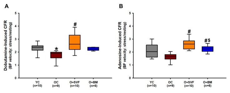
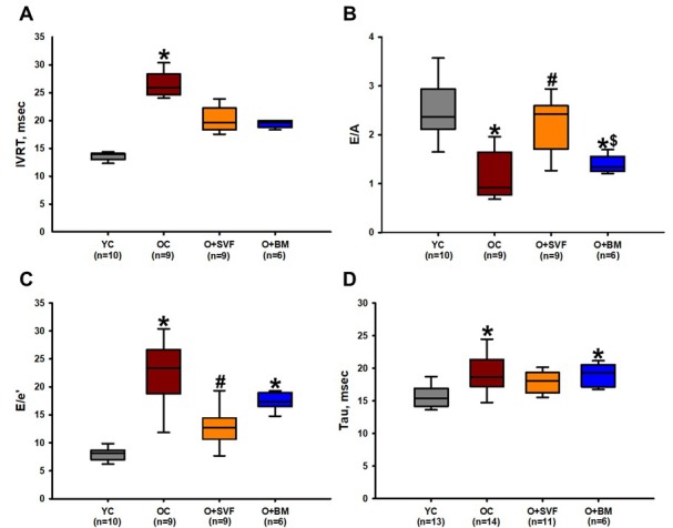
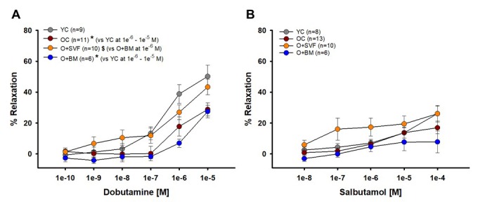
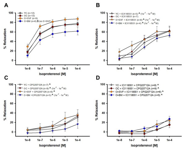
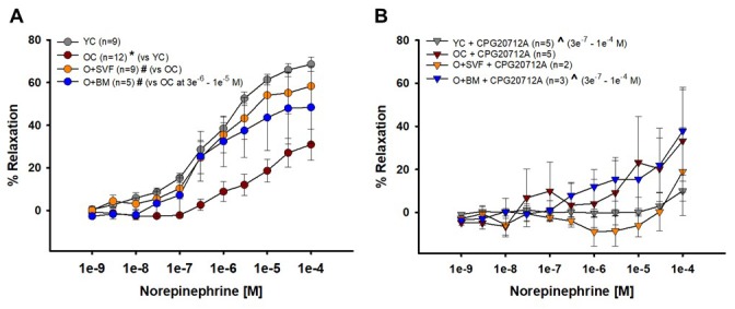
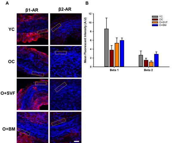
Our past study showed that a single tail vein injection of adipose-derived stromal vascular fraction (SVF) into old rats was associated with improved dobutamine-mediated coronary flow reserve. We hypothesize that i.v. injection of SVF improves coronary microvascular function in aged rats via alterations in beta adrenergic microvascular signaling. Female Fischer-344 rats aged young (3 months, n=32) and old (24 months, n=30) were utilized, along with two cell therapies intravenously injected in old rats four weeks prior to sacrifice: 1x10⁷ green fluorescent protein (GFP+) SVF cells (O+SVF, n=21), and 5x10⁶ GFP+ bone-marrow mesenchymal stromal cells (O+BM, n=6), both harvested from young donors. Cardiac ultrasound and pressure-volume measurements were obtained, and coronary arterioles were isolated from each group for microvessel reactivity studies and immunofluorescence staining. Coronary flow reserve decreased with advancing age, but this effect was rescued by the SVF treatment in the O+SVF group. Echocardiography showed an age-related diastolic dysfunction that was improved with SVF to a greater extent than with BM treatment. Coronary arterioles isolated from SVF-treated rats showed amelioration of the age-related decrease in vasodilation to a non-selective β-AR agonist. I.v. injected SVF cells improved β-adrenergic receptor-dependent coronary flow and microvascular function in a model of advanced age.

Keywords: adrenergic; aging; cell therapy; coronary; vasodilation.


Conflict of interest statement

CONFLICTS OF INTEREST: None to disclose.

Figures

Figure 1
Figure 1
Coronary flow reserve using Doppler echocardiography in rats. Stress test was performed on experimental groups using dobutamine (A) or adenosine (B) and CFR was calculated. O+SVF group exhibited increased CFR vs. OC in both adenosine and dobutamine conditions. P≤0.05 vs Young Control (*), vs Old Control (#), and vs Old+ SVF($); Data are presented as means±SD, analyzed with one-way ANOVA followed by post-hoc Dunn’s (A) or Holm-Sidak (B) test.
Figure 2
Figure 2
Diastolic function assessment using echocardiography and pressure- volume loop (PV-loop). Compared to YC rats, there was an age-related deterioration in diastolic function as measured echocardiographically by IVRT (A), E/A ratio (B), E/e’ ratio (C), and hemodynamically by Tau (D). Old rats treated with SVF significantly reversed this dysfunction in measures of E/A ratio (B) and E/e’ ratio (C) compared to OC, and normalized diastolic function to YC levels in IVRT (A) and Tau (D). P≤0.05 vs Young Control (*), vs Old Control (#), and vs Old+SVF ($); Data are presented as means±SD, analyzed with one-way ANOVA followed by post-hoc Holm-Sidak (B, D) or Dunn’s (A, C) test.
Figure 3
Figure 3
Isolated coronary arteriolar vasoreactivity to β1- and β2-AR agonists. Dobutamine, primarily a β1-AR agonist, induced vasorelaxation in all groups (A). Coronary arterioles from YC animals exhibited a significantly greater dilation compared to OC and O+BM at concentrations 1e-6 and 1e-5 [M] (*) (A). Salbutamol, a β2-AR agonist, induced mild vasorelaxation that was similar between all the groups (B). P≤0.05 vs Young Control (*), vs Old Control (#), and vs Old+SVF ($); data are presented as means±SEM and analyzed with two-way repeated measures ANOVA followed by post-hoc Bonferroni test.
Figure 4
Figure 4
Contribution of β1- and β2-AR to isoproterenol-induced vasodilation from isolated coronary arterioles. Vasorelaxation to isoproterenol, primarily a non-selective β1-, β2-, and β3-AR agonist, was significantly impaired in the O+BM group compared to O+SVF ($) (A). Isoproterenol with ICI118551, a β2-AR antagonist, eliminated differences between the groups. Compared to pre-incubation, all groups except OC (YC, O+SVF, and O+BM) had significant attenuation in the inhibited dose response (^) at several concentrations (B). Isoproterenol with CPG20712A, a β1-AR antagonist, also eliminated differences between the groups, and all groups exhibited significant attenuation in the response compared to pre-inhibition (^) (C). No group differences to isoproterenol were noted following inhibition with both ICI118551 and CPG20712A, and all groups exhibited significant attenuation in the response compared to pre-inhibition (^) at every concentration (D). P ≤ 0.05 vs Young Control (*), vs Old Control (#), vs Old+SVF, and pre- vs post-inhibition (^); data are presented as means±SEM and analyzed with two-way repeated measures ANOVA, paired for inhibitor analysis, followed by post-hoc Bonferroni test.
Figure 5
Figure 5
Contribution of β2-AR to norepinephrine-induced vasoreactivity in isolated coronary arterioles. Relaxation to NE was significantly impaired in OC animals compared to YC and O+SVF groups at all concentrations (A). NE with CPG20712A, a β1-AR antagonist, attenuated (^) the majority of the vasodilation response (3e-7 – 1e-4 [M]) in YC and O+BM treated groups (B). P≤0.05 vs Young Control (*), vs Old Control (#), vs Old+SVF ($), and pre- vs post-inhibition (^); data are presented as means±SEM and analyzed with two-way repeated measures ANOVA, paired for inhibitor analysis, followed by post-hoc Bonferroni test.
Figure 6
Figure 6
β1- and β2-AR immunofluorescence in isolated coronary arterioles. Representative images with ROI boxes used for fluorescent intensity analysis are shown on isolated coronary arterioles stained for β1- or β2-AR (A). There is no significant difference between the groups in the expression of β1- or β2-AR as measured by quantification of fluorescent intensity (B). Data are represented as means+SEM and analyzed with one-way ANOVA (n≥4). Scale bar is 50 micrometers.

References

    1. Hachamovitch R, Wicker P, Capasso JM, Anversa P. Alterations of coronary blood flow and reserve with aging in Fischer 344 rats. Am J Physiol. 1989; 256:H66–73. - PubMed
    1. Mendzef SD, Slovinski JR. Neurohormones and heart failure. Nurs Clin North Am. 2004; 39:845–61. 10.1016/j.cnur.2004.07.004 - DOI - PubMed
    1. Ferrara N, Komici K, Corbi G, Pagano G, Furgi G, Rengo C, Femminella GD, Leosco D, Bonaduce D. β-adrenergic receptor responsiveness in aging heart and clinical implications. Front Physiol. 2014; 4:396. 10.3389/fphys.2013.00396 - DOI - PMC - PubMed
    1. Davies CH, Ferrara N, Harding SE. Beta-adrenoceptor function changes with age of subject in myocytes from non-failing human ventricle. Cardiovasc Res. 1996; 31:152–56. - PubMed
    1. Pan HY, Hoffman BB, Pershe RA, Blaschke TF. Decline in beta adrenergic receptor-mediated vascular relaxation with aging in man. J Pharmacol Exp Ther. 1986; 239:802–07. - PubMed

Publication types