. 2019 Jul 11;10(1):3067.
doi: 10.1038/s41467-019-10932-4.

Zinc-binding to the cytoplasmic PAS domain regulates the essential WalK histidine kinase of Staphylococcus aureus

Affiliations

Zinc-binding to the cytoplasmic PAS domain regulates the essential WalK histidine kinase of Staphylococcus aureus

Ian R Monk et al. Nat Commun. .

Abstract

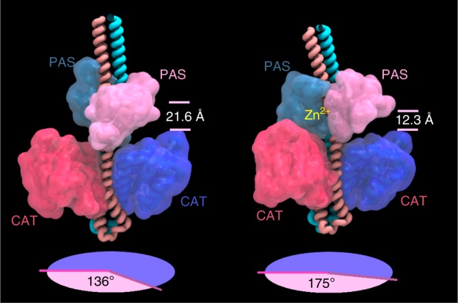
WalKR (YycFG) is the only essential two-component regulator in the human pathogen Staphylococcus aureus. WalKR regulates peptidoglycan synthesis, but this function alone does not explain its essentiality. Here, to further understand WalKR function, we investigate a suppressor mutant that arose when WalKR activity was impaired; a histidine to tyrosine substitution (H271Y) in the cytoplasmic Per-Arnt-Sim (PASCYT) domain of the histidine kinase WalK. Introducing the WalKH271Y mutation into wild-type S. aureus activates the WalKR regulon. Structural analyses of the WalK PASCYT domain reveal a metal-binding site, in which a zinc ion (Zn2+) is tetrahedrally-coordinated by four amino acids including H271. The WalKH271Y mutation abrogates metal binding, increasing WalK kinase activity and WalR phosphorylation. Thus, Zn2+-binding negatively regulates WalKR. Promoter-reporter experiments using S. aureus confirm Zn2+ sensing by this system. Identification of a metal ligand recognized by the WalKR system broadens our understanding of this critical S. aureus regulon.


Conflict of interest statement

The authors declare no competing interests.

Figures

Fig. 1
Fig. 1
Colony sectoring in a ∆yycHI leads to mutation in WalK. a Molecular model of the essential two-component histidine kinase WalK (dimer 608 residues) from S. aureus in a phospholipid bilayer. The amino acid boundaries of the various WalK domains (PASEC: extracellular PAS, HAMP: present in Histidine kinases, Adenylate cyclases, Methyl accepting proteins and Phosphatases, PASCYT: cytoplasmic PAS, DHp: dimerisation and histidine phosphorylation, CAT: catalytic/ATP binding) are highlighted. b Plating of NRS384∆yycHI onto Brain Heart Infusion agar after 48 h at 37 °C promotes colony sectoring (arrow heads). Red box inset shows enlarged view of one sectored colony. c The WalKH271Y mutation identified from whole-genome sequencing of a single sectored colony was introduced by allelic exchange into the NRS384 background, with the mutation increasing haemolysis on sheep blood agar. Also shown are the wild-type (NRS384), ∆yycHI, ∆yycHIWalKH271Y and the WalKH271Y-COMP, for reference
Fig. 2
Fig. 2
Phenotypic impact of WalKH271Y mutation on S. aureus. a Growth kinetics of the WalKH271Y mutant compared to the wild-type (WT) and complemented strains. Overnight cultures were diluted 1:100 into fresh and grown at 37 °C (200 rpm). The cultures were sampled at the indicated time points for OD600 and colony-forming unit (CFU) readings. The WalKH271Y strain exhibited altered growth kinetics compared to the WT and complemented strains, with the loss of a lag phase and a subsequent reduction in doubling time during exponential growth. Error bars indicate standard deviation (s.d.) from three biological replicates. b Analysis of the total exoproteome and Atl secretion. Supernatant proteins from exponential or stationary phase cultures were run on 10% sodium dodecyl sulfate-polyacrylamide gel electrophoresis gels for SYPRO Ruby staining or c zymogram analysis through the incorporation of 1.5% Micrococcus luteus cells. The lane contents are: (1) NRS384, (2) WalKH271Y, (3) WalKH271Y-COMP, and (4) Δatl, with ‘A’ and ‘G’ representing amidase and glucosaminidase, respectively. The protein profile of an atl mutant was included as a control for Atl activity. d Impact of the mutations on the sensitivity to lysostaphin. Star denotes ‘below the limit of detection’ (103 CFU ml−1). Depicted are the data points, mean and s.d. of triplicate biological experiments. The null hypothesis (no difference in mean lysostaphin sensitivity between WT and WalKH271Y at 0.2 µg ml−1 or ΔyycHI and ΔyycHIWalKH271Y at 1 µg ml−1) was rejected for P < 0.05* (unpaired, Student’s t test). e Impact on vancomycin sensitivity. Agar plates were made with a 0–2 µg ml−1 concentration gradient of vancomycin. Independent Etest and Vitek vancomycin MIC measurements are indicated next to the gradient plate
Fig. 3
Fig. 3
Crystal structure of S. aureus WalK-PASFULL. a Cartoon representation of the crystal structure of WalK-PASFULL. The α-helices are coloured red, β-strands yellow and loops green. The bound Zn2+ is shown as a grey sphere and its coordinating residues as cyan sticks. The N- and C-terminus of the structure are labelled. b Sequence and crystal structure-based secondary structure of WalK-PASFULL generated by Pro-origami. α-Helices are shown as red cylinders and β-strands as yellow arrows. c Surface electrostatic potential of WalK-PASFULL shown in the same orientation as in a. Positive and negative potentials are shown in blue and red, respectively, coloured continuously between –10 and 10 kT e−1. Surface electrostatic potential was calculated using APBS; the calculation included the Zn2+ ion. d The Zn2+-binding site of WalK-PASFULL. Metal-coordinating residues are shown as cyan sticks, with the atoms contributing to the interactions as spheres. The coordinating bonds are illustrated with black dashed lines
Fig. 4
Fig. 4
WalK autophosphorylation and WalR phosphorylation. a Autophosphorylation of WalK. Incubation of WalKCYTO and WalKCYTO-H271Y with [γ-32P]-ATP for 60 min shows that H271Y mutation significantly increases WalK autophosphorylation (**p = 0.0016, n = 10, error bars s.d.). Null hypothesis (no difference between means) was rejected for p < 0.01 (two-tailed Mann–Whitney U test). b Establishing phos-tag acrylamide for the analysis of WalR phosphorylation. Either pRAB11 (empty vector), WalRFLAG (wild-type WalR with 3×FLAG tag) or WalRD53A-FLAG (mutation to abolish D53 phosphorylation in WalRFLAG) were transformed into NRS384, WalKH271Y and ΔpknB or Δstp1. WalR was detected by western blot with anti-FLAG M2 monoclonal antibody. c Analysis of chromosomally FLAG-tagged WalR by phos-tag. The walR gene in either the wild-type or WalKH271Y background was tagged with a 3×FLAG tag on the C-terminus. A growth profile (grey circle/square) and the phosphorylation status (white circle/square) of WalR was examined at time points equating to early, mid, late log and early stationary phase. The ratio of phosphorylated to non-phosphorylated WalR presented in c was determined by densitometry of phos-tag electrophoresed/western blotted samples shown in d. e Mutation of WalR-binding sites to confirm that PisaA is regulated by WalR. Cumulative fluorescence expression from the native isaA promoter region or isaA with mutated WalR-binding sites (CCC 1 or CCC 2, Supplementary Fig. 1B) were introduced into the enhanced yellow fluorescent protein reporter plasmid and transformed into NRS384. Strains were grown in Luria Broth to stationary phase with the level of fluorescence determined from three independent experiments. f Impact of Zn2+ on the expression of isaA in metal defined media, with the addition of (i) 0 µM ZnS04, (ii) 25 µM ZnS04, or (iii) 25 µM ZnSO4 and 10 µM TPEN to S. aureus NRS384 grown to stationary phase (~24 h) with the level of fluorescence determined from three independent experiments (all shown). Depicted are the individual data points, mean and s.d. from these three replicates. The null hypothesis (no difference between means) was rejected for p < 0.05 (two-way analysis of variance with Tukey correction, *p < 0.05). Source data are provided as a Source Data file
Fig. 5
Fig. 5
Molecular modelling of WalK in the presence or absence of Zn2+. Still images taken from molecular dynamics simulation of full-length, membrane-bound S. aureus WalK in the a absence and b presence of Zn2+, showing the predicted conformational changes induced in the WalK Per-Arnt Sim (PAS) and catalytic (CAT) domain upon metal binding. Dihedral angles between the cytoplasmic PAS and CAT domains were measured over the course of the simulations (lower circular projections) and suggested that Zn2+ binding directly influences the relative positioning of the PAS and CAT domains. In the absence of Zn2+, the dihedral angle between the PASCYTO and CAT domains in each monomer was ~136° when viewed down the central axis of the kinase, while the average distance between the upper and lower helices of the cytoplasmic domains was 21.6 Å. In the presence of Zn2+, this angle changed to ~175° and the average distance between the upper and lower helices contracted to 12.3 Å
Fig. 6
Fig. 6
Structural comparison of WalK. a Comparison of metal-coordinating residues from a range of Gram-positive bacteria with WalK orthologues. Protein sequences were aligned with ClustalW using default parameters. Residues that match the S. aureus consensus are highlighted in grey. b Superposition of the crystal structure of WalK-PASFULL (green) with VicK (magenta, PDB: 4I5S) in cartoon representation. The Zn2+-coordinating residues in WalK-PASFULL are shown as sticks, with the homologous residues in VicK shown in magenta. The bound Zn2+ ion is shown as a sphere. c Superposition of WalK-PASFULL (green) with the VicK2 homodimer shown by transparent surface representation (magenta/grey, PDB: 4I5S). The crystal structure of WalK-PASFULL is shown by cartoon representation with the Zn2+-binding site metal-coordinating residues shown as sticks and the bound ion as a sphere

References

    1. Tong SY, Davis JS, Eichenberger E, Holland TL, Fowler VG., Jr. Staphylococcus aureus infections: epidemiology, pathophysiology, clinical manifestations, and management. Clin. Microbiol. Rev. 2015;28:603–661. doi: 10.1128/CMR.00134-14. - DOI - PMC - PubMed
    1. Howden BP, Davies JK, Johnson PD, Stinear TP, Grayson ML. Reduced vancomycin susceptibility in Staphylococcus aureus, including vancomycin-intermediate and heterogeneous vancomycin-intermediate strains: resistance mechanisms, laboratory detection, and clinical implications. Clin. Microbiol. Rev. 2010;23:99–139. doi: 10.1128/CMR.00042-09. - DOI - PMC - PubMed
    1. Kelley PG, Gao W, Ward PB, Howden BP. Daptomycin non-susceptibility in vancomycin-intermediate Staphylococcus aureus (VISA) and heterogeneous-VISA (hVISA): implications for therapy after vancomycin treatment failure. J. Antimicrob. Chemother. 2011;66:1057–1060. doi: 10.1093/jac/dkr066. - DOI - PubMed
    1. Chua K, Howden BP. Treating Gram-positive infections: vancomycin update and the whys, wherefores and evidence base for continuous infusion of anti-Gram-positive antibiotics. Curr. Infect. Dis. Rep. 2009;22:525–534. doi: 10.1097/QCO.0b013e328331fbcd. - DOI - PubMed
    1. Holmes NE, et al. Antibiotic choice may not explain poorer outcomes in patients with Staphylococcus aureus bacteremia and high vancomycin minimum inhibitory concentrations. J. Infect. Dis. 2011;204:340–347. doi: 10.1093/infdis/jir270. - DOI - PubMed

Publication types

MeSH terms

LinkOut - more resources